


Abstract
Strategies to promote angiogenesis can benefit cerebral ischemia. We determined whether liposomal delivery of angiogenic peptides with a known biologic activity of vascular endothelial growth factor benefitted cerebral ischemia. Also, the study examined the potential of 18F-FDG PET imaging in ischemic stroke treatment. Methods: Male Sprague–Dawley rats (n = 40) underwent 40 min of middle cerebral artery occlusion. After 15 min of reperfusion, the rats (n = 10) received angiogenic peptides incorporated into liposomes. Animals receiving phosphate-buffered solution or liposomes without peptides served as controls. One week later, 18F-FDG PET imaging was performed to examine regional changes in glucose utilization in response to the angiogenic therapy. The following day, 99mTc-hexamethylpropyleneamine oxime autoradiography was performed to determine changes in cerebral perfusion after angiogenic therapy. Corresponding changes in angiogenic markers, including von Willebrand factor and angiopoietin-1 and -2, were determined by immunostaining and polymerase chain reaction analysis, respectively. Results: A 40-min period of middle cerebral artery occlusion decreased blood perfusion in the ipsilateral ischemic cortex of the brain, compared with that in the contralateral cortex, as measured by 99mTc-hexamethylpropyleneamine oxime autoradiography. Liposomal delivery of angiogenic peptides to the ischemic hemisphere of the brain attenuated the cerebral perfusion defect compared with controls. Similarly, vascular density evidenced by von Willebrand factor–positive staining was increased in response to angiogenic therapy, compared with that of controls. This increase was accompanied by an early increase in angiopoietin-2 expression, a gene participating in angiogenesis. 18F-FDG PET imaging measured at 7 d after treatment revealed that liposomal delivery of angiogenic peptides facilitated glucose utilization in the ipsilateral ischemic cortex of the brain, compared with that in the controls. Furthermore, the change in regional glucose utilization was correlated with the extent of improvement in cerebral perfusion (r = 0.742, P = 0.035). Conclusion: Liposomal delivery of angiogenic peptides benefits cerebral ischemia. 18F-FDG PET imaging holds promise as an indicator of the effectiveness of angiogenic therapy in cerebral ischemia.
Cerebral ischemia caused by insufficient blood supply to the brain is a leading cause of morbidity and mortality, affecting 15 million people annually. Although efforts have been made to prevent at-risk tissues from infarction and worsening ischemic injury, the therapy to treat cerebral ischemia has had limited success. Angiogenesis is a crucial step underlying functional recovery after ischemia by restoring reduced blood flow. Literature has shown that patients with decreased cerebral blood flow and diminished angiogenic ability after cerebral ischemia tend to have worsening functional recovery and decreased survival (1–4), supporting the notion that strategies to promote angiogenesis can provide added benefits in the treatment of cerebral ischemia.
Drug delivery systems are commonly used to improve therapeutic effectiveness. Although liposomes have been recognized as a potentially powerful drug delivery system, the effectiveness of treating cerebral ischemia by therapeutic angiogenesis has not yet been demonstrated. Therefore, the primary aim of this study was to investigate the therapeutic effectiveness of liposomal angiogenic peptides in the treatment of cerebral ischemia. To achieve this goal, the present study used angiogenic peptides instead of proteins to treat cerebral ischemia because, unlike angiogenic proteins, peptides have low production costs with no immunogenicity and are better suited for clinical use (5,6).
18F-FDG, a glucose analog with a hydroxyl group in position 2′ replaced by a positron-emitting radioactive isotope 18F, is commonly used as a marker for tissue glucose uptake; as such, it provides a sensitive and reliable means of assessing glucose metabolism. PET imaging with 18F-FDG is widely used in the clinical setting of cancer and myocardial ischemia as a tool to monitor changes in glucose utilization. However, routine clinical use in a setting of cerebral ischemia is limited. Therefore, the purpose of the study was to determine whether liposomal delivery of angiogenic peptides with known biologic activity of vascular endothelial growth factor (VEGF) benefits cerebral ischemia and to examine the potential of 18F-FDG PET imaging in ischemic stroke treatment.
MATERIALS AND METHODS
Preparation of Liposomal Vesicles and Peptide Loading
Liposomes were prepared by a mixture of phosphatidylcholine, cholesterol, and phosphatidylethanolamine (the molar ratio of 5:4:1; Sigma) using the method of the lipid thin-film hydration/extrusion. Briefly, a mixture of phosphatidylcholine, cholesterol, and phosphatidylethanolamine was dissolved in chloroform/methanol (7:3; Duck-San Pure Chemical). The mixture was allowed to evaporate (51°C, 4 h) to obtain a thin film. A phosphate-buffered solution (PBS, pH 7.4) containing glutathione (liposomes, 40 mM) alone or glutathione with angiogenic peptides (l-peptides, 43 μM) was added to the film. The film was completely hydrated (1.5 h, 37°C). The peptides (MRIKPHQGQHI; Peptron) used were derived from VEGF (sequence 81–91) with the proven capability of mimicking the biologic activity protein (7,8).
To obtain small unilamellar vesicles, the liposomes were extruded over 20 times through polycarbonate film with decreasing pore sizes (400, 200, 100, and 50 nm) using the Avanti mini extruder (Avanti Polar Lipids). Free peptides were removed by size-exclusion chromatography on a PD Midi-Trap G-25 column (GE Healthcare) for l-peptides. The mean diameter of the liposomes and the size distribution were determined by a dynamic light scattering particle size analyzer (Malvern Instruments).
The loading efficiency of the angiogenic peptides was determined by comparing peptide concentrations in PBS before and after liposomal encapsulation using high-performance liquid chromatography (Thermo Scientific) with a C18 silica gel column (5 μm, 10 × 250 mm).
Liposome Labeling with 99mTc-Hexamethylpropyleneamine Oxime (99mTc-HMPAO)
Extravesicular glutathione was removed by centrifugation at 10,000 rpm 3 times, followed by size-exclusion chromatography on a PD Midi-Trap G-25 column. A mixture (1 mL) of liposomes and 99mTc-HMPAO was stirred and incubated for 25 min. Free 99mTc-HMPAO that was not bound to liposomes was excluded by size-exclusion chromatography on a PD Midi-Trap G-25 column with PBS as an eluent.
Stability of 99mTc-Labeled Liposomes
The stability of 99mTc-labeled liposomes was determined using a Sephadex G-25 column with PBS as an eluent solvent to separate free 99mTc-HMPAO from 99mTc-labeled liposomes. The same volume of liposomes and fetal bovine serum was mixed and incubated at 37°C. Aliquots of fraction were taken at different time points, and the radioactivity was counted. The stability of liposomes was expressed as percentage changes from the preincubation values.
Study Protocol
All experiments in this study were approved by the Institutional Animal Care and Use Committee of Chonbuk National University, Korea.
The therapeutic effectiveness of liposome-encapsulated angiogenic peptides was studied using a model of transient middle cerebral artery (MCA) occlusion (n = 40). The angiogenic potential of peptides used in this study was validated previously in vivo and in vitro (7,8).
Male Sprague–Dawley rats (∼300 g) underwent a transient 40-min period of MCA occlusion to induce mild ischemic injury (9). After 15 min of reperfusion, liposome-encapsulated angiogenic peptides (l-peptides, 5.4 nM) were administered (200 μL) intraarterially via the internal carotid artery. Animals receiving PBS (control) or liposomes without peptides (liposomes) served as controls. PET/CT brain scanning was performed at 7 d after surgery, and changes in cerebral perfusion were determined by measuring the activity of 99mTc-HMPAO in a single-blind manner. Existing evidence indicates that peak angiogenic responses to ischemia therapy occur within 7 d of treatment. On the basis of the evidence, we expected that controlled release of angiogenic peptides by our liposomal formulation could prolong the angiogenic benefits. Thus, changes in perfusion and vascular density were studied 8 d after treatment. von Willebrand factor–positive staining was performed to determine changes in vascular density due to the treatment of liposomal angiogenic peptides. Changes in expression of genes participating in angiogenesis and inflammation were also determined 24 h after surgery by quantitative real-time polymerase chain reaction (RT-PCR).
MCA Occlusion
Transient cerebral ischemia was produced by unilateral occlusion of the MCA. Briefly, rats (∼300 g) were anesthetized with a mixture of ketamine (50 mg/kg) and xylazine (10 mg/kg). Anesthesia was maintained with 2.5% isoflurane mixed with O2 (100%). A commercial 4-0 monofilament nylon suture with a silicone-coated tip (0.19-mm diameter; catalog no. 403756 [Doccol]) was introduced through the left common carotid artery into the internal carotid artery to occlude the origin of the left MCA. The monofilament was advanced until slight resistance was noted. Forty minutes later, blood flow to the left MCA was restored by pulling the filament out of the artery (9).
Acquisition of In Vivo Small-Animal PET/CT Images and Quantitative Analysis
Animals (n = 8/group) were anesthetized with isoflurane at 7 d after treatment. 18F-FDG (∼11.1 MBq) was injected in a volume of 700 μL via the tail vein. Thirty minutes later, PET/CT images were acquired using a FLEX X-PET/X-O small-animal imaging instrument (GE Healthcare). Briefly, CT systems were calibrated to acquire 512 projections (75 kVp, 64-mm detector center of rotation). The CT images were acquired with 256 projections over 2 min. Transaxial emission images were reconstructed using software (Exxim Computing Corp.) that implements a cone-beam reconstruction algorithm. This image reconstruction was followed by 20 min of 18F-FDG PET scanning. The radioactivity data of 18F-FDG were reconstructed using the 3-dimensional ordered-subsets expectation maximization method. Reconstructed data from PET and CT were coregistered and visualized using software (Visage Imaging), with an isotropic spatial resolution of 71 μm for CT and 2 mm for PET.
For quantitative interpretation, a volume-of-interest analysis was performed. Regions of interest of the ipsilateral ischemic and contralateral cortices were manually drawn around the lateral surface of the hemisphere covering MCA territories from triplet images of axial, coronal, and sagittal orientations. Volume-of-interest data were calculated from the region-of-interest data using the ordered-subsets expectation maximization method. 18F-FDG uptake in the nonischemic cortex was used as a reference to evaluate regional metabolic changes in the ipsilateral ischemic cortex. The results were expressed as a ratio.
Determination of Cerebral Perfusion by 99mTc-HMPAO Autoradiography
Rats (n = 10/group) were anesthetized with isoflurane (3%) at 8 d after treatment. 99mTc-HMPAO (∼37 MBq, intravenous) was administered to determine the extent of perfusion defects caused by ischemia and the changes caused by treatment of liposomal angiogenic formulation. Thirty minutes after injection, cervical dislocation was performed. The brains were rapidly excised and fixed with 10% phosphate-buffered formalin for 30 min. Later, the brains were sectioned (2-mm thickness) perpendicular to the long axis of the brain (4 slices) covering the MCA territories. The slices were exposed to imaging plates for 30 min. The images were scanned using a Typhoon image scanner (GE Healthcare) to determine the activity of 99mTc-HMPAO.
Quantitative image analysis was performed using Multi-Gauge software (Fuji Film). Activity of 99mTc-HMPAO in the ipsilateral ischemic cortex of the brain (left cortex) versus that in the contralateral cortex (right cortex) was estimated using region-of-interest analysis. Specifically, 99mTc-HMPAO activity in the ipsilateral cortex was normalized to that in the contralateral cortex. The results were expressed as a ratio. The ratio values were obtained from the 4 slices corresponding to the MCA territories. The mean of 4 sections is presented.
Immunohistochemical Staining and Quantitative Determination
Liposomal delivery of angiogenic peptides and the therapeutic effectiveness were also immunohistologically studied by quantitatively determining vascular density using von Willebrand factor–positive staining (10) and neuroprotection using glial fibrillary acidic protein (GFAP)–positive staining (n = 12/group, respectively). Animals treated with and without liposomal angiogenic peptides were sacrificed at 8 d after surgery. The brains were fixed with formalin (supplemental materials [available at http://jnm.snmjournals.org]). The coronal slices of the brains sectioned were incubated with the primary antibody against von Willebrand factor (DAKO) as a marker for endothelial phenotype and GFAP (Abcam) as a marker for astrocyte activation. The immunostained tissues were then photographed.
The total number of von Willebrand factor–positive vessels present in the ischemic border and GFAP-positive cells present in the ischemic hemisphere were counted in a double-blind manner. Six fields were randomly chosen from the areas of interest. The means from the 6 fields are presented.
RNA Isolation and RT-PCR Analysis
The total RNA (n = 4/group) was isolated from the ipsilateral cortex (24 h after treatment) using RNeasy mini kits (Qiagen). Complementary DNA was reverse-transcribed from the purified RNA followed by quantitative PCR amplification (RT-PCR). All reactions were performed in triplicate. Absolute expression levels were calculated after normalization to β actin. The levels of messenger RNA were expressed as the ratio of negative control without ischemia. The RT-PCR products were further visualized after electrophoresis migration in agarose gel (supplemental materials).
Statistical Analyses
Statistical analyses were performed with SPSS 12.0 (SPSS Inc.). Data of cerebral perfusion and 18F-FDG uptake were analyzed using the Kruskal–Wallis tests followed by the Mann–Whitney U tests corrected with Bonferroni for multiple comparisons. Data of vascular density, gene expressions, and glia activation were analyzed using the Mann–Whitney U test. A significance level of 0.05 with 2-sided P values was considered significant. Pearson correlation analysis was performed to determine a relationship between the extent of improved cerebral perfusion and change in 18F-FDG uptake. Data are reported as median and interquartile range (IQR).
RESULTS
Liposomal Formulation Size, Loading Efficiency, and Stability
The mean diameter of liposomes was 127.6 ± 48.0 nm. On labeling with 99mTc-HMPAO, there were no differences in the size distribution between groups (131.8 ± 45.3 nm vs. 127.6 ± 48.0 nm without radiolabeling). The loading efficiency of angiogenic peptides in liposomes was 62.1 ± 0.2%. The stability of the liposomal formulation studied was 92.2% (50% fetal bovine serum, at 37°C) at 24 h (Fig. 1).
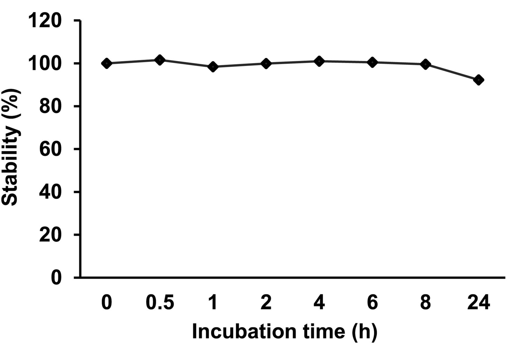
Stability of liposome formulation was determined at various time points in 50% fetal bovine serum.
Effects on Cerebral Perfusion Defects
The therapeutic effectiveness of liposomal angiogenic peptides was determined using 99mTc-HMPAO autoradiography 8 d after treatment. Figure 2 shows representative images of 99mTc-HMPAO autoradiography with and without liposomal delivery of angiogenic peptides. A 40-min period of ischemia reduced cerebral blood perfusion, shown by decreased 99mTc-HMPAO activity in the ipsilateral ischemic cortex of the brain. Delivery of liposomal angiogenic peptides to the ischemic hemisphere of the brain attenuated the cerebral perfusion defect (median, 1.01-fold of the contralateral; IQR, 0.96–1.22; P = 0.004 vs. control; P = 0.01 vs. liposomes), compared with that of control (median, 0.92-fold of the contralateral; IQR, 0.82–0.96) and liposomes (median, 0.91-fold of the contralateral; IQR, 0.90–0.95).

Autoradiographic perfusion images of cerebral sections with (top right) and without (top left) treatment with liposomal angiogenic peptides. Box plots (bottom) show quantification data. Fifteen minutes after occlusion, liposomal angiogenic peptides (200 μL) were administered intraarterially. 99mTc-HMPAO autoradiograms were obtained 8 d after treatment to quantify the radioactivity. Forty minutes of ischemia reduced cerebral perfusion in ipsilateral ischemic cortex. Liposomal delivery of angiogenic peptides attenuated extent of perfusion defect. Control = rats receiving PBS. *Significantly different vs. control.
Effects on Angiogenesis and Glia Activation
We studied whether the liposomal delivery of angiogenic peptides promoted angiogenesis and increased vascular density. Representative images depicting increased vascular density in response to the treatment of liposomal angiogenic peptides are shown in Figures 3A and 3C). In control animals without liposomal therapy, a median of 13 vessels per high-power field (IQR, 12–15) were counted in the ipsilateral penumbra. Treatment of liposomal angiogenic peptides increased the vascular density (median, 16 vessels per high-power field; IQR, 15.5–20; P = 0.039 vs. control). These findings are consistent with the hypothesis that liposomal delivery of angiogenic peptides promotes angiogenesis, which leads to improved cerebral perfusion.
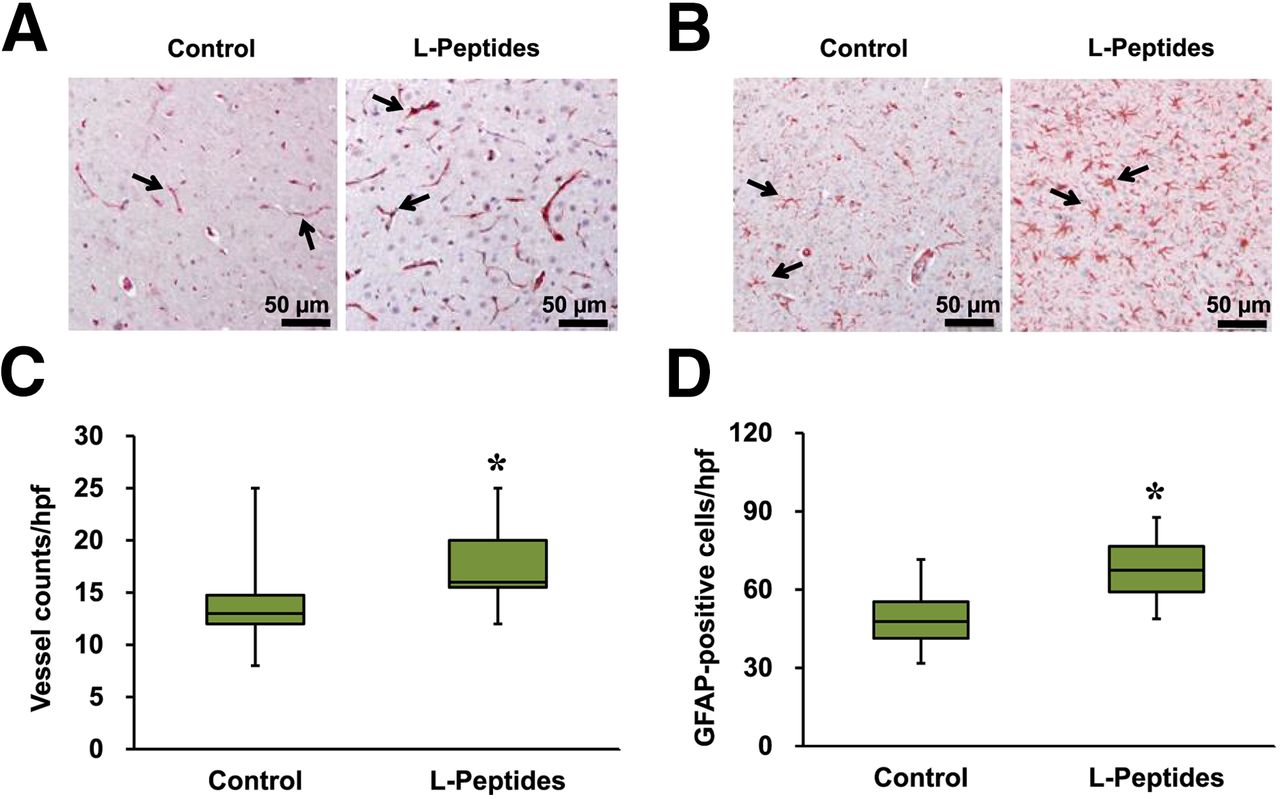
Immunohistologic images stained with antibodies against von Willebrand factor (A and C) and GFAP (B and D) with and without liposomal angiogenic therapy in ischemic periinfarct region (8 d after treatment, ×200). Liposomal delivery of angiogenic peptides increased vascular density and glia activation, compared with nontreated controls. (C and D) Quantification of immunohistologic images. Arrows in A and B indicate von Willebrand factor–positive vessels and GFAP-positive cells. Control = rats receiving PBS. *Significantly different vs. control.
Astrocytes (Figs. 3B and 3D) perform a variety of functions in the brain, such as cerebral protection and neuronal survival after ischemia. We analyzed whether the angiogenic therapy applied in this study affected GFAP activation. Compared with controls (median, 48 cells; IQR, 41–55), a substantial increase in GFAP immunoreactivity was detected in the ischemic hemisphere of the brains treated with liposomal angiogenic peptides (median, 67 cells; IQR, 59–77; P = 0.001 vs. control). These data support the notion that liposomal delivery of angiogenic peptides provides protective effects in the ischemic brain in this model.
Angiogenic and Antiinflammatory Gene Expressions
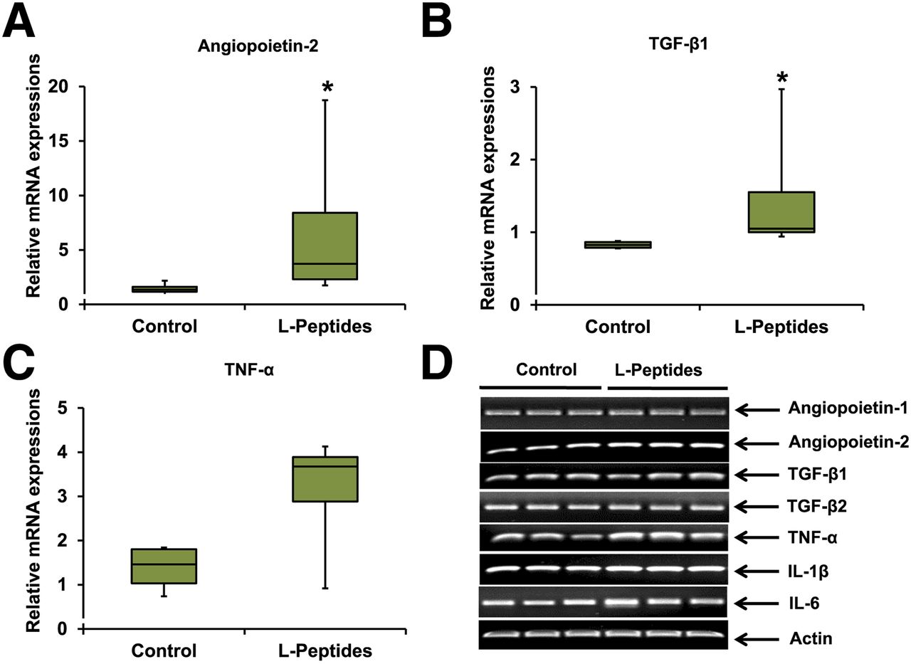
We anticipated that signals for angiogenesis would be activated in the early stage after liposomal angiogenic therapy (Figs. 4A and 4D). Angiopoietin-1 and -2 are important modulators of angiogenesis that affect endothelial cell functions. Thus, changes in angiopoietin gene expression were determined 24 h after treatment. Indeed, an increased expression of angiopoietin-2 (median, 1.1-fold of the normal without ischemia; IQR, 1.0–1.6; P = 0.043 vs. control; median, 0.83-fold of the normal; IQR, 0.79–0.87) was detectable in the ipsilateral ischemic cortex of the brain after treatment with liposomal angiogenic peptides. These results are in contrast to those of angiopoietin-1, which showed no appreciable changes after 24 h of treatment (median, 0.59-fold of the normal without ischemia; IQR, 0.48–0.63; P = 0.02 vs. control; median, 0.66-fold of the normal; IQR, 0.43–0.88). These results are compatible with previous studies of cerebral ischemia exhibiting differential patterns of temporal expression of angiopoietin-2 versus -1 (11,12).

Changes in messenger RNA (mRNA) expression of angiogenesis and inflammation-related genes with and without liposomal angiogenic therapy. Total RNA was extracted from cerebral cortex (24 h after surgery). mRNA expressions were examined by quantitative RT-PCR. Levels of mRNAs are expressed as ratios of negative controls without ischemia. Liposomal angiogenic peptides increased expression of angiopoietin-2 and TGF-1β. *Significantly different vs. control.
Inflammatory responses after cerebral ischemia damage the brain and worsen disease pathology. Thus, expression of inflammatory cytokines was also determined along with angiogenic markers (Figs. 4B–4D). At 24 h after treatment, no changes in proinflammatory genes (TGF-β2, TNF-α, IL-1β, IL-6) were detected in response to the angiogenic therapy. Notably, an elevated expression of TGF-β1, a gene that is involved in antiinflammatory functions and angiogenic responses, was observed (median, 1.05-fold of the normal without ischemia; IQR, 1.00–1.55; P = 0.05 vs. control; median, 0.83-fold of the normal; IQR, 0.79–0.87).
Liposomal Angiogenic Treatment and Changes in 18F-FDG Uptake
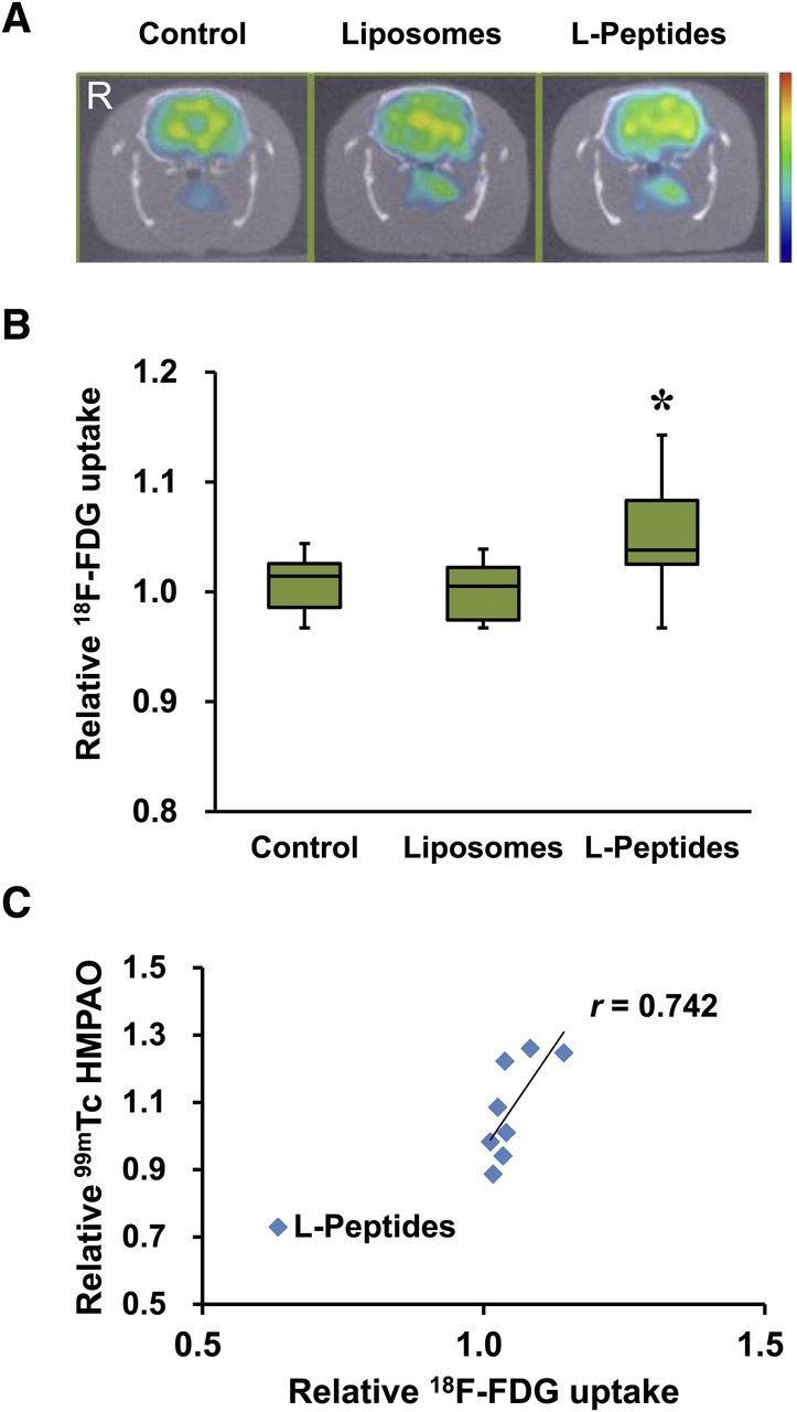
Regional changes in glucose utilization were studied to determine the potential utility of 18F-FDG PET imaging (Fig. 5). A 40-min period of ischemia did not itself alter 18F-FDG uptake in the ipsilateral ischemic cortex of the brain, compared with that in the contralateral cortex, measured at 7 d after surgery. In response to liposomal delivery of angiogenic peptides, cortical 18F-FDG uptake was increased (median, 1.04-fold of the contralateral; IQR, 1.03–1.08; P = 0.018 vs. control; P = 0.021 vs. liposomes), compared with that of control (median, 1.01-fold of the contralateral; IQR, 0.99–1.03) and liposomes (median, 1.01-fold of the contralateral; IQR, 0.97–1.02). These data suggest that liposomal delivery of angiogenic peptides enhances glucose utilization in the area of the ipsilateral ischemic cortex. More importantly, we found that the increase in 18F-FDG uptake by angiogenic therapy correlated with the extent of improvement in cerebral perfusion (r = 0.742, P = 0.035). Conversely, in control animals, no correlation between 18F-FDG uptake and change in cerebral perfusion was observed (r = 0.670, P = 0.100).

(A) 18F-FDG PET radioactivity images for coronal slices of brain with (right) and without (left) liposomal angiogenic therapy obtained 7 d after treatment. 18F-FDG uptake data in ipsilateral cortex show that relative radioactivity was higher in groups treated with liposomal angiogenic peptides than controls (B). Correlation study shows that increase in 18F-FDG uptake after angiogenic therapy was correlated with extent of improvement in cerebral perfusion. Control = rats receiving PBS; l-peptides = liposomal angiogenic peptides. *Significantly different vs. control.
DISCUSSION
The results of the present study showed that liposomal angiogenic peptides mimicking VEGF reduced cerebral perfusion defects caused by ischemia and increased vascular density, compared with controls, suggesting that liposomal delivery of angiogenic peptides benefits cerebral ischemia. Liposomal angiogenic peptides also enhanced neuroprotection, as evidenced by an increase in TGF-β1 expression and glia activation, compared with that in controls. Our study using 18F-FDG PET imaging also demonstrated changes in regional glucose utilization after liposomal therapy. Specifically, liposomal delivery of angiogenic peptides facilitated glucose utilization in the ipsilateral ischemic cortex of the brain, compared with that in controls. Furthermore, the extent of improvement correlated with increased cerebral perfusion. These findings suggest 18F-FDG PET imaging as a tool to study the effectiveness of angiogenic therapy to treat cerebral ischemia.
The brain receives oxygen and nutrients for survival via arteries that are exclusive to the brain and is thus highly vulnerable to injuries from ischemia caused by insufficient blood supply. For this reason, strategies to promote angiogenesis and improve cerebral blood supply hold promise for treating cerebral ischemia. In the present study, angiogenesis was facilitated by liposomal delivery of peptides exhibiting known biologic activity of VEGF, an angiogenic and neuroprotective factor (13).
Although liposomes are a potentially powerful drug delivery system, there are currently no liposomal angiogenic formulations that can effectively treat cerebral ischemia. The results of the present study showing that liposomal delivery of peptides with known angiogenic properties to the ischemic hemisphere of the brain ameliorated cerebral perfusion defects, increased vascular density, and induced antiinflammation and glia activation provide supportive evidence that the liposomal formulation used in this study is therapeutically effective. Future studies using multiple factors capable of angiogenesis, neuroprotection, and neurogenesis are warranted to further facilitate the therapeutic effect.
An effective drug treatment to enhance outcomes of cerebral ischemia remains a significant challenge. Specifically, the presence of the blood–brain barrier often limits therapeutic effectiveness by forming a physical barricade and restricting drug delivery across cells. Cerebral ischemia causes a broad spectrum of pathophysiologic changes, including blood–brain barrier disruptions and increased vascular permeability: the enhanced permeability and retention effect. Previous studies have shown that an increase in vascular permeability by ischemia may enable selective targeting of drugs at the critical site of action, allowing effective drug delivery to the ischemic hemisphere of the brain (14,15). Our results of γ imaging by 99mTc-HMPAO (supplemental materials) clearly demonstrated liposomal accumulation in the ischemic hemisphere of the brain. These data indicate that selective targeting of liposomal angiogenic peptides to the ipsilateral ischemic hemisphere of the brain is feasible. These data further support the hypothesis that the therapeutic effects demonstrated in this study resulted from the peptides that were delivered by liposomal vesicles.
In addition to the therapeutic effectiveness, a substantial increase in TGF-β1 gene expression, an antiinflammatory agent, as well as an angiogenic cytokine, was also observed in the absence of changes in proinflammatory responses. Proinflammatory responses to cerebral ischemia lead to secondary injury to the brain and worsen disease pathology. Experimental studies have shown benefits of antiinflammatory therapy by decreasing ischemic brain injury (16,17). On the basis of the existing evidence, it is probable that the increased antiinflammatory response seen in this study might have played a role in neuroprotection and attenuated the cerebral perfusion defect. The elevated level of glia activation shown in this study further supports the mechanism of neuroprotection.
A dominant source of energy in the brain is glucose. Glucose metabolism has a tightly controlled relationship with cerebral blood flow. Insufficient blood supply to the brain during ischemia affects glucose metabolism, the extent of which varies depending on the degree to which cerebral blood flow is diminished (18,19). The present study using a mild degree of ischemia showed an increase in 18F-FDG uptake in the ipsilateral ischemic cortex of the brain 7 d after treatment. Similar results were previously demonstrated in patients with stroke and in animals subjected to a brief period of ischemia (9,20,21). Our results extend the previous observation by establishing a liposomal angiogenic formulation that is potentially beneficial in treating cerebral ischemia.
PET scanning with 18F-FDG, which offers the advantage of noninvasively and repeatedly visualizing functional information of tissues, is commonly used to identify changes in glucose metabolism in clinical settings, especially in cancer and myocardial ischemia. Brain disorders are often associated with changes in regional cerebral blood flow and glucose metabolism (20,22,23). Despite this, 18F-FDG PET imaging has limited use, particularly in a clinical setting of cerebral ischemia. The present study attempted to explore the potential of 18F-FDG PET imaging in therapeutic angiogenesis for ischemic stroke treatment. The finding that angiogenic therapy increased 18F-FDG uptake in the ipsilateral cortex of the brain, the degree of which correlated with attenuated cerebral perfusion defects, indicates that glucose hypermetabolism during the subacute phase after angiogenic therapy may serve as an indicator of improved cerebral perfusion.
The present study has limitations. Changes in cerebral perfusion and vascular density were studied to present therapeutic effectiveness without assessment of the functional outcomes. The effect of peptides without liposomal encapsulation was not studied. Thus, it is not clear whether the therapeutic benefits shown in our study simply resulted from the provision of angiogenic peptides or the liposomal formulation. Although the results of the present study indicated that our liposomal angiogenic therapy improved cerebral perfusion and increased vessel density, it remains to be determined whether such changes will affect clinical outcomes.
CONCLUSION
We provide evidence that liposomal delivery of angiogenic peptides exhibiting biologic properties of VEGF benefits cerebral ischemia. Our results also show that cerebral 18F-FDG PET imaging can potentially have profound clinical implications by serving as a signature of improved cerebral perfusion.
DISCLOSURE
The costs of publication of this article were defrayed in part by the payment of page charges. Therefore, and solely to indicate this fact, this article is hereby marked “advertisement” in accordance with 18 USC section 1734. This research was funded by Radiation Technology R&D program 2012M2A2A7035779 and Basic Science Research Program 2014R1A1A2008959 through the National Research Foundation of Korea grant funded by the Ministry of Science ICT and Future Planning. No other potential conflict of interest relevant to this article was reported.
Footnotes
Published online May 14, 2015.
- © 2015 by the Society of Nuclear Medicine and Molecular Imaging, Inc.
REFERENCES
- Received for publication January 16, 2015.
- Accepted for publication April 9, 2015.


Jump to section
Related Articles
Cited By...
- No citing articles found.