


Article Figures & Data
Figures
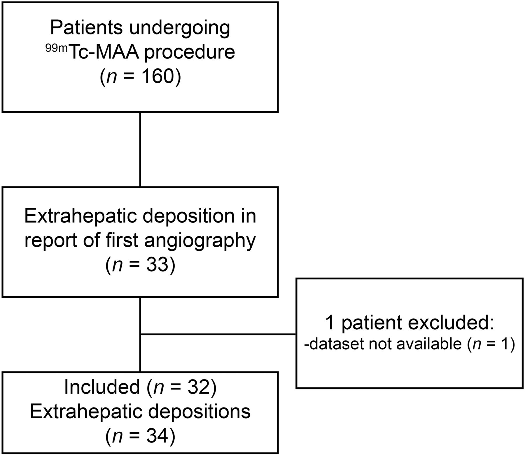
- FIGURE 1.
Volume estimation in phantom study of 6 spheres of different volumes (0.5–26.5 mL). Estimate of volume on SPECT is number of all voxels from threshold up to voxel with maximum value, multiplied by voxel volume; it decreases as threshold increases. True, known, volume of each sphere is indicated by black dot. Higher threshold leads to underestimation whereas lower threshold leads to overestimation. For all spheres, a threshold of 40% leads to underestimation of volume.
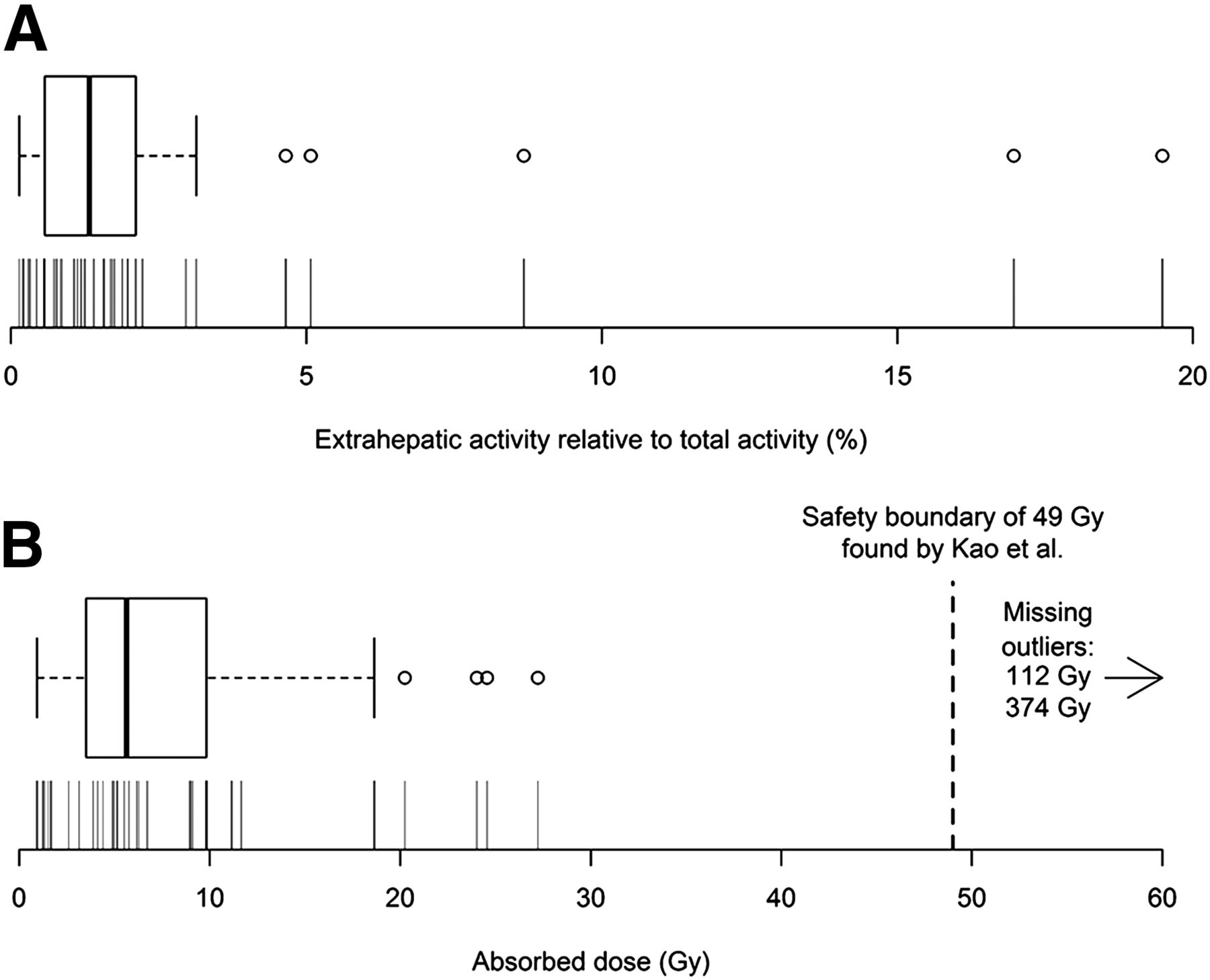
- FIGURE 2.
Absorbed dose estimation in phantom study using 6 spheres of different volumes (0.5–26.5 mL). Absorbed dose is displayed as ratio of true absorbed dose in sphere. Threshold needed to estimate true absorbed dose (dotted line) differs per sphere. Absorbed dose is overestimated in all spheres using threshold of 40% (▲).
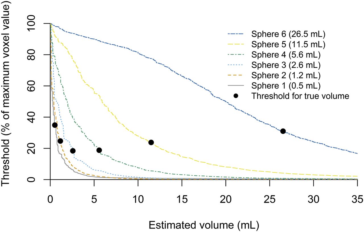
- FIGURE 3.
Flowchart of study design.
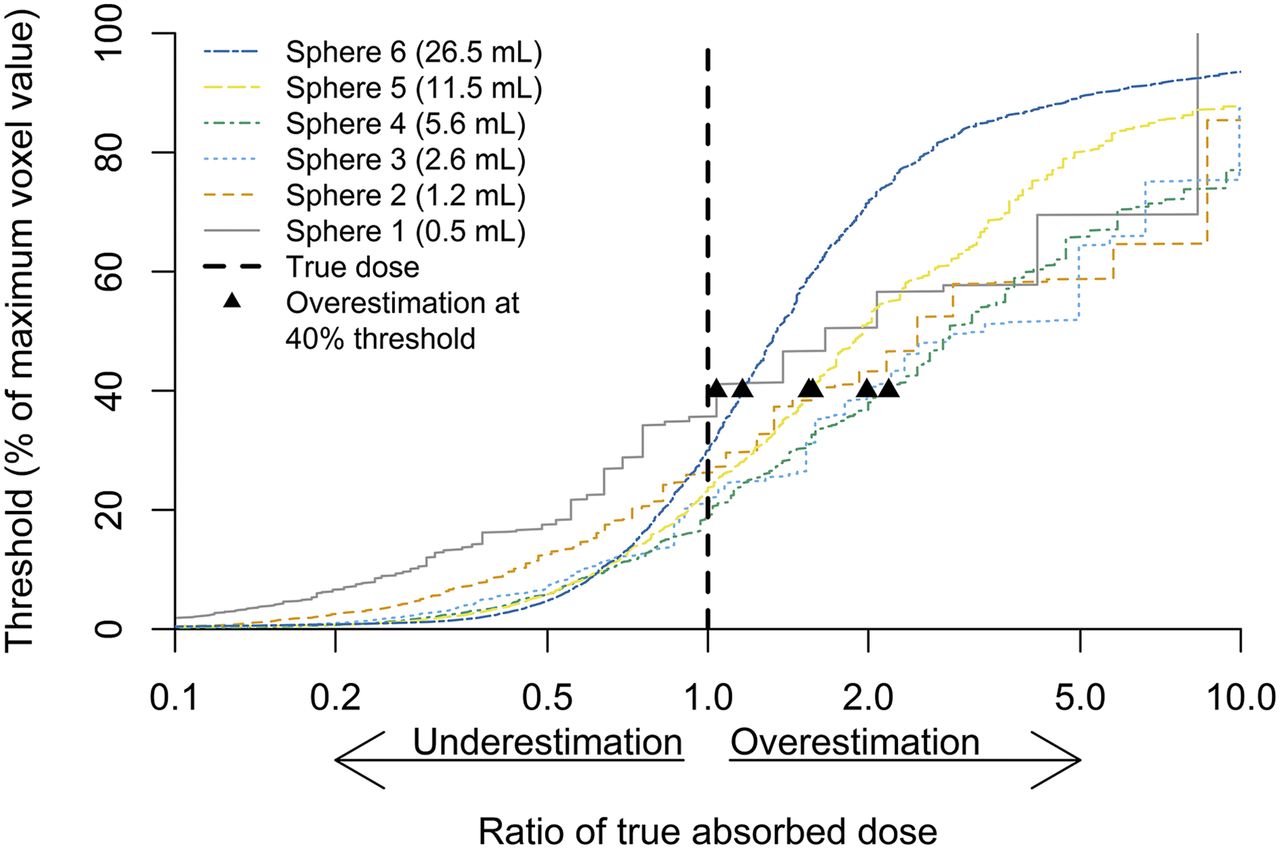
- FIGURE 4.
(A) Box plot and density plot of extrahepatic activity relative to total activity (liver and extrahepatic deposition). Distribution is right-skewed. Each vertical indicates observation. (B) Box plot and density plot of absorbed dose to extrahepatic tissue from 250-MBq 166Ho scout dose, plotted on log-scale (in Gy). Boundary from which complications occurred in study by Kao et al. of 49 Gy is displayed (17).
- FIGURE 5.
Extrahepatic deposition of 8.7% of administered activity near duodenum after injection in proper hepatic artery (A and B), which was corrected after selective lobar injection in right (C) and left (D) hepatic artery (E). Had 250-MBq 166Ho scout dose been injected, theoretic extrahepatic absorbed dose would have been 112 Gy.
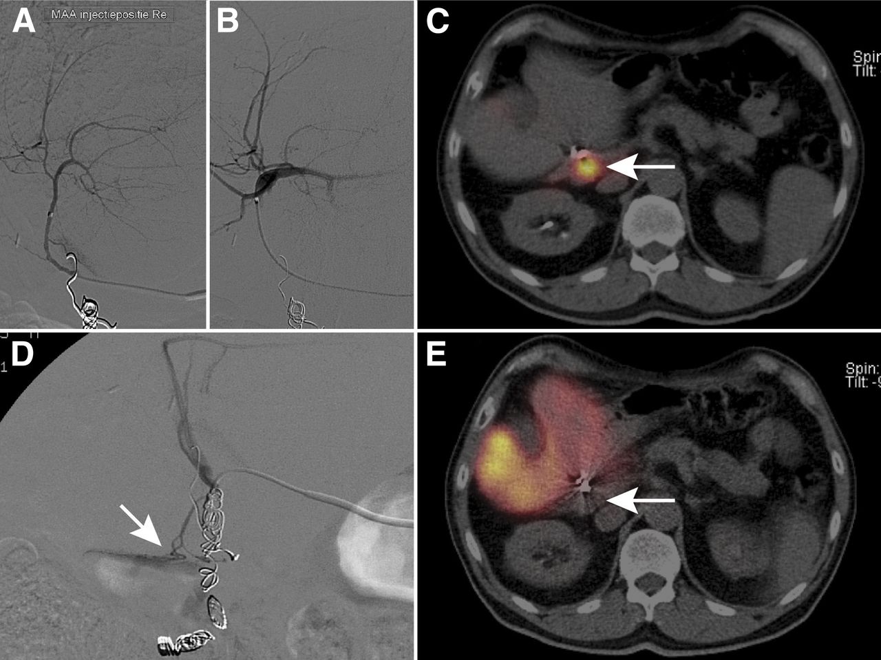
- FIGURE 6.
Extrahepatic deposition in a patient, who previously received right hemihepatectomy, after injection in right (A) and left (B) hepatic artery. Deposition (C, white arrow) represents 19.5% of total activity (little activity is seen in liver). During repeated angiography, culprit vessel originated from gastroduodenal artery (D, white arrow). After coil embolization and single injection in proper hepatic artery, no deposition was seen (E, white arrow). Had 250-MBq 166Ho scout dose been injected, theoretic absorbed dose would have been 374 Gy.


Tables
Characteristic n (%) or median (range) Patients/# cases 32/34 Sex Male 25 (78%) Female 7 (22%) Age (y) 67 (36–80) Primary tumor Colorectal carcinoma 19 (59%) Cholangiocellular carcinoma 3 (9%) Hepatocellular carcinoma 3 (9%) Ocular melanoma 2 (6%) Adenocarcinoma of unknown primary 1 (3%) Breast carcinoma 1 (3%) Gastric carcinoma 1 (3%) Neuroendocrine tumor 1 (3%) Ampullary carcinoma 1 (3%) Coil embolization Gastroduodenal and right gastric 17 (53%) Gastroduodenal 6 (19%) Gastroduodenal and cystic 1 (3%) Gastroduodenal and pancreatic 1 (3%) Gastroduodenal and duodenal 1 (3%) None 6 (19%) No. of injection positions 1 17 (53%) Common 4 Proper 11 Right 1 Replaced left 1 2 14 (44%) Common + replaced right 1 Common + replaced left 1 Right + left 9 Right + replaced left 1 Replaced right + left 1 Replaced right + left (from superior mesenteric artery) 1 3 1 (3%) Replaced right and selective (2×) left 1
Supplemental Data
Files in this Data Supplement:


Jump to section
Related Articles
Cited By...
- PET/CT and SPECT/CT imaging of 90Y hepatic radioembolization at therapeutic and diagnostic activity levels: anthropomorphic phantom study
- Lung Dose Measured on Postradioembolization 90Y PET/CT and Incidence of Radiation Pneumonitis
- First Evidence for a Dose-Response Relationship in Patients Treated with 166Ho Radioembolization: A Prospective Study
- Therapeutic Radiometals Beyond 177Lu and 90Y: Production and Application of Promising {alpha}-Particle, {beta}--Particle, and Auger Electron Emitters
- Insights into the Dose-Response Relationship of Radioembolization with Resin 90Y-Microspheres: A Prospective Cohort Study in Patients with Colorectal Cancer Liver Metastases