


Article Figures & Data
Figures
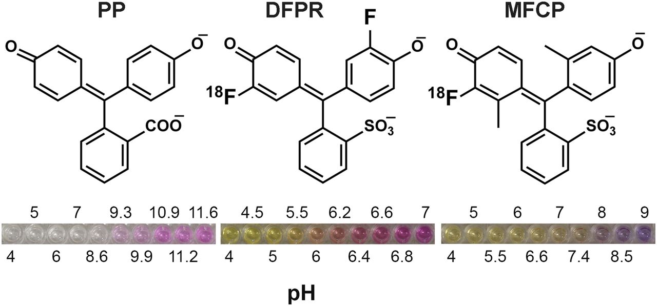
- FIGURE 1.
Chemical structures of pH indicators. (Left) PP. (Middle) DFPR. (Right) MFCP. pH-dependent changes in indicator color are shown underneath.
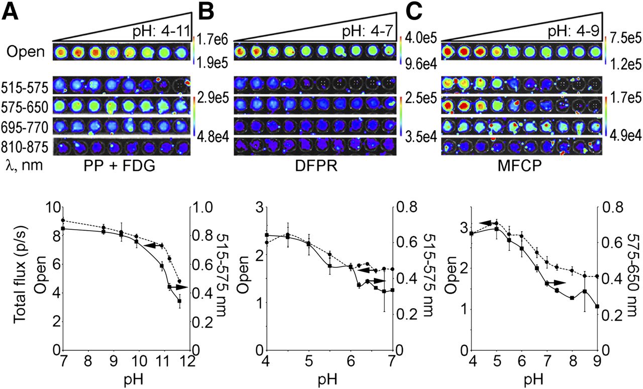
- FIGURE 2.
Inter- and intramolecular quenching of CR. (A) PP (25 mM) was adjusted to pH 4–11, mixed with 3.7 MBq (100 μCi) of 18F-FDG, and imaged using all filters on IVIS Lumina II. (Top) Images of plate are shown. (Bottom) Quantifications of total flux normalized to A-PET counts for open filter (dashed line) and optimal-quenching 515- to 575-nm filter (solid line) are shown. (B) 18F-DFPR (3.7 MBq [100 μCi]; 25 nmol) adjusted to pH 4–7, with open and 515- to 575-nm filter quantification shown. (C) 18F-MFCP (5.18 MBq [140 μCi]; 70 nmol) adjusted to pH 4–9, with open and 575- to 650-nm filters. For each pH indicator, filtered images were set to same scale. For all experiments, n = 3.
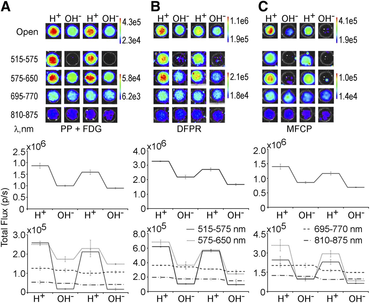
- FIGURE 3.
Optical switching of PP + 18F-FDG, 18F-DFPR, and 18F-MFCP. (A) Ten microliters of 1 M HCl (H+) or NaOH (OH−) were added to wells containing 25 mM PP + 7.4 MBq (200 μCi) of 18F-FDG. 18F-DFPR (7.4 MBq [200 μCi]; 50 nmol) (λmax = 570 nm) (B) and 18F-MFCP (3.7 MBq [100 μCi]; 50 nmol) (λmax = 582 nm) (C) and images acquired after acid or base addition. Images for open and selective filters are shown on top; quantification of each well is shown on bottom.
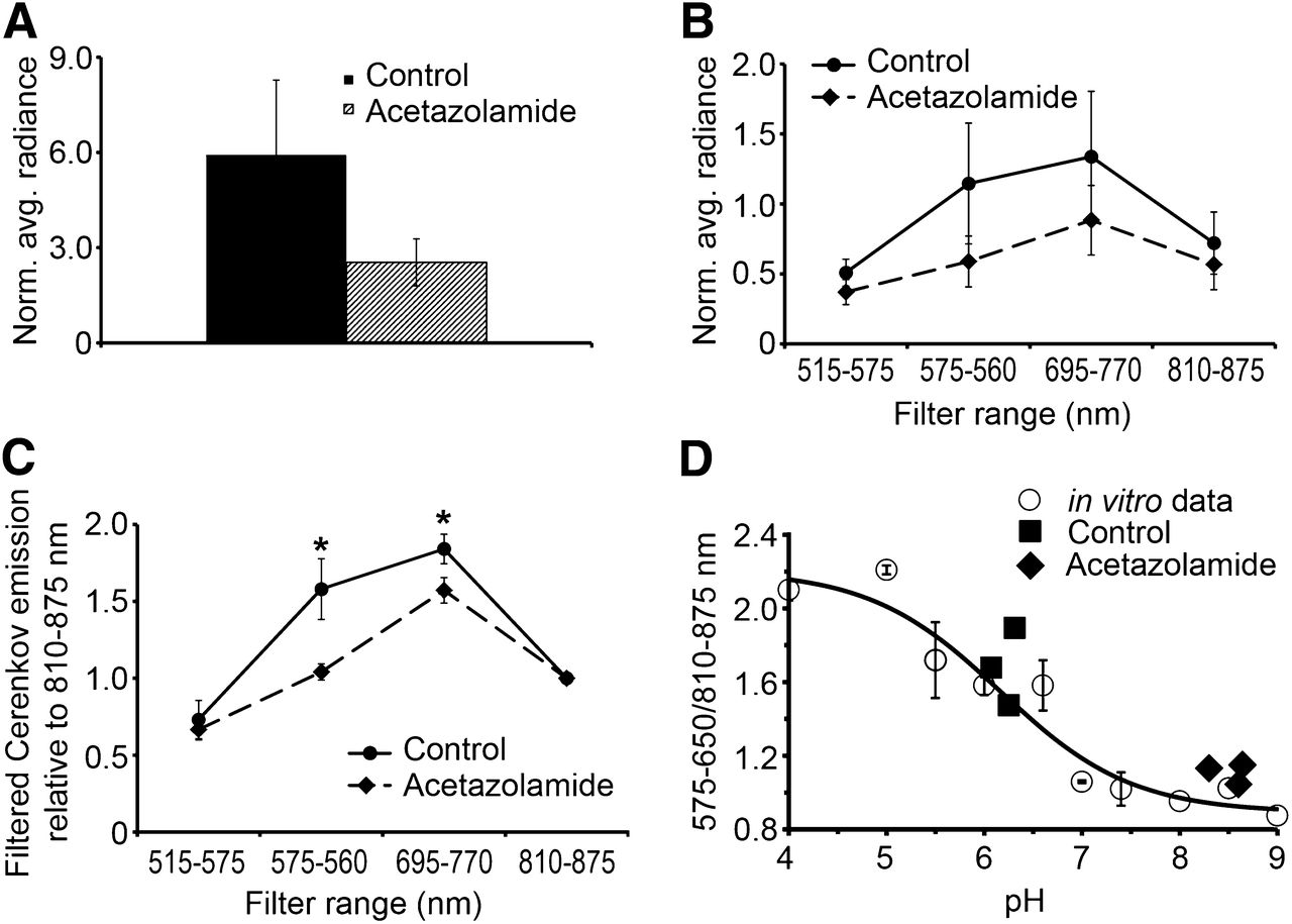
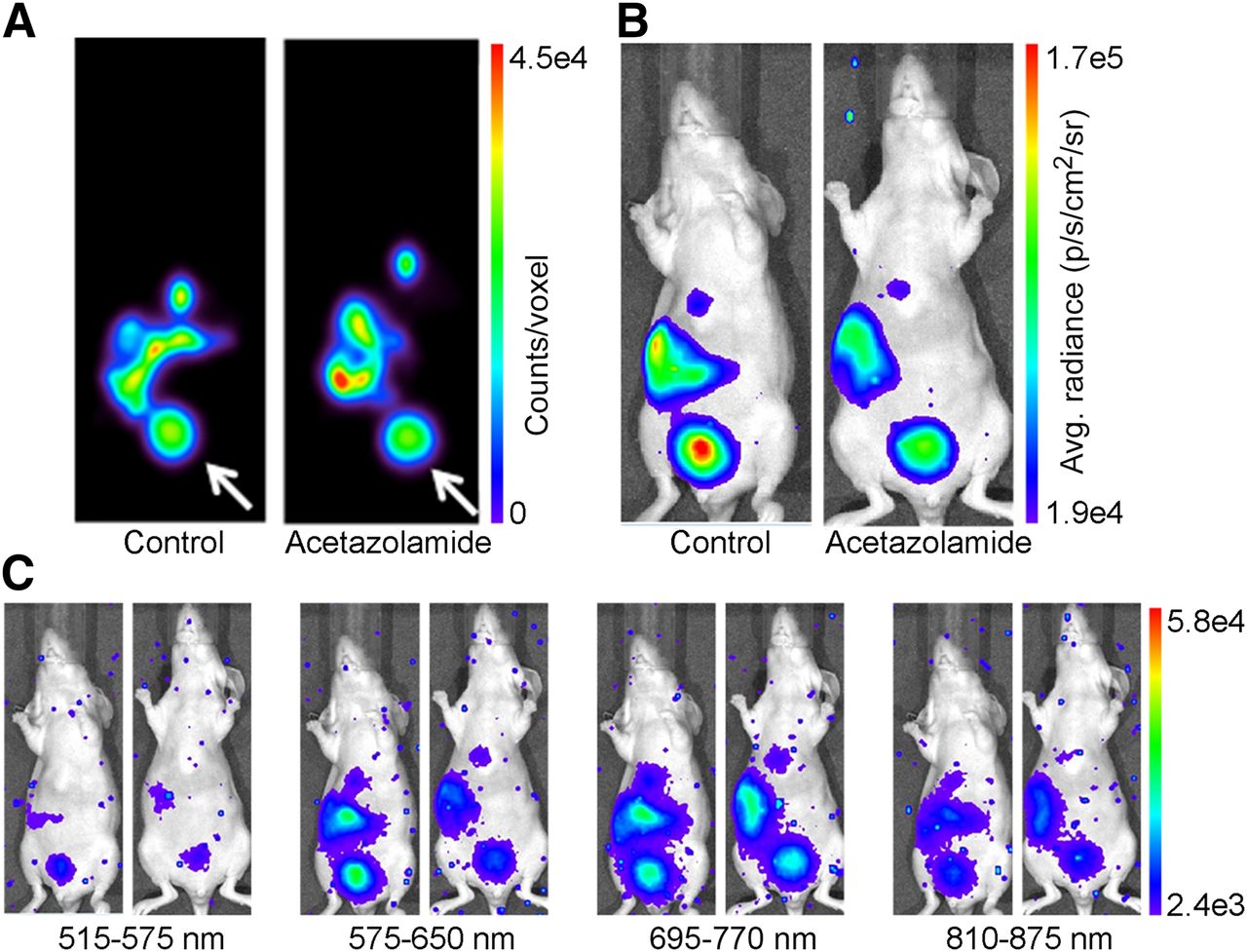
- FIGURE 4.
In vivo imaging of Cerenkov quenching. Mice were treated with either saline or acetazolamide (160 mg/kg) for 5 d before imaging with 18F-MFCP. (A) A-PET images of single coronal slice (0.5-mm thick) through center of mouse in control and acetazolamide-treated mice show 18F-MFCP accumulation in bladder (white arrow), gallbladder, liver, and intestine. Optical images of same mice were acquired using open filter (B) and 4 emission filters (C). Avg. = average.
- FIGURE 5.
Quantification of in vivo Cerenkov images. Average radiance from bladder using open filter (A), 4 selective filters (B), and ratio of emission at each filter relative to 810–875 nm (C). *Significant, P < 0.05, Student t test. (D) Ratiometric pH determination. ○ = ratio of Cerenkov emission at (575–650 nm)/(810–875 nm) for in vitro 18F-MFCP (data from Fig. 2C); solid line = fit to sigmoidal curve and gives pKa of 6.25, similar to 6.4 determined by open filter data; ◆ = Cerenkov emission from control mice plotted against actual urine pH; ■ = Cerenkov emission from acetazolamide-treated mice plotted against actual urine pH. avg. = average; Norm. = normal.


Tables
Parent compound Monofluorinated Difluorinated Indicator name pKa λmax pKa λmax pKa λmax Phenol red 7.9 558 7.3 564 6.4 570 Cresol red 8.2 572 7.6 576 6.8 582 Cresol purple 8.3 578 7.5 582 7.0 590 Phenolphthalein 9.7 553 9.3 558 8.7 564 Naptholphthalein 7.8 653 7.7 659 7.2 665 pKa = negative logarithm of acid dissociation constant (unitless); λmax = absorbance maximum of basic form in nm.
Supplemental Data
Files in this Data Supplement:


