


Article Figures & Data
Figures
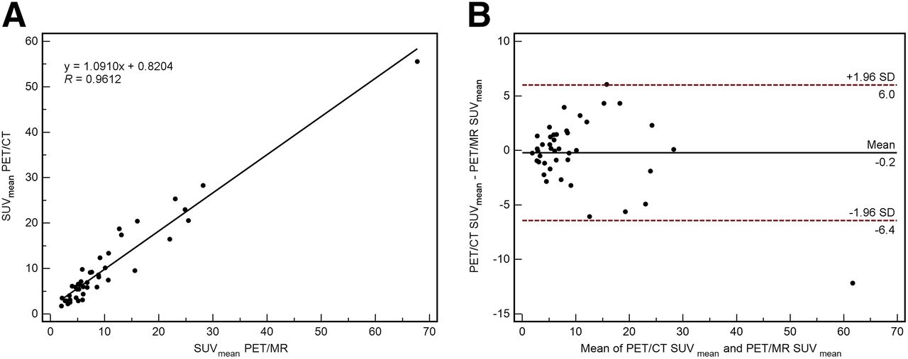
- FIGURE 1.
Analysis of tracer uptake between PET/CT and subsequent PET/MR as assessed by SUVmean in focal lesions reveals high correlation (R = 0.96) between the 2 modalities (A). Difference between the 2 SUV measurements is shown by Bland–Altman plot (B), in which difference between the 2 SUV measurements is plotted against their average. For SUVmean, mean difference is −0.2 SUV (95% confidence interval, +6.0 and −6.4 SUV), indicating nearly perfect quantitative agreement between SUVs from the 2 modalities.
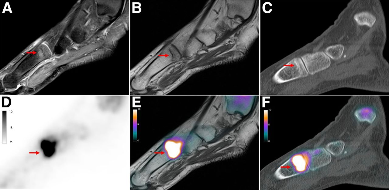
- FIGURE 2.
Simultaneously acquired 18F-fluoride PET/CT and PET/MR images of 49-y-old woman with pain over left metatarsal foot for several months without history of trauma. Sagittal MR images show BME in fat-saturated proton-density–weighted images at base of os metatarsale I (arrow, A) with presence of hypointense fracture line on T1-weighted image (arrow, B). In corresponding PET scan of PET/MR (D and E) and PET/CT (F), intense focal 18F-fluoride uptake at base of os metatarsale I is shown (arrow). However, in PET-positive region, corresponding CT scan shows only slight sclerotic band (arrow, C).
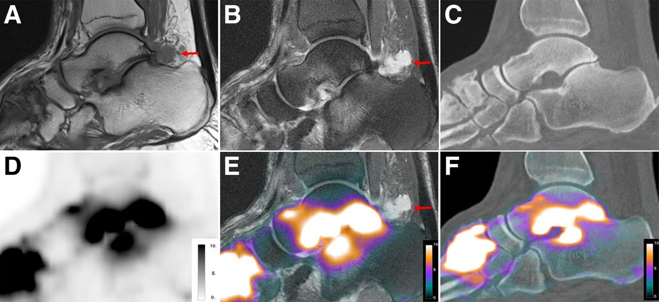
- FIGURE 3.
Sagittal 18F-fluoride PET/CT and PET/MR images of 55-y-old man with persistent pain and swelling of left foot for several years. There are advanced signs of osteoarthritis, particularly in tarsometatarsal joints and in subtalar joint, which can be observed on sagittal T1-weighted turbo spin echo MR sequence (A), sagittal proton-density fat-saturated MR sequence (B), and sagittal CT (C), including joint space narrowing, osteophytes, subchondral cysts, and subchondral BME (only on MR imaging). Additionally, only MR imaging could detect T1-weighted hypointense, proton-density fat-saturated hyperintense ganglion cyst originating from posterior subtalar joint (arrows). Corresponding PET scan of PET/MR (D and E) and PET/CT (F) shows intense focal 18F-fluoride uptake on both sides of tarsometatarsal and subtalar joint; however, in this case no additional information was provided by PET. Slight difference in slice positioning leads to different impression of 18F-fluoride uptake.


Tables
SUVmean SUVmax Parameter Mean ± SD Range R 95% CI Mean ± SD Range R 95% CI Pathologic lesions on PET/MR 10.4 ± 11.3 2.0–67.7 0.96* 0.93–0.98 15.6 ± 16.9 2.9–94.1 0.96* 0.93–0.98 Pathologic lesions on PET/CT 10.2 ± 9.9 1.8–55.6 16.3 ± 19.2 2.5–117.5 Regions of normal bone on PET/MR 0.67 ± 0.36 0.12–1.76 0.75* 0.59–0.85 1.00 ± 0.62 0.20–2.84 0.84* 0.73–0.91 Regions of normal bone on PET/CT 0.89 ± 0.53 0.12–2.55 1.17 ± 0.71 0.21–2.76 ↵* P < 0.0001.
CI = confidence interval.
Correlation coefficient and 95% confidence interval are for correlation between PET/MR and PET/CT.
Supplemental Data
Files in this Data Supplement:


