


Article Figures & Data
Figures
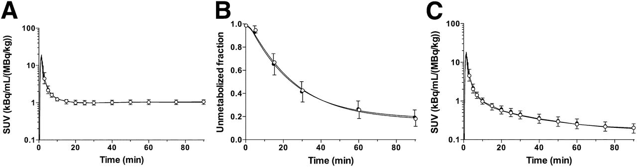
- FIGURE 1.
Mean ± SD of total plasma activity (A), parent fraction in plasma (B), and metabolite-corrected plasma activity (C) over time after injection of 11C-LY2795050 in test (closed circles, n = 16) and retest (open circles, n = 16) scans. A and C are displayed in SUV units (concentration/[injected dose/body weight]).
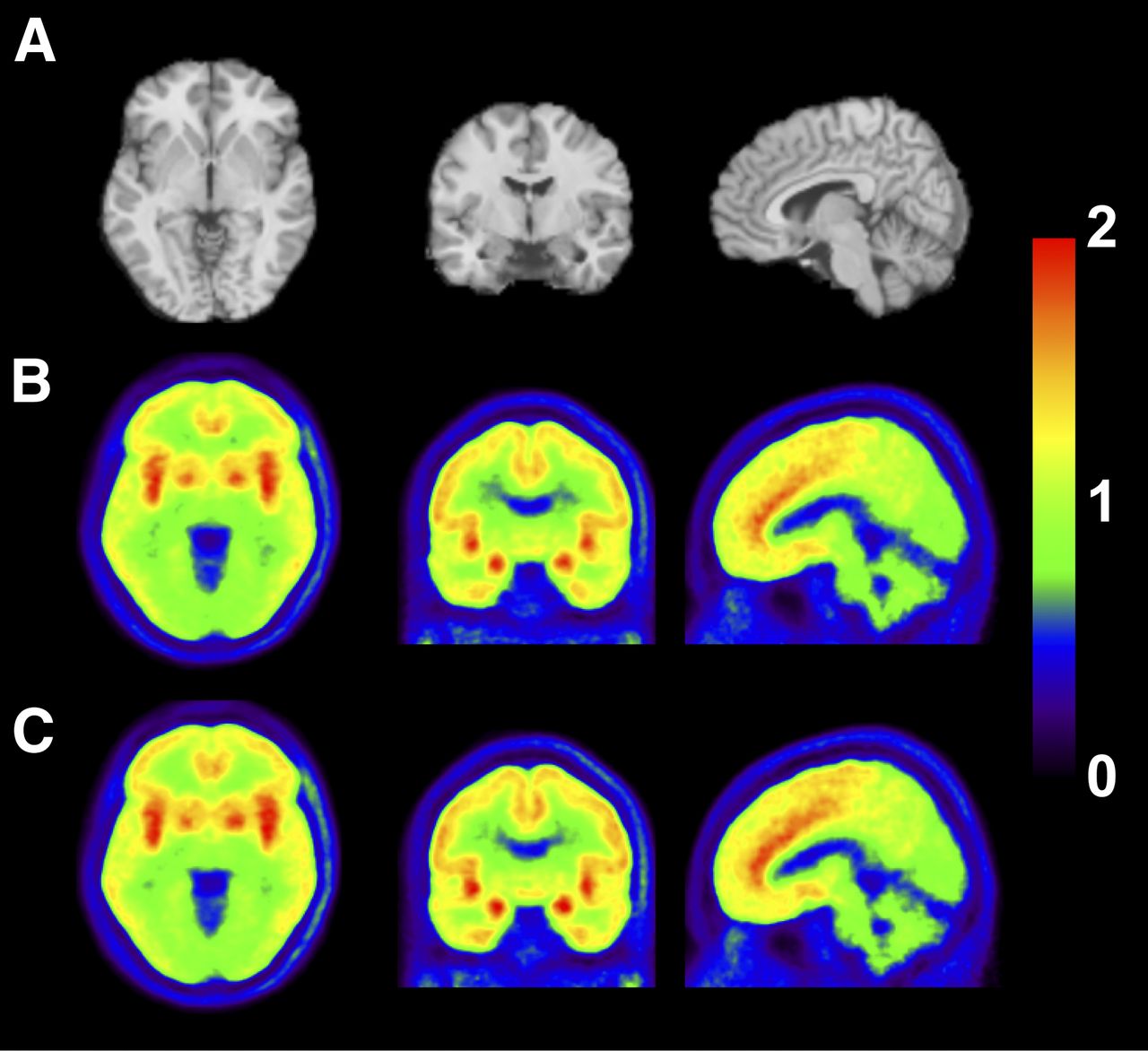
- FIGURE 2.
MR images and coregistered PET images summed from 30 to 90 min after injection of 11C-LY2795050: MR images (A), test scan (B), and retest scan (C). PET images are displayed in SUV units. SUV images were averaged across all subjects.
- FIGURE 3.
Time–activity curves from 1 subject for 6 selected regions after injection of 11C-LY2795050 under test (A) and retest (B) conditions. Fits with MA1 (t* = 30 min) and 2TC models are displayed as solid and dotted lines, respectively.


Tables
Parameter Test (n = 16) Retest (n = 16) Age (y) 28 ± 7 28 ± 7 Body weight (kg) 78.6 ± 11.4 78.6 ± 11.4 Injected dose (MBq) 390 ± 135 461 ± 178 Specific activity (MBq/nmol) 18.0 ± 6.4 21.8 ± 9.6 Injected mass (μg) 9.1 ± 1.2 9.1 ± 1.3 Data are mean ± SD.
VT (mL/cm3) VT/fP (mL/cm3) BPND BPP (mL/cm3) BPF (mL/cm3) Region Test Retest Test Retest Test Retest Test Retest Test Retest Amygdala 4.17 (13) 3.98 (13) 439 (18) 448 (26) 1.55 (19) 1.45 (21) 2.52 (15) 2.33 (17) 265 (21) 264 (30) Insula 3.40 (11) 3.38 (10) 357 (15) 379 (22) 1.07 (17) 1.08 (14) 1.74 (11) 1.74 (8) 183 (15) 195 (20) Ant. cingulate cortex 3.28 (12) 3.26 (11) 345 (16) 365 (23) 1.00 (14) 1.00 (15) 1.63 (12) 1.61 (11) 171 (17) 181 (22) Globus pallidus 3.13 (11) 3.11 (13) 329 (16) 349 (26) 0.92 (23) 0.91 (21) 1.47 (16) 1.46 (20) 156 (21) 164 (30) Putamen 2.90 (11) 2.91 (10) 305 (16) 326 (22) 0.77 (20) 0.79 (21) 1.25 (14) 1.27 (15) 132 (19) 142 (24) Temporal cortex 2.65 (11) 2.64 (10) 278 (15) 295 (21) 0.62 (24) 0.62 (22) 0.99 (17) 0.99 (14) 104 (20) 111 (20) Frontal cortex 2.64 (11) 2.63 (11) 278 (16) 294 (22) 0.61 (22) 0.61 (20) 0.98 (16) 0.98 (14) 104 (21) 110 (22) Occipital cortex 2.47 (11) 2.45 (10) 259 (15) 274 (21) 0.51 (30) 0.51 (27) 0.81 (22) 0.81 (19) 85 (24) 90 (23) Hippocampus 2.35 (11) 2.30 (11) 247 (15) 259 (25) 0.44 (30) 0.42 (34) 0.70 (25) 0.66 (30) 73 (26) 75 (38) Caudate 2.28 (11) 2.28 (12) 240 (16) 256 (26) 0.40 (45) 0.40 (47) 0.62 (40) 0.63 (42) 66 (43) 72 (51) Post. cingulate cortex 2.19 (12) 2.17 (13) 230 (17) 244 (24) 0.33 (43) 0.34 (47) 0.53 (36) 0.53 (43) 56 (38) 59 (46) Thalamus 2.19 (9) 2.17 (9) 230 (15) 244 (23) 0.34 (43) 0.34 (47) 0.53 (37) 0.53 (43) 57 (39) 60 (45) Centrum semiovale 2.19 (11) 2.14 (16) 230 (15) 241 (27) 0.34 (46) 0.31 (48) 0.54 (38) 0.50 (43) 56 (40) 56 (47) Cerebellum 1.94 (16) 1.92 (16) 203 (19) 216 (26) 0.17 (0) 0.17 (0) 0.28 (16) 0.28 (16) 30 (19) 31 (26) Ant. = anterior; Post. = posterior.
Data are mean of 16 subjects, with each value measured twice in test and retest scans. Data in parentheses are percentage coefficient of variation (intersubject variability).
2TC, VT (mL/cm3) MA1, VT (mL/cm3) MA1, VT/fP (mL/cm3) Region aTRV TRV ICC aTRV TRV ICC aTRV TRV ICC Amygdala 9% 3% ± 11% 0.39 (-0.18;0.77) 12% 5% ± 14% 0.41 (-0.08;0.74) 18% −1% ± 22% 0.55 (0.11;0.82) Insula 7% −1% ± 9% 0.58 (0.13;0.84) 7% 0% ± 9% 0.68 (0.31;0.87) 14% −5% ± 18% 0.46 (-0.02;0.77) Ant. cingulate cortex 8% 0% ± 10% 0.61 (0.19;0.84) 6% 0% ± 8% 0.78 (0.50;0.92) 14% −5% ± 16% 0.60 (0.17;0.84) Globus pallidus 8% 0% ± 10% 0.70 (0.32;0.89) 8% 1% ± 9% 0.74 (0.41;0.90) 15% −4% ± 18% 0.55 (0.11;0.82) Putamen 7% −2% ± 8% 0.73 (0.37;0.90) 6% 0% ± 7% 0.81 (0.55;0.93) 13% −6% ± 16% 0.57 (0.14;0.83) Temporal cortex 9% 0% ± 11% 0.50 (0.04;0.79) 7% 0% ± 8% 0.70 (0.33;0.88) 13% −5% ± 17% 0.50 (0.04;0.79) Frontal cortex 8% 0% ± 10% 0.61 (0.20;0.84) 7% 0% ± 8% 0.74 (0.41;0.90) 13% −5% ± 17% 0.54 (0.09;0.81) Occipital cortex 9% 0% ± 11% 0.52 (0.06;0.80) 6% 1% ± 7% 0.77 (0.46;0.91) 13% −5% ± 16% 0.52 (0.07;0.80) Hippocampus 9% 2% ± 10% 0.63 (0.22;0.85) 8% 2% ± 9% 0.67 (0.28;0.87) 15% −4% ± 18% 0.54 (0.10;0.81) Caudate 7% −2% ± 8% 0.77 (0.42;0.92) 6% 0% ± 7% 0.82 (0.56;0.93) 13% −5% ± 16% 0.60 (0.18;0.84) Post. cingulate cortex 9% 1% ± 11% 0.72 (0.37;0.89) 7% 1% ± 9% 0.76 (0.45;0.91) 14% −5% ± 17% 0.61 (0.19;0.84) Thalamus 8% 0% ± 9% 0.50 (0.04;0.79) 7% 1% ± 9% 0.54 (0.09;0.81) 15% −5% ± 18% 0.51 (0.05;0.79) Centrum semiovale 10% 4% ± 13% 0.57 (0.12;0.83) 10% 3% ± 13% 0.57 (0.14;0.83) 18% −3% ± 20% 0.52 (0.06;0.80) Cerebellum 7% 0% ± 9% 0.85 (0.63;0.94) 6% 1% ± 8% 0.89 (0.71;0.96) 13% −5% ± 17% 0.64 (0.23;0.85) Ant. = anterior; Post. = posterior.
2TC VT estimates with relative SE > 10% or k4 < 0.001 min−1 were excluded from calculation. Data within parentheses are 95% confidence intervals.
BPND BPP (mL/cm3) BPF (mL/cm3) Region aTRV TRV ICC aTRV TRV ICC aTRV TRV ICC Amygdala 13% 7% ± 17% 0.56 (0.12;0.82) 16% 8% ± 20% 0.24 (−0.26;0.65) 22% 2% ± 27% 0.53 (0.07;0.80) Insula 7% −1% ± 8% 0.88 (0.71;0.96) 10% 0% ± 11% 0.36 (−0.14;0.71) 15% −6% ± 19% 0.33 (−0.17;0.70) Ant. cingulate cortex 5% 0% ± 7% 0.91 (0.76;0.97) 8% 1% ± 9% 0.67 (0.29;0.87) 14% −5% ± 16% 0.61 (0.20;0.84) Globus pallidus 9% 1% ± 11% 0.89 (0.71;0.96) 10% 1% ± 13% 0.76 (0.45;0.91) 18% −4% ± 20% 0.62 (0.20;0.85) Putamen 7% −2% ± 8% 0.93 (0.82;0.98) 7% −2% ± 8% 0.85 (0.62;0.94) 14% −7% ± 15% 0.66 (0.27;0.87) Temporal cortex 6% −1% ± 8% 0.95 (0.87;0.98) 9% −1% ± 10% 0.77 (0.47;0.91) 15% −6% ± 18% 0.59 (0.16;0.83) Frontal cortex 5% −1% ± 7% 0.94 (0.83;0.98) 8% 0% ± 10% 0.76 (0.45;0.91) 15% −6% ± 18% 0.63 (0.22;0.85) Occipital cortex 9% −1% ± 13% 0.95 (0.87;0.98) 10% 0% ± 12% 0.86 (0.66;0.95) 14% −6% ± 18% 0.71 (0.36;0.89) Hippocampus 15% 7% ± 17% 0.89 (0.72;0.96) 17% 7% ± 18% 0.79 (0.51;0.92) 22% 2% ± 25% 0.70 (0.33;0.88) Caudate 10% −2% ± 16% 0.98 (0.95;0.99) 10% −1% ± 14% 0.96 (0.90;0.99) 15% −7% ± 17% 0.88 (0.69;0.95) Post. cingulate cortex 22% 9% ± 44% 0.92 (0.80;0.97) 24% 10% ± 44% 0.86 (0.66;0.95) 27% 5% ± 45% 0.82 (0.57;0.93) Thalamus 31% 22% ± 81% 0.96 (0.90;0.99) 36% 24% ± 86% 0.91 (0.77;0.97) 45% 22% ± 98% 0.83 (0.59;0.94) Centrum semiovale 33% 16% ± 59% 0.82 (0.58;0.93) 35% 15% ± 60% 0.70 (0.34;0.88) 42% 11% ± 64% 0.67 (0.29;0.87) Ant. = anterior; Post. = posterior.
Data within parentheses are 95% confidence intervals.


Jump to section
Related Articles
Cited By...
- First-in-Human Assessment of 11C-LSN3172176, an M1 Muscarinic Acetylcholine Receptor PET Radiotracer
- Kinetic Modeling and Test-Retest Reproducibility of 11C-EKAP and 11C-FEKAP, Novel Agonist Radiotracers for PET Imaging of the {kappa}-Opioid Receptor in Humans
- Impact of Pharmacological Manipulation of the {kappa}-Opioid Receptor System on Self-grooming and Anhedonic-like Behaviors in Male Mice
- Novel 18F-Labeled {kappa}-Opioid Receptor Antagonist as PET Radiotracer: Synthesis and In Vivo Evaluation of 18F-LY2459989 in Nonhuman Primates
- Receptor Occupancy of the {kappa}-Opioid Antagonist LY2456302 Measured with Positron Emission Tomography and the Novel Radiotracer 11C-LY2795050