


Article Figures & Data
Figures
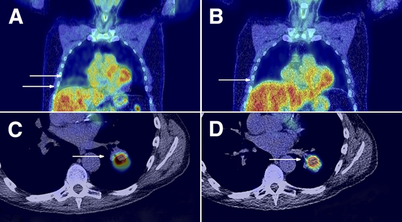
- FIGURE 1.
Two patients with non–small cell lung cancer, 1 with squamous cell carcinoma (A and B) and 1 with adenocarcinoma (C and D). First column (A and C) depicts coronal (A) and axial (C) plane of standard CT fused with the respiration-gated PET image, reconstructed with 35% duty cycle. Second column (B and D) depicts same coronal (B) and axial (D) plane of triggered CT fused with corresponding gated PET images, reconstructed with 35% duty cycle. Both patients were scanned on Biograph 40 mCT scanner. Both patients show improvement in match between PET and CT when PET and triggered CT is compared with PET and standard CT. PET and triggered CT (B and D) show perfect match as indicated by single arrows, whereas there is mismatch for PET and standard CT group (A and C). For coronal PET and standard CT scan (A), 2 arrows were used to indicate lung–liver boundary on PET and CT.
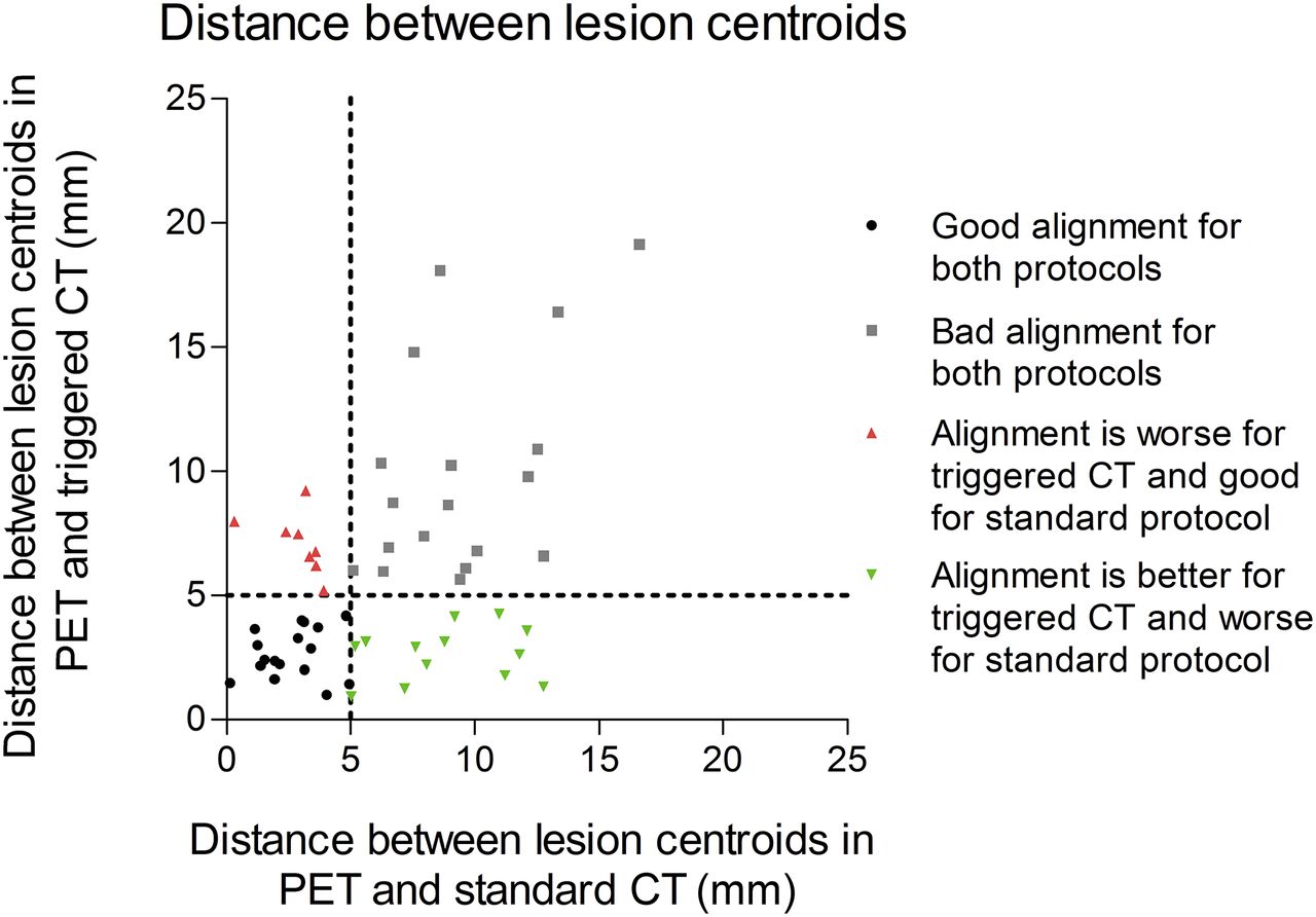
- FIGURE 2.
Scatterplot showing results of distance between centroids for lesions between triggered CT and standard CT. Lesions with good alignment for both protocols—lesions with a distance between centroids less than 5 mm—are indicated in black. Lesions that showed bad alignment with standard CT protocol (>5 mm) and improved with triggered CT protocol (<5 mm) are shown in green. Red indicates lesions where triggered CT became worse (standard CT < 5 mm, triggered CT > 5 mm).
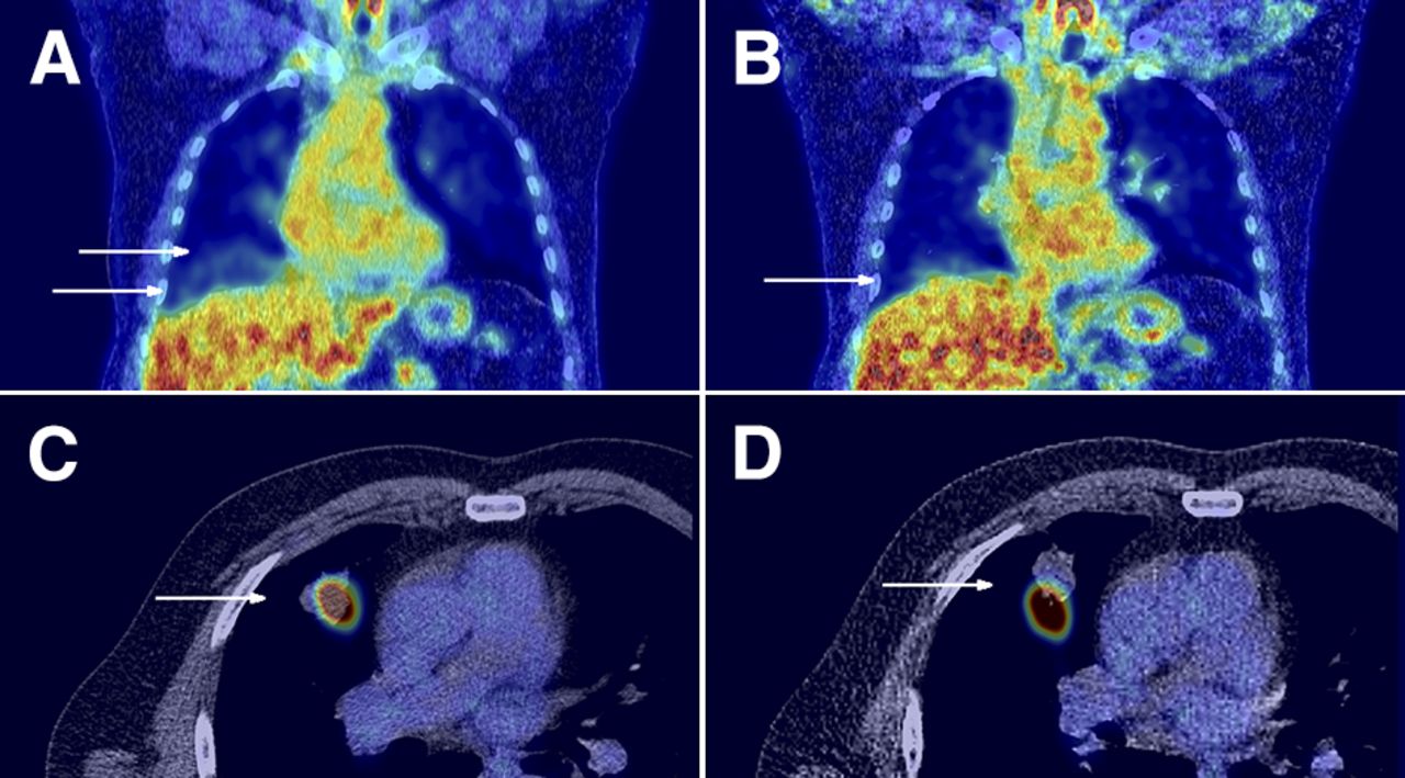
- FIGURE 3.
Patient with non–small cell lung cancer squamous cell carcinoma in right upper lobe. On left are coronal (A) and axial (C) planes of standard CT and corresponding respiration-gated PET image, on right coronal (B) and axial (D) planes of triggered CT and corresponding PET. Improved match for lung–liver boundary can be seen when triggered CT is used (A and B), but spatial match for PET and CT of lung lesion is worse (C and D). For first PET and standard CT scan (A), 2 arrows were used to indicate lung–liver boundary on PET and CT. Patient was scanned on Biograph 40 mCT scanner.
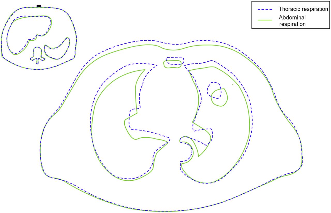
- FIGURE 4.
Illustration of mismatch in 2 scans caused by difference in breathing. One scan was made during thoracic respiration (dashed line) and 1 during abdominal respiration (solid line). Pressure sensor is located at same position, indicating that the 2 scans were acquired at similar time during respiratory cycle (left corner). Excursion of abdominal wall at location of pressure sensor differs from that at position at thoracic wall, suggesting less effective tracking of respiratory motion. Even though scans were made during same respiratory phase, it did not result in better match for lung lesion.

Tables
Characteristic Patients scanned with step-and-shoot method (Biograph 40 mCT scanner) (value ± SD) Patients scanned with continuous-bed-motion method (Biograph 64 mCT Flow scanner) (value ± SD) Sex Males 19 5 Females 11 3 Age (y) 68.7 ± 8.6 66.0 ± 6.2 Weight (kg) 81.5 ± 17.3 68.6 ± 18.7 Location of lesion Upper lobes 19 4 Middle and lower lobes 13 9 Hilum 8 3 Duration of triggered CT (min) 3.1 ± 1.6 1.7 ± 0.7 Length of triggered CT (cm) 46.8 ± 7.0 32.0 ± 3.9 Lesions showing invasive growth into or attachment to large structures (arteries, veins, and main bronchi) of lung hilum were assigned to hilum group.
Parameter Standard CT and PET Triggered CT and PET P Mismatch of lung–liver boundary (mm) 9.2 ± 8.1 4.5 ± 6.7 <0.001* Average distance between lesion centroids (mm) 6.3 ± 4.0 5.6 ± 4.2 0.424 Jaccard similarity coefficient 0.30 ± 0.21 0.32 ± 0.20 0.609 SUVmax (g/cm3) 10.5 ± 6.7 10.9 ± 6.7 <0.001* SUVmean (g/cm3) 6.1 ± 4.0 6.4 ± 4.0 0.001* ↵* Significant difference between 2 groups.
Numbers are averages and SD for all patients (i.e., scanned on Biograph 40 mCT and on Biograph 64 mCT Flow scanner).


