


Article Figures & Data
Figures
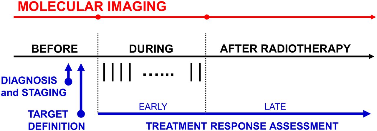
- FIGURE 1.
Use of molecular imaging in radiation oncology follows all steps of treatment process but with some specific challenges, particularly for defining treatment targets (e.g., requiring accurate spatial localization) and in treatment response assessment (e.g., requiring special attention to radiation-induced inflammation). Vertical bars during radiotherapy indicate individual treatment fractions.
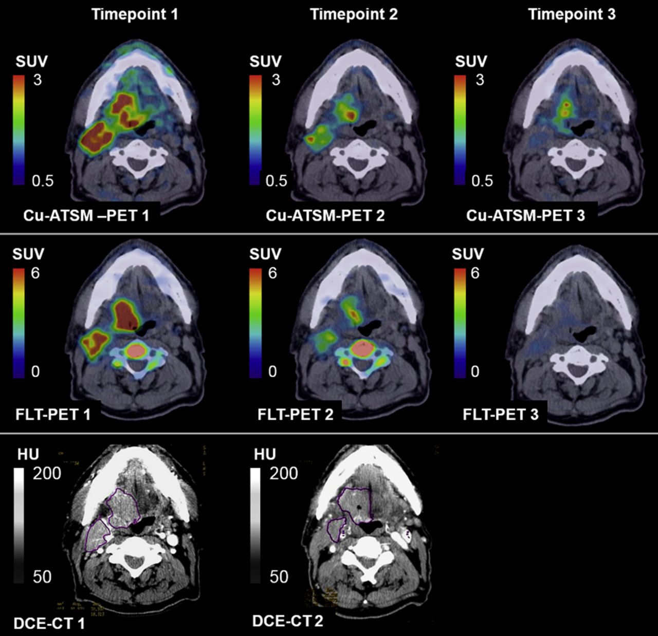
- FIGURE 2.
Multimodality multitracer molecular images of 61Cu-diacetyl-bis(N4-methylthiosemicarbazone) (Cu-ATSM) PET, 18F-fluorothymidine (FLT) PET, and DCE CT in patient before bevacizumab monotherapy (time point 1), after 3 wk of bevacizumab (time point 2), and after 1–2 wk of chemoradiation therapy (time point 3), indicating potential of molecular imaging to assess complex radiation treatment regimens. (Reprinted with permission of (116).)

Tables
- TABLE 1
Uncertainties and Quality Control Measures for PET/CT in Radiation Therapy Planning
Category Procedure Uncertainties Quality control Scanning protocol Patient preparation Metabolism levels (18F-FDG) Limit physical activity Blood glucose levels (18F-FDG) Measure fasting blood glucose with exclusion criteria Bowel size/positioning Use fasting protocol Radiotracer injection Residual activity in syringe Measure/correct for residual activity Decay correction errors Synchronize scanner clock Acquisition Patient positioning Spatial offset between PET and treatment-planning CT Ensure consistent patient positioning using identical positioning devices Quantitative uncertainties from attenuating objects Avoid placing objects outside image field of view Scanning Patient motion Implement motion management strategies Attenuation correction uncertainties from iodine contrast material Acquire separate low-dose CT scan or apply corrections Equipment failure or electronic drift Calibrate detector and equipment frequently Increased SUV because of longer uptake period Apply strict protocol for uptake period Reconstruction Reconstruction Selection of optimal image reconstruction method/parameters Benchmark algorithms using phantoms (task-specific) Randoms, scatter, attenuation, detector sensitivity, and partial-volume effect Apply appropriate calibrations and corrections Analysis Segmentation Differentiation of normal tissue and tumor uptake Know radiotracer’s normal biodistribution Segmentation uncertainties Develop segmentation protocol; benchmark algorithms with phantoms Limited spatial resolution and sensitivity Include margins Quantification Quantitative accuracy Calibrate PET scanner to dose calibrator Selection of relevant quantitative measures Compare semiquantitative metrics with kinetic analysis–derived parameters; consult literature Quantitative differences between scanners/institutions Quantitatively harmonize scanners Treatment planning Target definition Registration errors Benchmark algorithms using physical or digital phantoms; crop images Motion Use same motion management method as was used during imaging Similar uncertainties exist for all other molecular imaging modalities, which require specific quality control measures.
- TABLE 2
Methods to Incorporate Molecular Images into Radiation Therapy Planning and Their Respective Degrees of Complexity
Complexity Registration Segmentation Target definition Motion management Basic Manual alignment Manual Boundary definition None (phase-averaged) Rigid/affine Threshold-based Subvolume boost Gated imaging and delivery Advanced Deformable Automatic algorithms Voxelwise dose painting 4D imaging and delivery - TABLE 3
Selected References on Early Response Assessment and Treatment Adaption, Late Response Assessment, and Normal-Tissue Response Evaluation
Cancer type Early response assessment and treatment Late response assessment Normal-tissue response evaluation Lung 18F-FDG PET (82–85,135) 18F-FDG PET (81) 18F-FDG PET (136,139–141) Non–18F-FDG PET (107,110) SPECT (75,136–138) MRI (136,138) Head and neck 18F-FDG PET (68,90) 18F-FDG PET (87–89) 18F-FDG PET (142) Non–18F-FDG PET (108,109) Non–18F-FDG PET (138) SPECT (118) SPECT (138) MRI (124) MRI (138) Rectal 18F-FDG PET (91,92) 18F-FDG PET (93) MRI (126) Esophageal 18F-FDG PET (95) 18F-FDG PET (94) 18F-FDG PET (143) Cervical 18F-FDG PET (98) 18F-FDG PET (96) 18F-FDG PET (144) MRI (119,123) Non–18F-FDG PET (145) Brain MRI (122) Non–18F-FDG PET (112) 18F-FDG PET (136,146,147) Non–18F-FDG PET (146,148) MRI (136,147) Liver MRI (125) Non–18F-FDG PET (138) SPECT (138) MRI (138) - TABLE 4
NCCN Recommendations on Use of 18F-FDG PET/CT for Target Definition and Treatment Response Evaluation in Radiotherapy
Cancer type Target definition Response evaluation Cervical Recommended Optional Esophageal Recommended Recommended Head and neck — Optional Hodgkin lymphoma Optional Recommended Non-Hodgkin lymphoma Optional Recommended Non–small cell lung Recommended — Small cell lung Recommended — Pancreatic Recommended — Recommendations are as of 2015. No significant evidence to support use of 18F-FDG PET/CT at this point is indicated with a dash, which does not mean that use of 18F-FDG PET/CT may not be beneficial but merely that evidence at this point is still insufficient.


