
Main menu

  • Home
  • Content
    • Current
    • Ahead of print
    • Past Issues
    • JNM Supplement
    • SNMMI Annual Meeting Abstracts
    • Continuing Education
    • JNM Podcasts
  • Subscriptions
    • Subscribers
    • Institutional and Non-member
    • Rates
    • Journal Claims
    • Corporate & Special Sales
  • Authors
    • Submit to JNM
    • Information for Authors
    • Assignment of Copyright
    • AQARA requirements
  • Info
    • Reviewers
    • Permissions
    • Advertisers
  • About
    • About Us
    • Editorial Board
    • Contact Information
  • More
    • Alerts
    • Feedback
    • Help
    • SNMMI Journals
  • SNMMI
    • JNM
    • JNMT
    • SNMMI Journals
    • SNMMI

User menu

  • Subscribe
  • My alerts
  • Log in
  • My Cart

Search

  • Advanced search
Journal of Nuclear Medicine
  • SNMMI
    • JNM
    • JNMT
    • SNMMI Journals
    • SNMMI
  • Subscribe
  • My alerts
  • Log in
  • My Cart
Journal of Nuclear Medicine

Advanced Search

  • Home
  • Content
    • Current
    • Ahead of print
    • Past Issues
    • JNM Supplement
    • SNMMI Annual Meeting Abstracts
    • Continuing Education
    • JNM Podcasts
  • Subscriptions
    • Subscribers
    • Institutional and Non-member
    • Rates
    • Journal Claims
    • Corporate & Special Sales
  • Authors
    • Submit to JNM
    • Information for Authors
    • Assignment of Copyright
    • AQARA requirements
  • Info
    • Reviewers
    • Permissions
    • Advertisers
  • About
    • About Us
    • Editorial Board
    • Contact Information
  • More
    • Alerts
    • Feedback
    • Help
    • SNMMI Journals
  • View or Listen to JNM Podcast
  • Visit JNM on Facebook
  • Join JNM on LinkedIn
  • Follow JNM on Twitter
  • Subscribe to our RSS feeds
Research ArticlePET/MR Imaging: Potential Clinical and Research Applications

Principles of PET/MR Imaging

Jonathan A. Disselhorst, Ilja Bezrukov, Armin Kolb, Christoph Parl and Bernd J. Pichler
Journal of Nuclear Medicine June 2014, 55 (Supplement 2) 2S-10S; DOI: https://doi.org/10.2967/jnumed.113.129098
Jonathan A. Disselhorst
1Werner Siemens Imaging Center, Department of Preclinical Imaging and Radiopharmacy, Eberhard Karls University of Tübingen, Tübingen, Germany; and
  • Find this author on Google Scholar
  • Find this author on PubMed
  • Search for this author on this site
Ilja Bezrukov
1Werner Siemens Imaging Center, Department of Preclinical Imaging and Radiopharmacy, Eberhard Karls University of Tübingen, Tübingen, Germany; and
2Max-Planck-Institute for Intelligent Systems, Tübingen, Germany
  • Find this author on Google Scholar
  • Find this author on PubMed
  • Search for this author on this site
Armin Kolb
1Werner Siemens Imaging Center, Department of Preclinical Imaging and Radiopharmacy, Eberhard Karls University of Tübingen, Tübingen, Germany; and
  • Find this author on Google Scholar
  • Find this author on PubMed
  • Search for this author on this site
Christoph Parl
1Werner Siemens Imaging Center, Department of Preclinical Imaging and Radiopharmacy, Eberhard Karls University of Tübingen, Tübingen, Germany; and
  • Find this author on Google Scholar
  • Find this author on PubMed
  • Search for this author on this site
Bernd J. Pichler
1Werner Siemens Imaging Center, Department of Preclinical Imaging and Radiopharmacy, Eberhard Karls University of Tübingen, Tübingen, Germany; and
  • Find this author on Google Scholar
  • Find this author on PubMed
  • Search for this author on this site
  • Article
  • Figures & Data
  • Supplemental
  • Info & Metrics
  • PDF
Loading

Article Figures & Data

Figures

  • Tables
  • Additional Files
  • FIGURE 1.
    • Download figure
    • Open in new tab
    • Download powerpoint
    FIGURE 1.

    Effect of increasing magnetic field strength on crystal maps of PMT-based PET detector at 1 mT (A), 3 mT (B), and 5 mT (C). (Reprinted with permission of (79).)

  • FIGURE 2.
    • Download figure
    • Open in new tab
    • Download powerpoint
    FIGURE 2.

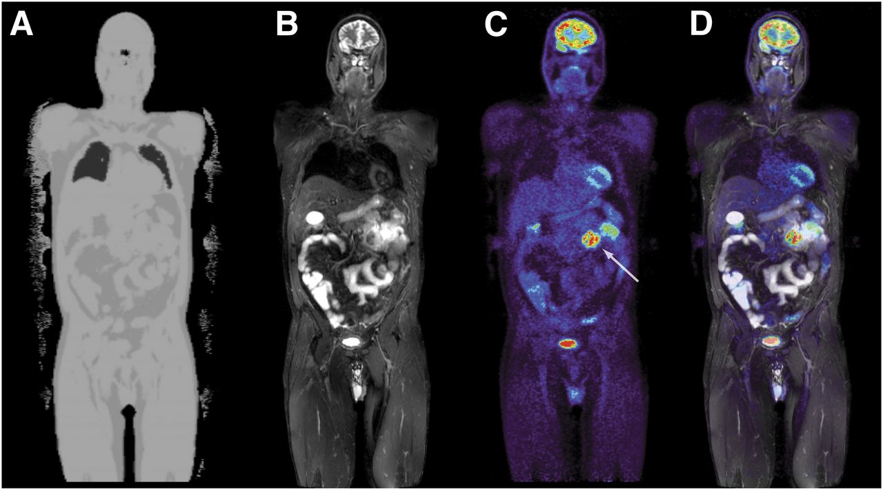
    PET/MR image of patient with enteric melanoma metastasis (arrow): MR-based attenuation map (A), T2-weighted short-τ inversion recovery sequence (B), 18F-FDG PET image (C), and PET overlaid on short-τ inversion recovery image (D). Data were acquired on a Biograph mMR system.

  • FIGURE 3.
    • Download figure
    • Open in new tab
    • Download powerpoint
    FIGURE 3.

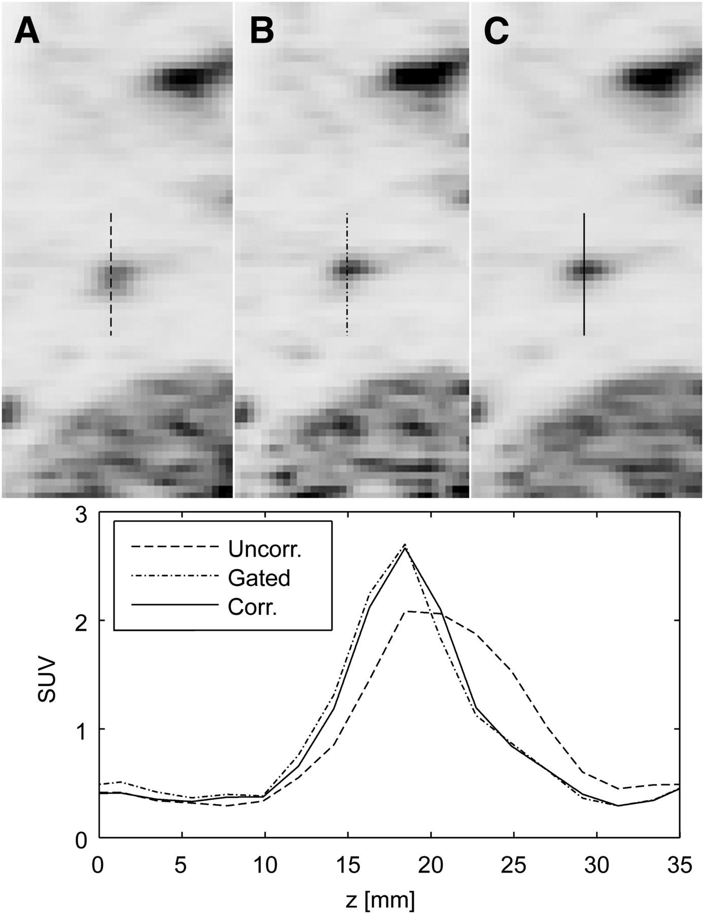
    PET images and profiles through lung lesion: uncorrected (A), gated (B), and motion-corrected (C). (Reprinted with permission of (64).)

Tables

  • Figures
  • Additional Files
    • View popup
    TABLE 1

    Abbreviated* Overview of PET/MR Systems Described in Literature

    Company or universityYear (ref)Usage†MR field strength (T)Scintillator, detector‡Crystal ringsBlocks/ringCrystal block (size [mm3])Axial, transaxial FOV (cm)
    University of California,1997P-R-Sim0.2LSO14816§1.0
    Los Angeles, CA (US)(6)MC PMT + fibers(2×2×10)<3.8
    University of California,1997P-R-Sim0.2 and 9.4LSO17224§0.2
    Los Angeles, CA (US)(7)MC PMT + fibers(2×2×5)<5.4
    Kings College,2005P-R-Sim3LSO181×4×40.3
    London (UK)(80)PS PMT + fibers(2×3×5)5.6
    University of Cambridge,2006P-R-Sim1LSO482412×127.2
    Cambridge (UK)(28)(split magnet)PS PMT+ fibers(1.52×1.52×10)<14.7
    West Virginia University,2007P-R-Sim3LSO20220×205.0
    Morgantown, WV (US)(81)PS PMT + fibers(2.5×2.5×15)8.0
    Kobe City College of2009P-R-Sim0.15MLS3322×2×20.5
    Technology, Kobe (JP)(25)PS PMT + fibers(2.5×3.5×3.5)NA
    Western Ontario,2009P-R-Seq0.3BGO828×8∼5.0
    London, ON (CA)(29)(field-cycled)PS PMT + fibers(6.2×5.6×30)NA
    Kobe City College of2010P-R-Sim0.3LGSO111611×9×22.1
    Technology, Kobe (JP)(13)PS PMT + fibers(1.9×2.2×6/7)8.0
    Nagoya University,2012P-R-Sim0.3LGSO131611×13×22.1
    Nagoya (JP)(82)PS PMT + fibers(0.9×1.3×5/6)5.6
    Mediso Ltd.,2013P-Co-Seq1LYSO811239×819.4
    Budapest (HU)(12)PS PMT(1.12×1.12×13)4.5-12.0
    University of California,2006P-R-Sim7LSO8168×81.2
    Davis, CA (US)(15)PS APD + fibers(1.43×1.43×6)3.5
    University of Tübingen,2007P-R-Sim7LSO121012×121.9
    Tübingen (DE)(43)APD(1.6×1.6×4.5)4.0
    ‖Brookhaven National2011P-R-Sim9.4LSO8124×81.8
    Laboratory, Upton, NY (US)(26)APD(2.2×2.2×5)3.8
    University of Tübingen,2013P-R-Sim7LSO451615×157.2
    Tübingen (DE)(42)APD(1.5×1.5×10)7.2
    Sogang University,2011P-R-Sim3LYSO4164×41.3
    Seoul (KR)(83)SiPM(3×3×10)<7.0
    Seoul National University,2012P-R-Sim3LGSO201220×183.2
    Seoul (KR)(84)SiPM(1.5×1.5×7)13.6
    RWTH Aachen,2012P-R-Sim3LYSO221022×223.0
    Aachen (DE)(19)Digital SiPM(1.3×1.3×10)16.0
    Eulji University,2012P-R-Sim3LYSO6126×6NA
    Gyeonggi (KR)(85)SiPM + fibers(2.47×2.74×20)
    Kobe City College of2012P-R-Sim0.15LGSO111611×9×213.2
    Technology, Kobe (JP)(86)SiPM phoswich(1.1×1.2×5/6)8.0
    Sogang University,2013P-R-Sim3LYSO4724×412.9
    Seoul (KR)(87)SiPM(3×3×20)25.0
    Koninklijke Philips NV,2011C-Co-Seq3PMT442823×4418.0
    Eindhoven (NL)(10)(4×4×22)60.0
    Siemens AG,2008C-R-Sim3 and 9.4APD723212×1219.3
    München (DE)(8,27)(2.5×2.5×20)32.0
    Siemens AG,2012C-Co-Sim3APD64568×825.8
    München (DE)(9)(4×4×20)59.4
    ¶GE Healthcare,2014C-Co-Sim3SiPM451124×925.0
    Waukesha, WI (US)(11)(3.95×5.3×25)60.0
    • ↵* Full version of this table can be found as supplemental file at http://jnm.snmjournals.org.

    • ↵† Code specifying field (P = preclinical; C = clinical), status (R = research; Co = commercial), and operation (Sim = simultaneous; Seq = sequential).

    • ↵‡ APD = avalanche photodiode; BGO = bismuth germanate; LGSO = lutetium gadolinium oxyorthosilicate; LSO = lutetium oxyorthosilicate; LYSO = lutetium yttrium oxyorthosilicate; MLS = mixed lutetium silicates; MC = multichannel; PMT = photomultiplier tube; PS = position-sensitive; SiPM = silicon photomultiplier.

    • ↵§ Describes number of crystals per MC-PMT.

    • ↵‖ A variant of this scanner has recently been introduced commercially by MR Solutions Ltd.

    • ↵¶ This scanner has been presented at Radiological Society of North America and Society of Nuclear Medicine and Molecular Imaging conferences but is not yet commercially available.

Additional Files

  • Figures
  • Tables
  • Supplemental Data

    Files in this Data Supplement:

    • Supplemental Data
PreviousNext
Back to top

In this issue

Journal of Nuclear Medicine: 55 (Supplement 2)
Journal of Nuclear Medicine
Vol. 55, Issue Supplement 2
June 1, 2014
  • Table of Contents
  • Table of Contents (PDF)
  • About the Cover
  • Index by author
Print
Download PDF
Article Alerts
Sign In to Email Alerts with your Email Address
Email Article

Thank you for your interest in spreading the word on Journal of Nuclear Medicine.

NOTE: We only request your email address so that the person you are recommending the page to knows that you wanted them to see it, and that it is not junk mail. We do not capture any email address.

Enter multiple addresses on separate lines or separate them with commas.
Principles of PET/MR Imaging
(Your Name) has sent you a message from Journal of Nuclear Medicine
(Your Name) thought you would like to see the Journal of Nuclear Medicine web site.
Citation Tools
Principles of PET/MR Imaging
Jonathan A. Disselhorst, Ilja Bezrukov, Armin Kolb, Christoph Parl, Bernd J. Pichler
Journal of Nuclear Medicine Jun 2014, 55 (Supplement 2) 2S-10S; DOI: 10.2967/jnumed.113.129098

Citation Manager Formats

  • BibTeX
  • Bookends
  • EasyBib
  • EndNote (tagged)
  • EndNote 8 (xml)
  • Medlars
  • Mendeley
  • Papers
  • RefWorks Tagged
  • Ref Manager
  • RIS
  • Zotero
Share
Principles of PET/MR Imaging
Jonathan A. Disselhorst, Ilja Bezrukov, Armin Kolb, Christoph Parl, Bernd J. Pichler
Journal of Nuclear Medicine Jun 2014, 55 (Supplement 2) 2S-10S; DOI: 10.2967/jnumed.113.129098
Twitter logo Facebook logo LinkedIn logo Mendeley logo
  • Tweet Widget
  • Facebook Like
  • Google Plus One
Bookmark this article

Jump to section

  • Article
    • Abstract
    • SYSTEM DESIGNS
    • CORRECTION METHODS AND RECONSTRUCTION
    • DATA ANALYSIS
    • CONCLUSION
    • DISCLOSURE
    • Footnotes
    • REFERENCES
  • Figures & Data
  • Supplemental
  • Info & Metrics
  • PDF

Related Articles

  • PubMed
  • Google Scholar

Cited By...

  • PET/MRI, Part 2: Technologic Principles
  • Linking imaging to omics utilizing image-guided tissue extraction
  • Comparison of the Accuracy of FMT/CT and PET/MRI for the Assessment of Antibody Biodistribution in Squamous Cell Carcinoma Xenografts
  • A Customizable Multimodality Imaging Compound That Relates External Landmarks to Internal Structures
  • Shine-Through in PET/MR Imaging: Effects of the Magnetic Field on Positron Range and Subsequent Image Artifacts
  • Introduction
  • Google Scholar

More in this TOC Section

  • Potential Clinical Applications of PET/MR Imaging in Neurodegenerative Diseases
  • PET/MR Imaging: A Critical Appraisal
  • Potential Applications of PET/MR Imaging in Cardiology
Show more PET/MR Imaging: Potential Clinical and Research Applications

Similar Articles

Keywords

  • Positron Emission Tomography
  • magnetic resonance imaging
  • multimodal imaging
  • instrumentation
SNMMI

© 2025 SNMMI

Powered by HighWire