
Main menu

  • Home
  • Content
    • Current
    • Ahead of print
    • Past Issues
    • JNM Supplement
    • SNMMI Annual Meeting Abstracts
    • Continuing Education
    • JNM Podcasts
  • Subscriptions
    • Subscribers
    • Institutional and Non-member
    • Rates
    • Journal Claims
    • Corporate & Special Sales
  • Authors
    • Submit to JNM
    • Information for Authors
    • Assignment of Copyright
    • AQARA requirements
  • Info
    • Reviewers
    • Permissions
    • Advertisers
  • About
    • About Us
    • Editorial Board
    • Contact Information
  • More
    • Alerts
    • Feedback
    • Help
    • SNMMI Journals
  • SNMMI
    • JNM
    • JNMT
    • SNMMI Journals
    • SNMMI

User menu

  • Subscribe
  • My alerts
  • Log in
  • Log out
  • My Cart

Search

  • Advanced search
Journal of Nuclear Medicine
  • SNMMI
    • JNM
    • JNMT
    • SNMMI Journals
    • SNMMI
  • Subscribe
  • My alerts
  • Log in
  • Log out
  • My Cart
Journal of Nuclear Medicine

Advanced Search

  • Home
  • Content
    • Current
    • Ahead of print
    • Past Issues
    • JNM Supplement
    • SNMMI Annual Meeting Abstracts
    • Continuing Education
    • JNM Podcasts
  • Subscriptions
    • Subscribers
    • Institutional and Non-member
    • Rates
    • Journal Claims
    • Corporate & Special Sales
  • Authors
    • Submit to JNM
    • Information for Authors
    • Assignment of Copyright
    • AQARA requirements
  • Info
    • Reviewers
    • Permissions
    • Advertisers
  • About
    • About Us
    • Editorial Board
    • Contact Information
  • More
    • Alerts
    • Feedback
    • Help
    • SNMMI Journals
  • View or Listen to JNM Podcast
  • Visit JNM on Facebook
  • Join JNM on LinkedIn
  • Follow JNM on Twitter
  • Subscribe to our RSS feeds
Research ArticlePET/MR Imaging: Potential Clinical and Research Applications

PET/MR in Oncology: Non–18F-FDG Tracers for Routine Applications

Isabel Rauscher, Matthias Eiber, Michael Souvatzoglou, Markus Schwaiger and Ambros J. Beer
Journal of Nuclear Medicine June 2014, 55 (Supplement 2) 25S-31S; DOI: https://doi.org/10.2967/jnumed.113.129536
Isabel Rauscher
1German Cancer Consortium (DKTK), Heidelberg, Germany
2Department of Nuclear Medicine, Technische Universität München, Munich, Germany
3German Cancer Research Center (DKFZ), Heidelberg, Germany; and
  • Find this author on Google Scholar
  • Find this author on PubMed
  • Search for this author on this site
Matthias Eiber
2Department of Nuclear Medicine, Technische Universität München, Munich, Germany
4Department of Diagnostic and Interventional Radiology, Technische Universität München, Munich, Germany
  • Find this author on Google Scholar
  • Find this author on PubMed
  • Search for this author on this site
Michael Souvatzoglou
2Department of Nuclear Medicine, Technische Universität München, Munich, Germany
  • Find this author on Google Scholar
  • Find this author on PubMed
  • Search for this author on this site
Markus Schwaiger
2Department of Nuclear Medicine, Technische Universität München, Munich, Germany
  • Find this author on Google Scholar
  • Find this author on PubMed
  • Search for this author on this site
Ambros J. Beer
2Department of Nuclear Medicine, Technische Universität München, Munich, Germany
  • Find this author on Google Scholar
  • Find this author on PubMed
  • Search for this author on this site
  • Article
  • Figures & Data
  • Supplemental
  • Info & Metrics
  • PDF
Loading

Article Figures & Data

Figures

  • Additional Files
  • FIGURE 1.
    • Download figure
    • Open in new tab
    • Download powerpoint
    FIGURE 1.

    Simultaneously acquired 68Ga-DOTATOC PET/CT and PET/MR images in 41-y-old patient with biopsy-proven NET of pancreas (dotted arrow) and liver metastasis in lateral fifth segment of liver (solid arrow) are shown. PET part of PET/MR (A), PET/MR (B), and PET/CT (F) images show intense focal 68Ga-DOTATOC uptake in pancreatic head and location of liver metastasis. In corresponding CT scan, liver metastasis can be clearly identified, with its peripheral arterial contrast enhancement (C), whereas identification of primary tumor in pancreatic head is difficult. In corresponding MR images, there is intense arterial hyperperfusion of liver metastasis and pancreatic head in dynamic GRE sequence (D) and low apparent diffusion coefficient values both in pancreatic head and in location of liver metastasis, indicating diffusion restriction (E).
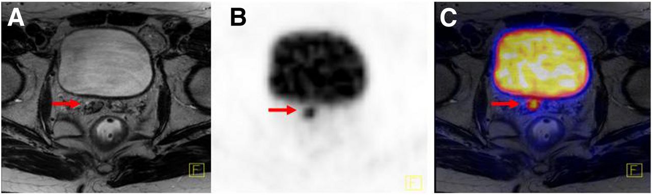
  • FIGURE 2.
    • Download figure
    • Open in new tab
    • Download powerpoint
    FIGURE 2.

    Axial 68Ga-PSMA PET/MR images of 63-y-old patient with recurrent prostate cancer with hypointense signal alterations in T2w images (A, arrow) and intense focal 68Ga-PSMA uptake in right seminal vesicle of PET part of PET/MR (B, arrow) and in fused PET with T2w images (C arrow).
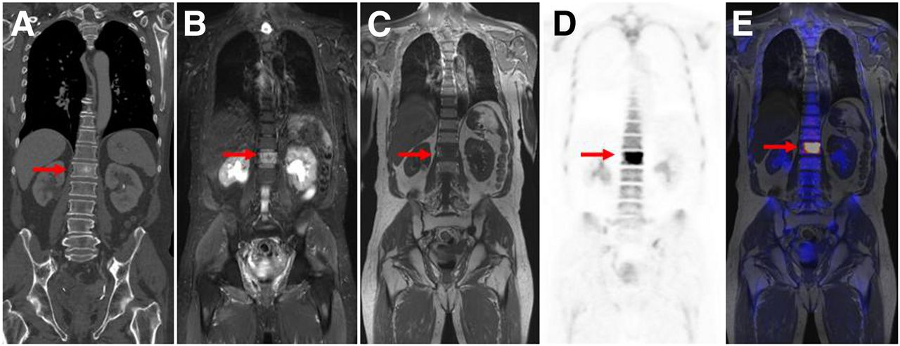
  • FIGURE 3.
    • Download figure
    • Open in new tab
    • Download powerpoint
    FIGURE 3.

    CT and 18F-fluoride PET/MR images of 64-y-old patient with prostate cancer and bone metastases with coronal CT images (A, arrow), coronal T2 short τ inversion recovery (B, arrow), and T1 TSE (C, arrow). PET of PET/MR shows intense focal 18F-fluoride uptake (D, arrow) whereas fused 18F-fluoride PET image with simultaneously acquired T1 TSE image (E) demonstrates concordance of metabolic and anatomic imaging.

Additional Files

  • Figures
  • Supplemental Data

    Files in this Data Supplement:

    • Supplemental Data
PreviousNext
Back to top

In this issue

Journal of Nuclear Medicine: 55 (Supplement 2)
Journal of Nuclear Medicine
Vol. 55, Issue Supplement 2
June 1, 2014
  • Table of Contents
  • Table of Contents (PDF)
  • About the Cover
  • Index by author
Print
Download PDF
Article Alerts
Sign In to Email Alerts with your Email Address
Email Article

Thank you for your interest in spreading the word on Journal of Nuclear Medicine.

NOTE: We only request your email address so that the person you are recommending the page to knows that you wanted them to see it, and that it is not junk mail. We do not capture any email address.

Enter multiple addresses on separate lines or separate them with commas.
PET/MR in Oncology: Non–18F-FDG Tracers for Routine Applications
(Your Name) has sent you a message from Journal of Nuclear Medicine
(Your Name) thought you would like to see the Journal of Nuclear Medicine web site.
Citation Tools
PET/MR in Oncology: Non–18F-FDG Tracers for Routine Applications
Isabel Rauscher, Matthias Eiber, Michael Souvatzoglou, Markus Schwaiger, Ambros J. Beer
Journal of Nuclear Medicine Jun 2014, 55 (Supplement 2) 25S-31S; DOI: 10.2967/jnumed.113.129536

Citation Manager Formats

  • BibTeX
  • Bookends
  • EasyBib
  • EndNote (tagged)
  • EndNote 8 (xml)
  • Medlars
  • Mendeley
  • Papers
  • RefWorks Tagged
  • Ref Manager
  • RIS
  • Zotero
Share
PET/MR in Oncology: Non–18F-FDG Tracers for Routine Applications
Isabel Rauscher, Matthias Eiber, Michael Souvatzoglou, Markus Schwaiger, Ambros J. Beer
Journal of Nuclear Medicine Jun 2014, 55 (Supplement 2) 25S-31S; DOI: 10.2967/jnumed.113.129536
Twitter logo Facebook logo LinkedIn logo Mendeley logo
  • Tweet Widget
  • Facebook Like
  • Google Plus One
Bookmark this article

Jump to section

  • Article
    • Abstract
    • ENDOCRINE
    • PROSTATE
    • BONE METASTASES
    • CONCLUSION
    • DISCLOSURE
    • Footnotes
    • REFERENCES
  • Figures & Data
  • Supplemental
  • Info & Metrics
  • PDF

Related Articles

  • PubMed
  • Google Scholar

Cited By...

  • Functional Genetic Screening Enables Theranostic Molecular Imaging in Cancer
  • 18F-FDG PET/CT and PET/MRI Perform Equally Well in Cancer: Evidence from Studies on More Than 2,300 Patients
  • Introduction
  • Google Scholar

More in this TOC Section

  • Potential Clinical Applications of PET/MR Imaging in Neurodegenerative Diseases
  • PET/MR Imaging: A Critical Appraisal
  • Potential Applications of PET/MR Imaging in Cardiology
Show more PET/MR Imaging: Potential Clinical and Research Applications

Similar Articles

Keywords

  • PET/MR
  • non–18F-FDG
  • oncology
SNMMI

© 2025 SNMMI

Powered by HighWire