


Article Figures & Data
Figures
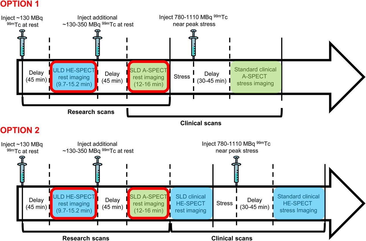
- FIGURE 1.
Study protocol. Sites had option to perform standard clinically indicated imaging using either A-SPECT camera (option 1) or HE SPECT camera (option 2). Images obtained using HE SPECT camera are denoted with blue shading and using A-SPECT camera with green shading. Comparison is made in each of the 2 options between images circled in red; subsequent images were obtained solely for clinical purposes and not analyzed in this study.
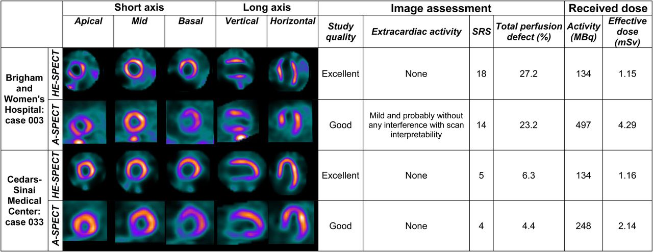
- FIGURE 2.
Comparison of representative images between ULD HE SPECT and SLD A-SPECT imaging.
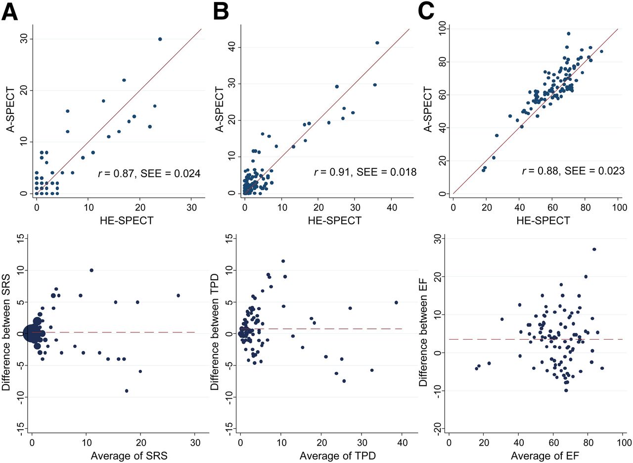
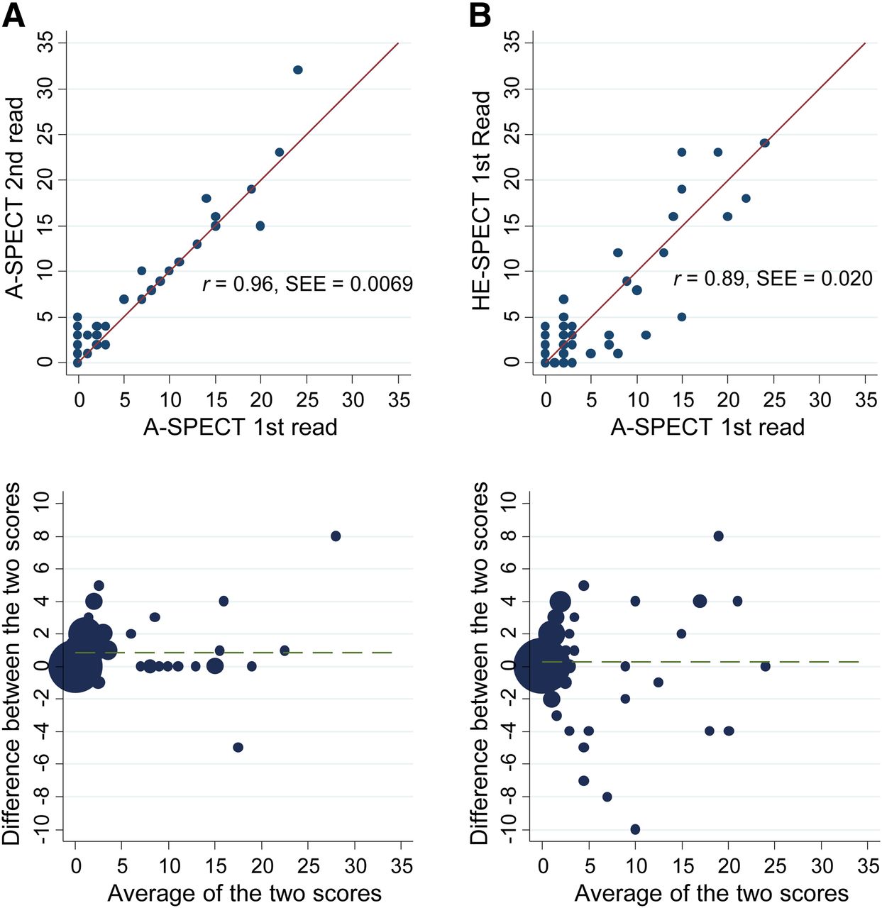
- FIGURE 3.
Comparison of SRS from consensus reading, TPD, and resting EF between SLD A-SPECT and ULD HE SPECT imaging. (A) SRS. (B) TPD (%). (C) EF (%). Top are scatterplots, and bottom are Bland–Altman plots. SEE denotes standard error of the estimate.
- FIGURE 4.
Comparison of intrareader agreement in SRS of SLD-A-SPECT to agreement between SLD A-SPECT and ULD HE SPECT imaging. (A) Intrareader agreement in SRS comparing 2 SLD A-SPECT reads, spaced 3 mo apart, for first reader. (B) Between-method agreement in SRS comparing SLD A-SPECT with ULD HE SPECT, for first reader. Top are scatterplots, and bottom are Bland–Altman plots.


Tables
Characteristic Value Total subjects 101 Group 1: intermediate/high likelihood of CAD 55 (54.5) 2: prior myocardial infarction 46 (45.5) Site Cedars-Sinai 49 (48.5) Sacred Heart 39 (38.6) Brigham and Women’s 13 (12.9) Mean age ± SD (y) 63.8 ± 11.3 Women 47 (46.5) BMI (kg/m2) 26.1 ± 2.8 Range 17.1–30.9 Diabetes mellitus 26 (25.7) On insulin 7 (6.9) On oral medications 17 (16.8) Hypertension 74 (73.3) Hyperlipidemia 77 (76.2) Current smoking 13 (12.9) Family history of premature heart disease 31 (30.7) No risk factors 3 (3.0) Stress type Exercise: Bruce Protocol 46 (45.5) Exercise: Modified Bruce Protocol 4 (4.0) Adenosine 25 (24.8) Regadenoson 25 (24.8) Dobutamine 1 (1.0) Data in parentheses are percentages.
Quantity Mean SD Range ULD injection Administered activity in MBq 198 (5.35) 37 (1.00) 127–275 (3.44–7.44) Residual activity in MBq 64 (1.74) 26 (0.71) 6.3–144 (0.17–3.89) Received activity in MBq 134 (3.62) 28 (0.75) 67–189 (1.80–5.10) Effective dose, received (mSv) 1.15 0.24 0.57–1.63 Supplemental injection, before SLD imaging Administered Activity in MBq 229 (6.21) 105 (2.84) 120–588 (3.23–15.89) Residual activity in MBq 65 (1.75) 46 (1.24) 5.9–270 (0.16–7.29) Received activity in MBq 165 (4.46) 68 (1.83) 75–383 (2.04–10.34) Effective dose, received (mSv) 1.42 0.58 0.65–3.30 Total SLD (supplemental injection + decayed ULD injection) Received activity in MBq 277 (7.50) 74 (1.99) 147–497 (3.98–13.43) Effective dose, received (mSv) 2.39 0.64 1.27–4.29 Data in parentheses are mCi.
Quantity ULD HE SPECT SLD A-SPECT IQ Excellent 48 24 Good 41 48 Fair 5 22 Poor 7 7 Mean score ± SD 4.29 ± 0.85 3.88 ± 0.85 P (Wilcoxon) <0.0001 Extracardiac activity None 55 40 Minimal 27 35 Mild 10 19 Moderate 3 2 Severe 6 5 Mean score ± SD 0.79 ± 1.12 0.98 ± 1.06 P (Wilcoxon) 0.05 Values are for consensus read.
All studies Abnormal studies (SRS > 1) Reads compared SLD A-SPECT ULD HE SPECT SLD A-SPECT ULD HE SPECT Intrareader agreement Reader 1, two reads 0.79 (95.7) 0.83 (96.7) 0.74 (92.9) 0.78 (93.7) Interreader agreement Reader 1 first read vs. Reader 2 0.82 (96.5) 0.84 (97.1) 0.74 (92.2) 0.79 (93.4) Reader 1 second read vs. Reader 2 0.78 (95.5) 0.85 (96.8) 0.73 (92.5) 0.80 (93.7) Between-method agreement Reader 1 first read SLD A-SPECT vs. reader 1 first read ULD HE SPECT 0.69 (94.4) 0.58 (87.8) Reader 1 second read SLD A-SPECT vs. reader 1 second read ULD HE SPECT 0.63 (92.0) 0.54 (87.8) Reader 1 first read SLD A-SPECT vs. reader 1 second read ULD HE SPECT 0.65 (93.2) 0.57 (87.4) Reader 1 second read SLD A-SPECT vs. reader 1 first read ULD HE SPECT 0.62 (92.4) 0.54 (87.6) Reader 2 SLD A-SPECT vs. reader 2 ULD HE SPECT 0.61 (92.8) 0.46 (83.3) Consensus read SLD A-SPECT vs. consensus read ULD HE SPECT 0.62 (93.0) 0.52 (87.4) ↵* Entries denote linearly weighted κ, with percentage agreement in parentheses.


Jump to section
Related Articles
Cited By...
- A Clinical Tool to Identify Candidates for Stress-First Myocardial Perfusion Imaging
- Solid-State Detector SPECT Myocardial Perfusion Imaging
- Imaging of the Thyroid and Parathyroid Using a Cardiac Cadmium-Zinc-Telluride Camera: Phantom Studies
- Accuracy of Computed Tomographic Angiography and Single-Photon Emission Computed Tomography-Acquired Myocardial Perfusion Imaging for the Diagnosis of Coronary Artery Disease
- Approaches to Reducing Radiation Dose from Radionuclide Myocardial Perfusion Imaging
- Radiation Dose and Prognosis of Ultra-Low-Dose Stress-First Myocardial Perfusion SPECT in Patients with Chest Pain Using a High-Efficiency Camera
- Minimizing Patient-Specific Tracer Dose in Myocardial Perfusion Imaging Using CZT SPECT
- IQ SPECT Allows a Significant Reduction in Administered Dose and Acquisition Time for Myocardial Perfusion Imaging: Evidence from a Phantom Study