
Main menu

  • Home
  • Content
    • Current
    • Ahead of print
    • Past Issues
    • JNM Supplement
    • SNMMI Annual Meeting Abstracts
    • Continuing Education
    • JNM Podcasts
  • Subscriptions
    • Subscribers
    • Institutional and Non-member
    • Rates
    • Journal Claims
    • Corporate & Special Sales
  • Authors
    • Submit to JNM
    • Information for Authors
    • Assignment of Copyright
    • AQARA requirements
  • Info
    • Reviewers
    • Permissions
    • Advertisers
  • About
    • About Us
    • Editorial Board
    • Contact Information
  • More
    • Alerts
    • Feedback
    • Help
    • SNMMI Journals
  • SNMMI
    • JNM
    • JNMT
    • SNMMI Journals
    • SNMMI

User menu

  • Subscribe
  • My alerts
  • Log in
  • My Cart

Search

  • Advanced search
Journal of Nuclear Medicine
  • SNMMI
    • JNM
    • JNMT
    • SNMMI Journals
    • SNMMI
  • Subscribe
  • My alerts
  • Log in
  • My Cart
Journal of Nuclear Medicine

Advanced Search

  • Home
  • Content
    • Current
    • Ahead of print
    • Past Issues
    • JNM Supplement
    • SNMMI Annual Meeting Abstracts
    • Continuing Education
    • JNM Podcasts
  • Subscriptions
    • Subscribers
    • Institutional and Non-member
    • Rates
    • Journal Claims
    • Corporate & Special Sales
  • Authors
    • Submit to JNM
    • Information for Authors
    • Assignment of Copyright
    • AQARA requirements
  • Info
    • Reviewers
    • Permissions
    • Advertisers
  • About
    • About Us
    • Editorial Board
    • Contact Information
  • More
    • Alerts
    • Feedback
    • Help
    • SNMMI Journals
  • View or Listen to JNM Podcast
  • Visit JNM on Facebook
  • Join JNM on LinkedIn
  • Follow JNM on Twitter
  • Subscribe to our RSS feeds
Research ArticleClinical Investigations

PET/CT with 18F-FDG–Labeled Autologous Leukocytes for the Diagnosis of Infected Fluid Collections in Acute Pancreatitis

Anish Bhattacharya, Rakesh Kochhar, Sarika Sharma, Pallab Ray, Naveen Kalra, Niranjan Khandelwal and Bhagwant R. Mittal
Journal of Nuclear Medicine August 2014, 55 (8) 1267-1272; DOI: https://doi.org/10.2967/jnumed.114.137232
Anish Bhattacharya
1Department of Nuclear Medicine, Postgraduate Institute of Medical Education and Research, Chandigarh, India
  • Find this author on Google Scholar
  • Find this author on PubMed
  • Search for this author on this site
Rakesh Kochhar
2Department of Gastroenterology, Postgraduate Institute of Medical Education and Research, Chandigarh, India
  • Find this author on Google Scholar
  • Find this author on PubMed
  • Search for this author on this site
Sarika Sharma
1Department of Nuclear Medicine, Postgraduate Institute of Medical Education and Research, Chandigarh, India
  • Find this author on Google Scholar
  • Find this author on PubMed
  • Search for this author on this site
Pallab Ray
3Department of Medical Microbiology, Postgraduate Institute of Medical Education and Research, Chandigarh, India; and
  • Find this author on Google Scholar
  • Find this author on PubMed
  • Search for this author on this site
Naveen Kalra
4Department of Radiodiagnosis, Postgraduate Institute of Medical Education and Research, Chandigarh, India
  • Find this author on Google Scholar
  • Find this author on PubMed
  • Search for this author on this site
Niranjan Khandelwal
4Department of Radiodiagnosis, Postgraduate Institute of Medical Education and Research, Chandigarh, India
  • Find this author on Google Scholar
  • Find this author on PubMed
  • Search for this author on this site
Bhagwant R. Mittal
1Department of Nuclear Medicine, Postgraduate Institute of Medical Education and Research, Chandigarh, India
  • Find this author on Google Scholar
  • Find this author on PubMed
  • Search for this author on this site
  • Article
  • Figures & Data
  • Info & Metrics
  • PDF
Loading

Article Figures & Data

Figures

  • Tables
  • FIGURE 1.
    • Download figure
    • Open in new tab
    • Download powerpoint
    FIGURE 1.

    18F-FDG–labeled leukocyte PET/CT images of patient with noninfected peripancreatic fluid collection. (A) Maximum-intensity-projection image showing no tracer uptake in central abdomen. (B–D) Coronal fused PET/CT (B), transaxial CT (C), and corresponding transaxial fused PET/CT (D) images showing large cystic non–tracer-avid peripancreatic fluid collection extending into lesser sac region. Fluid aspirated from collection was sterile on culture.

  • FIGURE 2.
    • Download figure
    • Open in new tab
    • Download powerpoint
    FIGURE 2.

    18F-FDG–labeled leukocyte PET/CT images of patient with peripancreatic fluid collection showing mild inflammation. (A) Maximum-intensity-projection image showing minimal tracer uptake in central abdomen, with nonhomogeneous uptake in liver and patchy uptake in basal segment of left lung. (B–D) Coronal fused PET/CT (B), transaxial CT (C), and corresponding transaxial fused PET/CT (D) images showing fluid collection with mildly tracer-avid margins (SUVmax, 3.0) in neck, body, and tail of pancreas. Fluid aspirated from collection was sterile on culture.
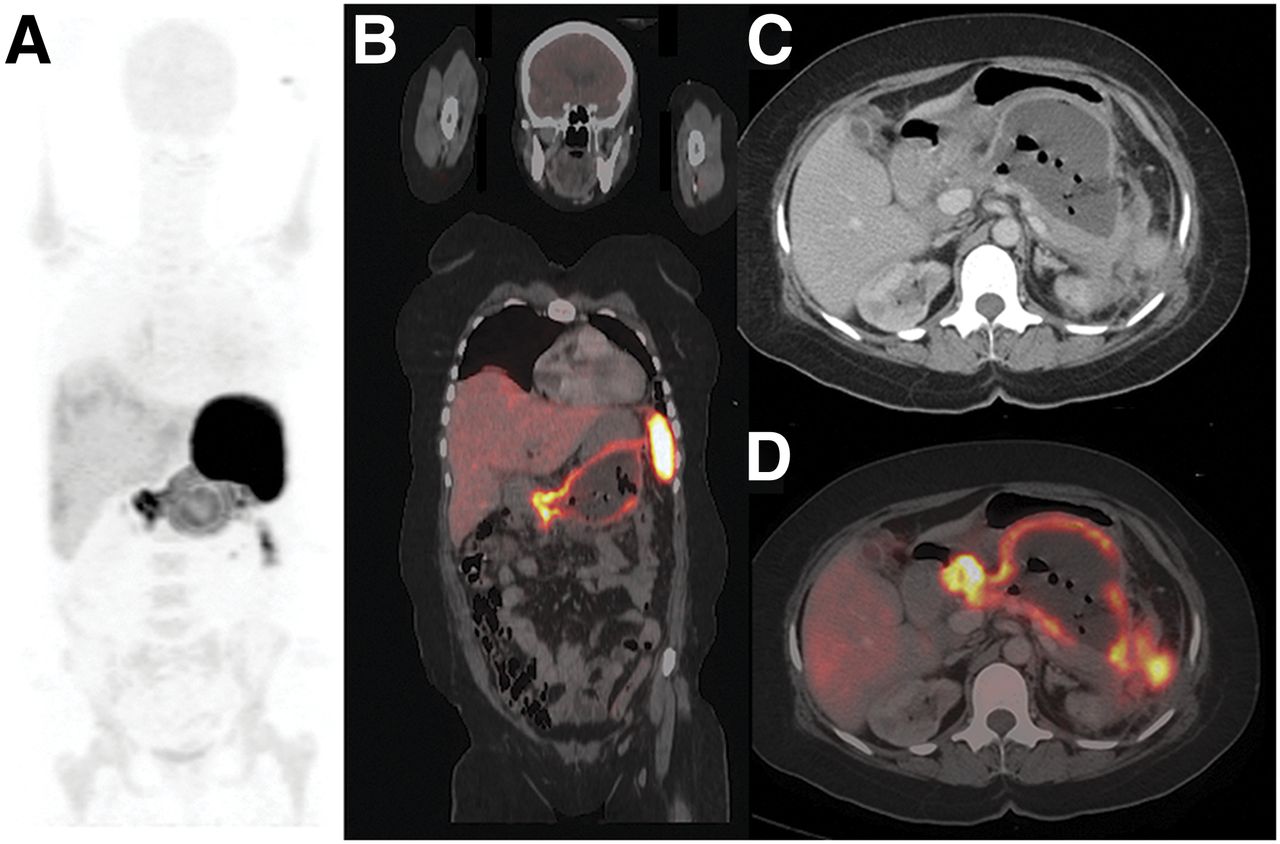
  • FIGURE 3.
    • Download figure
    • Open in new tab
    • Download powerpoint
    FIGURE 3.

    18F-FDG–labeled leukocyte PET/CT images of patient with infected peripancreatic fluid collection. (A) Maximum-intensity-projection image showing intense tracer uptake in central abdomen (SUVmax, 35), extending to spleen and left paracolic region. Mild irregular tracer uptake can be seen in both lungs. (B) Coronal fused PET/CT image showing large fluid collection with markedly tracer-avid margins replacing body and tail of pancreas. (C) Transaxial CT image at level of pancreas showing air bubbles in fluid collection. (D) Corresponding transaxial fused PET/CT image showing intense tracer uptake along margins of fluid collection. Fluid aspirated from collection showed growth of E. coli on culture.
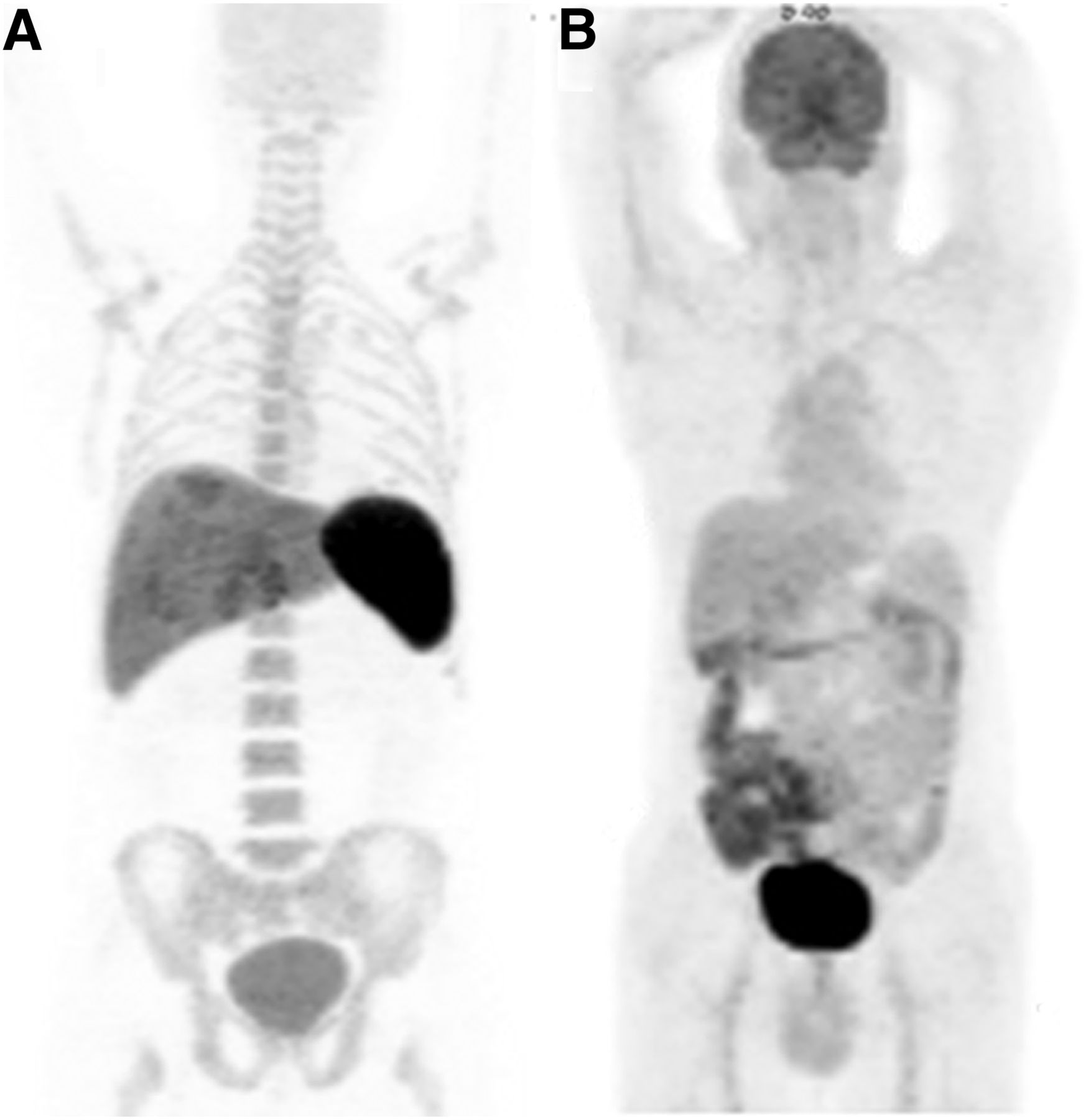
  • FIGURE 4.
    • Download figure
    • Open in new tab
    • Download powerpoint
    FIGURE 4.

    Maximum-intensity-projection images showing normal physiologic distribution of 18F-FDG–labeled autologous leukocytes in liver, spleen, and bone marrow, with minimal uptake in brain (A), and normal physiologic distribution of 18F-FDG alone (B).

Tables

  • Figures
    • View popup
    TABLE 1

    Characteristics of Patients

    Patients
    CharacteristicTotalWith positive scan resultsWith negative scan results
    No. of patients411229
    No. of febrile patients219 (75%)12 (41%)
    Blood glucose level (mg/100 mL)
     Mean ± SD118 ± 30140 ± 42109 ± 19
     Range83–21294–21283–175
    TLC/mm3
     Mean ± SD11,648 ± 5,37613,064 ± 5,30511,062 ± 5,387
     Range4,600–24,2005,600–20,9004,600–24,200
    Percentage of neutrophils
     Mean ± SD73 ± 1075 ± 772 ± 10
     Range55–9066–8555–90
    18F-FDG used (MBq)
     Mean ± SD429.2 ± 62.9462.5 ± 77.7418.1 ± 51.8
     Range314.5–555314.5–555336.7–529.1
    Radiotracer injected (MBq)
     Mean ± SD262.7 ± 70.3266.4 ± 66.6259 ± 70.3
     Range96.2–473.696.2–344.1111–473.6
    Percentage of labeling efficiency
     Mean ± SD81 ± 1784 ± 1980 ± 17
     Range31–9732–9731–96
    • TLC = total leukocyte count.

    • View popup
    TABLE 2

    Details of Fluid Collections with Positive Scan and Culture Results

    No. of patients
    Type of collectionTotalWith positive scan resultsWith positive culture results
    Acute necrotic collection2131*
    Walled-off necrosis1899
    Pseudocyst200
    • ↵* Aspirate was inadequate for culturing in 2 patients.

PreviousNext
Back to top

In this issue

Journal of Nuclear Medicine: 55 (8)
Journal of Nuclear Medicine
Vol. 55, Issue 8
August 1, 2014
  • Table of Contents
  • Table of Contents (PDF)
  • About the Cover
  • Index by author
Print
Download PDF
Article Alerts
Sign In to Email Alerts with your Email Address
Email Article

Thank you for your interest in spreading the word on Journal of Nuclear Medicine.

NOTE: We only request your email address so that the person you are recommending the page to knows that you wanted them to see it, and that it is not junk mail. We do not capture any email address.

Enter multiple addresses on separate lines or separate them with commas.
PET/CT with 18F-FDG–Labeled Autologous Leukocytes for the Diagnosis of Infected Fluid Collections in Acute Pancreatitis
(Your Name) has sent you a message from Journal of Nuclear Medicine
(Your Name) thought you would like to see the Journal of Nuclear Medicine web site.
Citation Tools
PET/CT with 18F-FDG–Labeled Autologous Leukocytes for the Diagnosis of Infected Fluid Collections in Acute Pancreatitis
Anish Bhattacharya, Rakesh Kochhar, Sarika Sharma, Pallab Ray, Naveen Kalra, Niranjan Khandelwal, Bhagwant R. Mittal
Journal of Nuclear Medicine Aug 2014, 55 (8) 1267-1272; DOI: 10.2967/jnumed.114.137232

Citation Manager Formats

  • BibTeX
  • Bookends
  • EasyBib
  • EndNote (tagged)
  • EndNote 8 (xml)
  • Medlars
  • Mendeley
  • Papers
  • RefWorks Tagged
  • Ref Manager
  • RIS
  • Zotero
Share
PET/CT with 18F-FDG–Labeled Autologous Leukocytes for the Diagnosis of Infected Fluid Collections in Acute Pancreatitis
Anish Bhattacharya, Rakesh Kochhar, Sarika Sharma, Pallab Ray, Naveen Kalra, Niranjan Khandelwal, Bhagwant R. Mittal
Journal of Nuclear Medicine Aug 2014, 55 (8) 1267-1272; DOI: 10.2967/jnumed.114.137232
Twitter logo Facebook logo LinkedIn logo Mendeley logo
  • Tweet Widget
  • Facebook Like
  • Google Plus One
Bookmark this article

Jump to section

  • Article
    • Abstract
    • MATERIALS AND METHODS
    • RESULTS
    • DISCUSSION
    • CONCLUSION
    • DISCLOSURE
    • Acknowledgments
    • Footnotes
    • REFERENCES
  • Figures & Data
  • Info & Metrics
  • PDF

Related Articles

  • This Month in JNM
  • PubMed
  • Google Scholar

Cited By...

  • No citing articles found.
  • Google Scholar

More in this TOC Section

  • Feasibility of Ultra-Low-Activity 18F-FDG PET/CT Imaging Using a Long–Axial-Field-of-View PET/CT System
  • Cardiac Presynaptic Sympathetic Nervous Function Evaluated by Cardiac PET in Patients with Chronotropic Incompetence Without Heart Failure
  • Validation and Evaluation of a Vendor-Provided Head Motion Correction Algorithm on the uMI Panorama PET/CT System
Show more Clinical Investigations

Similar Articles

Keywords

  • acute pancreatitis
  • infection
  • PET/CT
  • 18F-FDG
  • labeled leukocytes
SNMMI

© 2025 SNMMI

Powered by HighWire