


Article Figures & Data
Figures
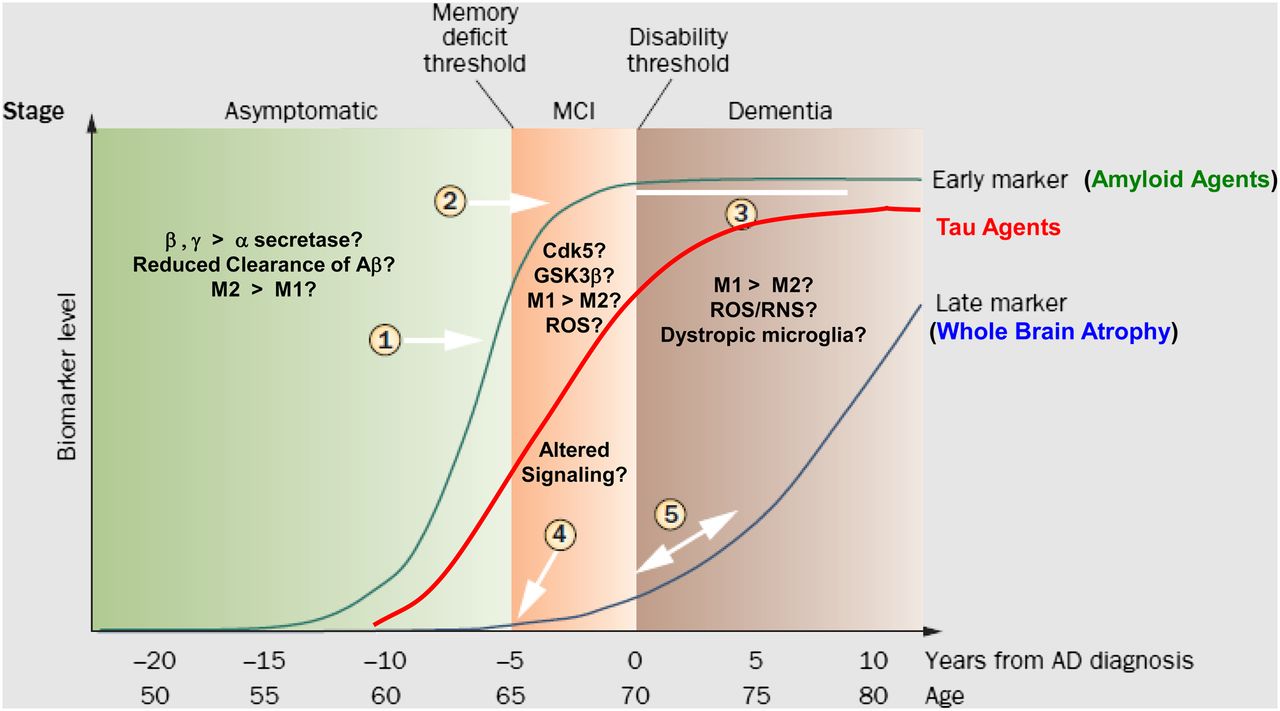
- FIGURE 1.
Molecular mechanism leading to formation of Aβ1–42 and subsequent formation of hyperphosphorylated tau. AβPP = Aβ precursor protein; AICD = amyloid precursor protein intracellular domain; BACE = β-site APP cleaving enzyme; Cdk5 = cyclin-dependent kinase 5; CHIP = C-terminus of heat-shock protein 70–interacting protein; CRE = cyclic adenosine monophosphate (cAMP) response element; CREB = cAMP response element-binding protein; CTF = C-terminal fragment; GRE = glucocorticoid response element; GSK3β = glycogen synthase kinase 3β; IL = interleukin; LTP = long-term potentiation; p3 = 3-kDa β-amyloid peptide; sAβPPα = soluble Aβ precursor protein alpha; sAβPPβ = soluble Aβ precursor protein beta; TNF-α = tumor necrosis factor α. (Reprinted with permission of (7).)
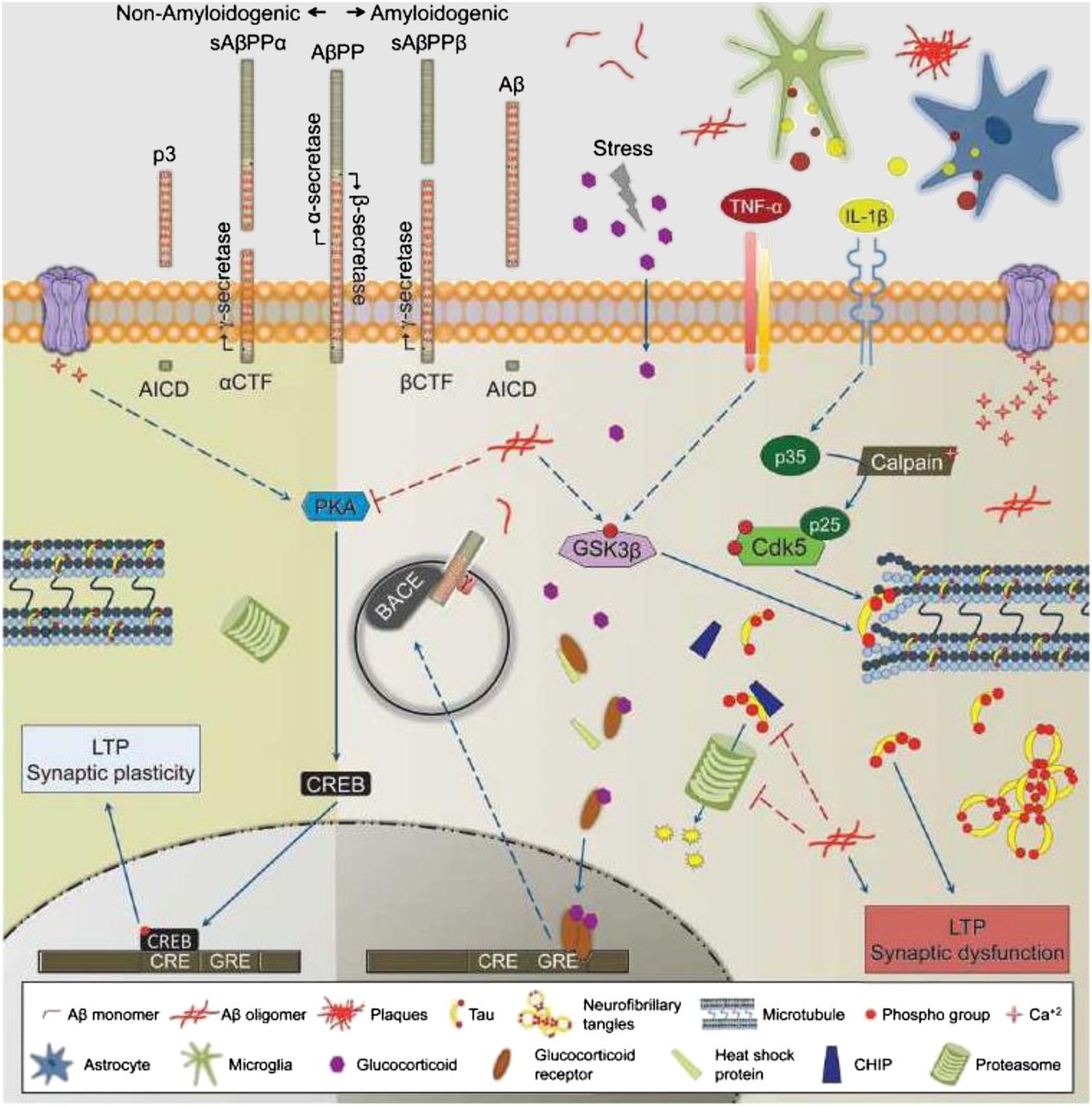
- FIGURE 2.
Microglial activation leading to neuroprotective (M2) and proinflammatory, neurotoxic (M1) microglia. Second wave of inflammation comes from infiltration of peripheral monocyte-derived macrophages from blood. AP-1 = activator protein 1; COX-2 = cyclooxygenase 2; DR6 = death receptor 6; IL = interleukin; iNOS = inducible macrophage-type nitric oxide synthase; NADPH = reduced nicotinamide adenine dinucleotide phosphate; NF-xB = nuclear factor xB; NO = nitric oxide; P2X7 = purinogenic 2X7 receptor; TGF-β = tumor growth factor-β; TLR = toll-like receptor; TNF-α = tumor necrosis factor α; (Reprinted with permission of (22).)
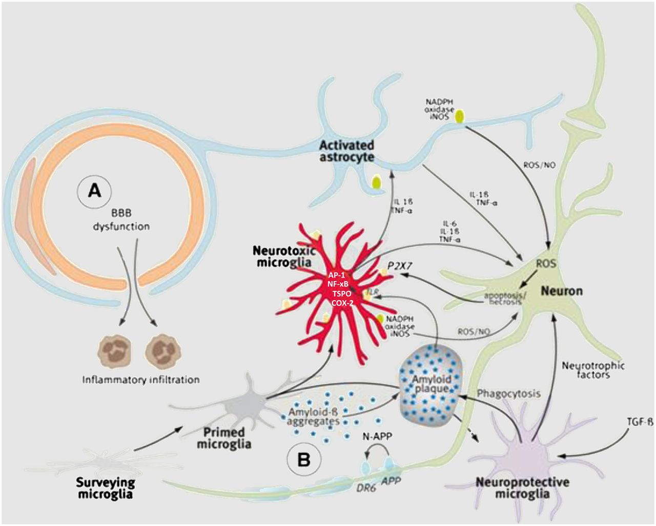
- FIGURE 3.
The 3 neurochemical phases of AD. Asymptomatic or prodromal phase is characterized by increased formation of Aβ plaques by either overproduction or reduced clearance of Aβ1–42. PET imaging studies with 11C-PiB confirm presence of Aβ plaques in this prodromal phase (1) and leveling off in MCI (2). Ceiling effect is shown (3). Red line shows theoretic time course for imaging with tau imaging agents or 18F-FDG. Late markers such as whole-brain atrophy are represented by blue line (4, 5) and can be followed with MR imaging. Cdk5 = cyclin-dependent kinase 5; GSK3β = glycogen synthase kinase 3β; MCI = mild cognitive impairment. (Adapted with permission of (25).)


