


Article Figures & Data
Figures
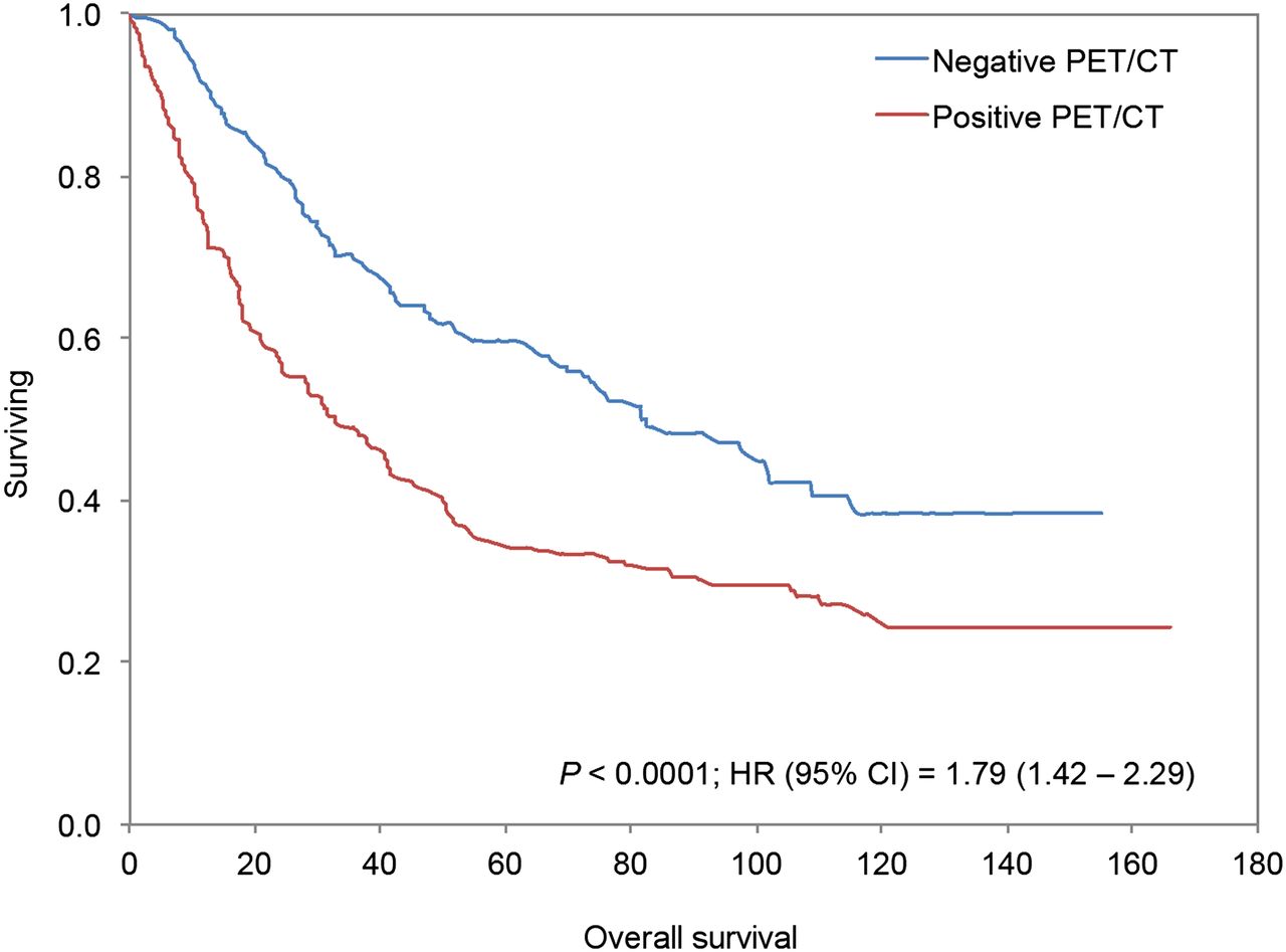
- FIGURE 1.
Kaplan–Meier survival plot for all scans (n = 488) in our study. OS between PET/CT scans positive for lung tumor and scans negative for tumor differed significantly.
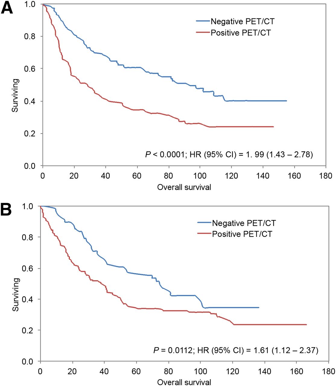
- FIGURE 2.
Kaplan–Meier survival plots showing OS for patients with scans performed 6–24 mo after treatment (A) and scans performed more than 24 mo after treatment (B). OS differed significantly between patients with PET/CT scans positive for lung tumor and patients with PET/CT scans negative for lung tumor in both periods.
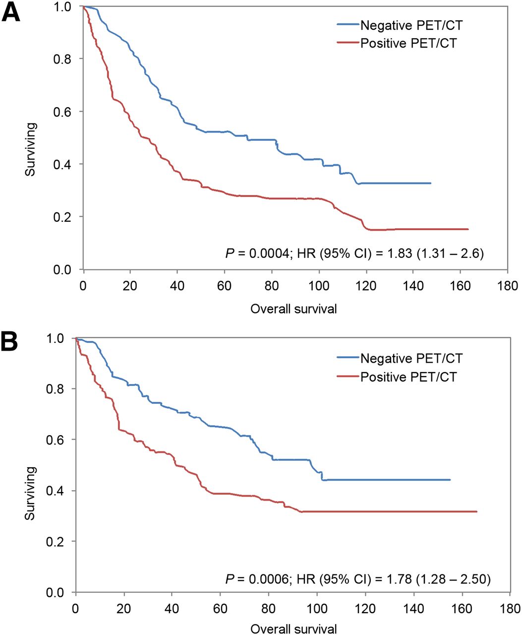
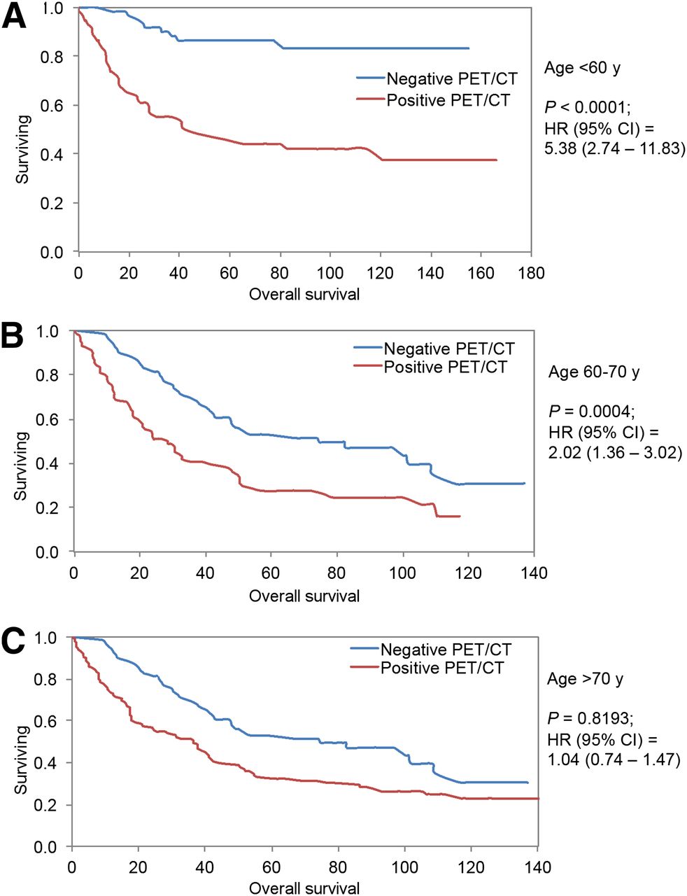
- FIGURE 3.
Kaplan–Meier survival plots showing OS for patients less than 60 y old (A), 60–70 y old (B), and more than 70 y old (C). OS differed significantly between PET/CT scans positive for lung tumor and PET/CT scans negative for lung tumor in patients < 60 y old and those 60–70 y old. No significant difference was observed in patients > 70 y old.
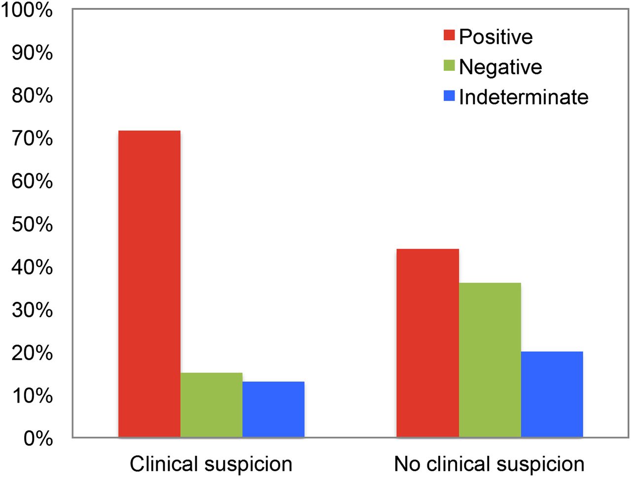
- FIGURE 4.
Added value of PET/CT to clinical assessment. PET/CT was helpful in excluding tumor in 15.2% (37/243) of patients with clinical suspicion of recurrence and in identifying recurrence in 43.7% (107/245) of patients with no prior clinical suspicion.
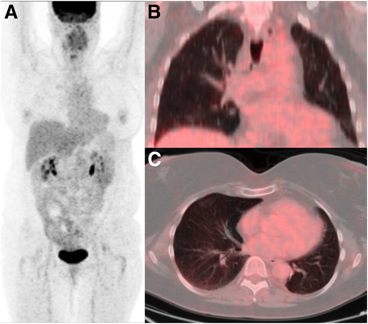
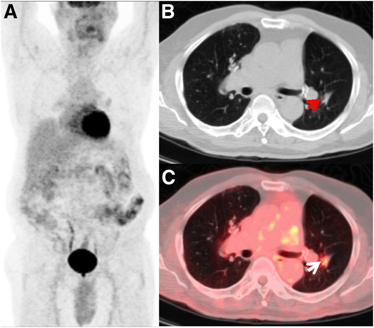
- FIGURE 5.
No clinical suspicion but positive PET results. Anterior maximum-intensity-projection (A), axial CT (B), and axial PET/CT (C) images of 76-y-old man with T1N0 non–small cell lung carcinoma after right upper lobectomy and adjuvant chemotherapy. Clinically, patient was comfortable, with no complaints during follow-up at 3 y after completion of treatment. Restaging PET/CT study showed hypermetabolic focus (arrows) within left lower-lobe nodule, consistent with disease recurrence. Patient completed additional chemotherapy based on the results of this study.
- FIGURE 6.
Clinical suspicion but negative PET results. Anterior maximum-intensity-projection (A), coronal PET/CT (B), and axial PET/CT (C) images of 51-y-old woman with limited-stage small cell carcinoma of left lung after left lower lobectomy and chemoradiation. Three years after completion of treatment, she presented with neurologic deficits including weakness of upper and lower extremities. Paraneoplastic syndrome was clinically suspected. Restaging PET/CT study showed no abnormal foci of metabolic activity to suggest active disease.
- FIGURE 7.
Kaplan–Meier survival plots for scans performed under clinical suspicion (A) and scans performed as routine surveillance (B). OS differed significantly between patients with PET/CT scans positive for lung tumor and patients with PET/CT scans negative for lung tumor under both routine and clinically suggestive settings.


Tables
Characteristic n % Age (mean ± SD, 66 ± 12 y) <60 y 71 27.2 60–70 y 97 37.2 >70 y 93 35.6 Sex Female 143 54.8 Male 118 45.2 Race White 185 70.9 Black 62 23.7 Other 14 5.4 Smoking Yes 221 84.7 No 33 12.6 Unknown 7 2.7 Histology Adenocarcinoma 79 30.3 Bronchioalveolar carcinoma 7 2.7 Bronchogenic carcinoid 1 0.4 Carcinoid 7 2.7 Epithelioid neoplasm 1 0.4 Mesothelioma 8 3.1 NSCLC 100 38.7 SCC 55 21.1 Unknown 2 0.8 Stage I 90 34.5 II 23 8.8 III 81 31.0 IV 17 6.5 Unknown 50 19.2 Last treatment Surgery 111 42.5 Radiation 74 28.4 Chemotherapy 76 29.1 PET/CT outcome Negative 63 24.1 Positive 198 75.9 NSCLC = non–small cell lung cancer; SCC = squamous cell carcinoma.
Characteristic Estimate 95% CI P Age 0.040 0.02, 0.05 <0.0001* Smoking −0.200 −0.39, −0.03 0.0218* Sex 0.150 0.03, 0.26 0.0112* Race 0.1134 White 0.056 −0.17, 0.32 Black 0.273 0.01, 0.556 Histology 0.1735 Adenocarcinoma −0.051 −0.26, 0.15 NSCLC 0.072 −0.11, 0.26 SCC 0.203 −0.01, 0.41 Stage −0.070 −0.19, 0.05 0.263 Treatment 0.0154* Surgery −0.100 −0.2, 0.05 Radiation 0.250 0.08, 0.42 Time to scan −0.001 −0.01, 0.002 0.4647 Clinical suspicion −0.060 −0.18, 0.05 0.2669 PET result −0.290 −0.41, −0.18 <0.0001* ↵* Significant variables.
NSCLC = non–small cell lung cancer; SCC = squamous cell carcinoma.


Jump to section
Related Articles
Cited By...
- 18F-FDG PET/CT: Therapy Response Assessment Interpretation (Hopkins Criteria) and Survival Outcomes in Lung Cancer Patients
- 18F-FDG PET/CT and Colorectal Cancer: Value of Fourth and Subsequent Posttherapy Follow-up Scans for Patient Management
- 18F-FDG PET/CT and Lung Cancer: Value of Fourth and Subsequent Posttherapy Follow-up Scans for Patient Management
- Relationship Between 18F-FDG Accumulation and Lactate Dehydrogenase A Expression in Lung Adenocarcinomas