
Main menu

  • Home
  • Content
    • Current
    • Ahead of print
    • Past Issues
    • JNM Supplement
    • SNMMI Annual Meeting Abstracts
    • Continuing Education
    • JNM Podcasts
  • Subscriptions
    • Subscribers
    • Institutional and Non-member
    • Rates
    • Journal Claims
    • Corporate & Special Sales
  • Authors
    • Submit to JNM
    • Information for Authors
    • Assignment of Copyright
    • AQARA requirements
  • Info
    • Reviewers
    • Permissions
    • Advertisers
  • About
    • About Us
    • Editorial Board
    • Contact Information
  • More
    • Alerts
    • Feedback
    • Help
    • SNMMI Journals
  • SNMMI
    • JNM
    • JNMT
    • SNMMI Journals
    • SNMMI

User menu

  • Subscribe
  • My alerts
  • Log in
  • My Cart

Search

  • Advanced search
Journal of Nuclear Medicine
  • SNMMI
    • JNM
    • JNMT
    • SNMMI Journals
    • SNMMI
  • Subscribe
  • My alerts
  • Log in
  • My Cart
Journal of Nuclear Medicine

Advanced Search

  • Home
  • Content
    • Current
    • Ahead of print
    • Past Issues
    • JNM Supplement
    • SNMMI Annual Meeting Abstracts
    • Continuing Education
    • JNM Podcasts
  • Subscriptions
    • Subscribers
    • Institutional and Non-member
    • Rates
    • Journal Claims
    • Corporate & Special Sales
  • Authors
    • Submit to JNM
    • Information for Authors
    • Assignment of Copyright
    • AQARA requirements
  • Info
    • Reviewers
    • Permissions
    • Advertisers
  • About
    • About Us
    • Editorial Board
    • Contact Information
  • More
    • Alerts
    • Feedback
    • Help
    • SNMMI Journals
  • View or Listen to JNM Podcast
  • Visit JNM on Facebook
  • Join JNM on LinkedIn
  • Follow JNM on Twitter
  • Subscribe to our RSS feeds
Research ArticleBasic Science Investigations

Dual-Modality Image-Guided Surgery of Prostate Cancer with a Radiolabeled Fluorescent Anti-PSMA Monoclonal Antibody

Susanne Lütje, Mark Rijpkema, Gerben M. Franssen, Giulio Fracasso, Wijnand Helfrich, Annemarie Eek, Wim J. Oyen, Marco Colombatti and Otto C. Boerman
Journal of Nuclear Medicine June 2014, 55 (6) 995-1001; DOI: https://doi.org/10.2967/jnumed.114.138180
Susanne Lütje
1Department of Radiology and Nuclear Medicine, Radboud University Medical Center, Nijmegen, The Netherlands
  • Find this author on Google Scholar
  • Find this author on PubMed
  • Search for this author on this site
Mark Rijpkema
1Department of Radiology and Nuclear Medicine, Radboud University Medical Center, Nijmegen, The Netherlands
  • Find this author on Google Scholar
  • Find this author on PubMed
  • Search for this author on this site
Gerben M. Franssen
1Department of Radiology and Nuclear Medicine, Radboud University Medical Center, Nijmegen, The Netherlands
  • Find this author on Google Scholar
  • Find this author on PubMed
  • Search for this author on this site
Giulio Fracasso
2Department of Pathology and Diagnostics, University of Verona, Verona, Italy; and
  • Find this author on Google Scholar
  • Find this author on PubMed
  • Search for this author on this site
Wijnand Helfrich
3Department of Surgery, University of Groningen, University Medical Centre Groningen, Groningen, The Netherlands
  • Find this author on Google Scholar
  • Find this author on PubMed
  • Search for this author on this site
Annemarie Eek
1Department of Radiology and Nuclear Medicine, Radboud University Medical Center, Nijmegen, The Netherlands
  • Find this author on Google Scholar
  • Find this author on PubMed
  • Search for this author on this site
Wim J. Oyen
1Department of Radiology and Nuclear Medicine, Radboud University Medical Center, Nijmegen, The Netherlands
  • Find this author on Google Scholar
  • Find this author on PubMed
  • Search for this author on this site
Marco Colombatti
2Department of Pathology and Diagnostics, University of Verona, Verona, Italy; and
  • Find this author on Google Scholar
  • Find this author on PubMed
  • Search for this author on this site
Otto C. Boerman
1Department of Radiology and Nuclear Medicine, Radboud University Medical Center, Nijmegen, The Netherlands
  • Find this author on Google Scholar
  • Find this author on PubMed
  • Search for this author on this site
  • Article
  • Figures & Data
  • Info & Metrics
  • PDF
Loading

Article Figures & Data

Figures

  • FIGURE 1.
    • Download figure
    • Open in new tab
    • Download powerpoint
    FIGURE 1.

    (A) Biodistribution of 111In-DTPA-D2B-IRDye800CW (0.55 MBq, 2.0 μg/mouse) in mice with subcutaneous PSMA-positive LNCaP and PSMA-negative PC3 xenografts at several time points after injection. Excess of unlabeled D2B IgG (300 μg/mouse) was coinjected in one group of mice as negative control. LNCaP-to-PC3 tumor ratios at 48 h after injection were 2.5 ± 0.7. (B) PSMA-positive and PSMA-negative tumor-to-blood ratios at several time points after injection of 111In-DTPA-D2B-IRDye800CW. (C) For illustration purposes, NIRF image of a mouse bearing a PSMA-positive LNCaP tumor on right flank (green arrow) and a PSMA-negative PC3 tumor on the left flank (blue arrow) at 48 h after intravenous injection of dual-labeled 111In-DTPA-D2B-IRDye800CW. *P < 0.0001 on t testing. **P < 0.0001 on t testing. ***P < 0.0001 on t testing. p = photons; p.i. = after injection; sr = steradian; xs = excess.

  • FIGURE 2.
    • Download figure
    • Open in new tab
    • Download powerpoint
    FIGURE 2.

    Dual-modality fluorescence and micro-SPECT/CT imaging with dual-labeled 111In-DTPA-D2B-IRDye800CW at 48 h after injection in a mouse with an intraperitoneally growing LS174T-PSMA tumor. Shown are NIRF image (acquisition time, 5 min) (A), corresponding micro-SPECT/CT image with mouse supine (B), and NIRF image of muscle sample (top) and tumor (bottom) after resection (C). Green arrows indicate the PSMA-positive tumor. p = photons; sr = steradian.
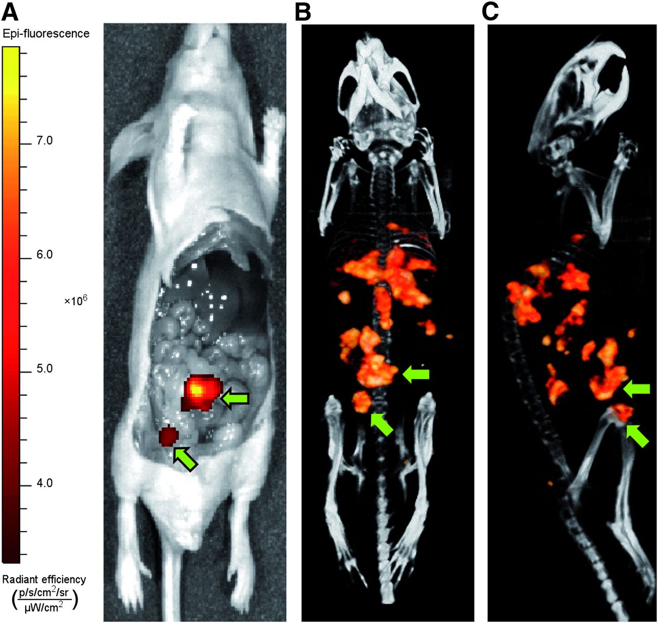
  • FIGURE 3.
    • Download figure
    • Open in new tab
    • Download powerpoint
    FIGURE 3.

    Dual-modality fluorescence and micro-SPECT/CT imaging with dual-labeled 111In-DTPA-D2B-IRDye800CW at 48 h after injection in a mouse with several intraperitoneal LS174T-PSMA tumors located at different depths in the peritoneal cavity. NIRF image (acquisition time, 5 min) (A) and corresponding micro-SPECT/CT images in supine (B) and left lateral (C) positions show limited penetration depth of NIRF imaging. Arrows indicate 2 tumors superficial enough to be visualized with fluorescence imaging. Tumors deeper inside peritoneal cavity are not visualized.
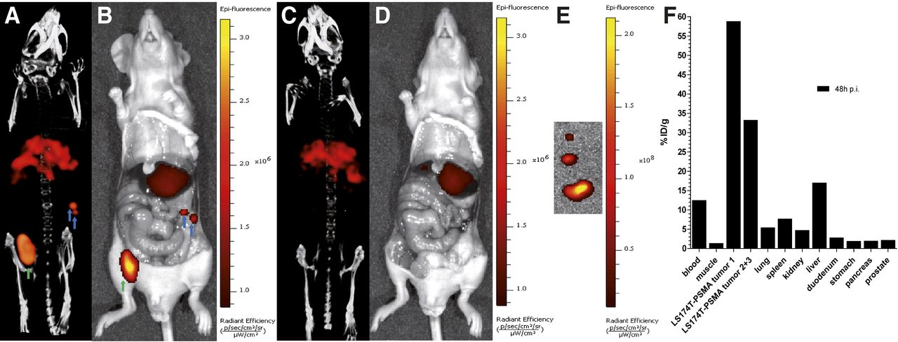
  • FIGURE 4.
    • Download figure
    • Open in new tab
    • Download powerpoint
    FIGURE 4.

    Dual-modality fluorescence and micro-SPECT/CT imaging with dual-labeled 111In-DTPA-D2B-IRDye800CW at 48 h after injection in mouse with intraperitoneally growing LS174T-PSMA tumors. Shown are micro-SPECT/CT image of 3 intraperitoneal PSMA-positive tumors (green and blue arrows) (A), merged photograph and NIRF image of mouse before resection of 3 tumors (B), micro-SPECT/CT image after resection of tumors (C), merged photograph and NIRF image of mouse after resection of tumors (D), merged photograph and optical fluorescence image of tumors after resection (E), and biodistribution of 111In-DTPA-D2B-IRDye800CW in mice with intraperitoneal LS174T-PSMA tumors (F). p = photons; p.i. = after injection; sr = steradian.

PreviousNext
Back to top

In this issue

Journal of Nuclear Medicine: 55 (6)
Journal of Nuclear Medicine
Vol. 55, Issue 6
June 1, 2014
  • Table of Contents
  • Table of Contents (PDF)
  • About the Cover
  • Index by author
Print
Download PDF
Article Alerts
Sign In to Email Alerts with your Email Address
Email Article

Thank you for your interest in spreading the word on Journal of Nuclear Medicine.

NOTE: We only request your email address so that the person you are recommending the page to knows that you wanted them to see it, and that it is not junk mail. We do not capture any email address.

Enter multiple addresses on separate lines or separate them with commas.
Dual-Modality Image-Guided Surgery of Prostate Cancer with a Radiolabeled Fluorescent Anti-PSMA Monoclonal Antibody
(Your Name) has sent you a message from Journal of Nuclear Medicine
(Your Name) thought you would like to see the Journal of Nuclear Medicine web site.
Citation Tools
Dual-Modality Image-Guided Surgery of Prostate Cancer with a Radiolabeled Fluorescent Anti-PSMA Monoclonal Antibody
Susanne Lütje, Mark Rijpkema, Gerben M. Franssen, Giulio Fracasso, Wijnand Helfrich, Annemarie Eek, Wim J. Oyen, Marco Colombatti, Otto C. Boerman
Journal of Nuclear Medicine Jun 2014, 55 (6) 995-1001; DOI: 10.2967/jnumed.114.138180

Citation Manager Formats

  • BibTeX
  • Bookends
  • EasyBib
  • EndNote (tagged)
  • EndNote 8 (xml)
  • Medlars
  • Mendeley
  • Papers
  • RefWorks Tagged
  • Ref Manager
  • RIS
  • Zotero
Share
Dual-Modality Image-Guided Surgery of Prostate Cancer with a Radiolabeled Fluorescent Anti-PSMA Monoclonal Antibody
Susanne Lütje, Mark Rijpkema, Gerben M. Franssen, Giulio Fracasso, Wijnand Helfrich, Annemarie Eek, Wim J. Oyen, Marco Colombatti, Otto C. Boerman
Journal of Nuclear Medicine Jun 2014, 55 (6) 995-1001; DOI: 10.2967/jnumed.114.138180
Twitter logo Facebook logo LinkedIn logo Mendeley logo
  • Tweet Widget
  • Facebook Like
  • Google Plus One
Bookmark this article

Jump to section

  • Article
    • Abstract
    • MATERIALS AND METHODS
    • RESULTS
    • DISCUSSION
    • CONCLUSION
    • DISCLOSURE
    • Acknowledgments
    • Footnotes
    • REFERENCES
  • Figures & Data
  • Info & Metrics
  • PDF

Related Articles

  • This Month in JNM
  • PubMed
  • Google Scholar

Cited By...

  • Precision Surgery Guided by Intraoperative Molecular Imaging
  • Rational Linker Design to Accelerate Excretion and Reduce Background Uptake of Peptidomimetic PSMA-Targeting Hybrid Molecules
  • A Dual-Modality Linker Enables Site-Specific Conjugation of Antibody Fragments for 18F-Immuno-PET and Fluorescence Imaging
  • In Vitro and In Vivo Characterization of an 18F-AlF-Labeled PSMA Ligand for Imaging of PSMA-Expressing Xenografts
  • Synthesis and Preclinical Characterization of the PSMA-Targeted Hybrid Tracer PSMA-I&F for Nuclear and Fluorescence Imaging of Prostate Cancer
  • PSMA-11-Derived Dual-Labeled PSMA Inhibitors for Preoperative PET Imaging and Precise Fluorescence-Guided Surgery of Prostate Cancer
  • Characterization of Site-Specifically Conjugated Monomethyl Auristatin E- and Duocarmycin-Based Anti-PSMA Antibody-Drug Conjugates for Treatment of PSMA-Expressing Tumors
  • Detection of Micrometastases Using SPECT/Fluorescence Dual-Modality Imaging in a CEA-Expressing Tumor Model
  • Targeted Dual-Modality Imaging in Renal Cell Carcinoma: An Ex Vivo Kidney Perfusion Study
  • Fluorescent Image-Guided Surgery with an Anti-Prostate Stem Cell Antigen (PSCA) Diabody Enables Targeted Resection of Mouse Prostate Cancer Xenografts in Real Time
  • Pretargeted Dual-Modality Immuno-SPECT and Near-Infrared Fluorescence Imaging for Image-Guided Surgery of Prostate Cancer
  • Google Scholar

More in this TOC Section

  • 11C-Methionine PET of Myocardial Inflammation in a Rat Model of Experimental Autoimmune Myocarditis
  • Counting Rate Characteristics and Image Distortion in Preclinical PET Imaging During Radiopharmaceutical Therapy
  • Design and Fabrication of Kidney Phantoms for Internal Radiation Dosimetry Using 3D Printing Technology
Show more Basic Science Investigations

Similar Articles

Keywords

  • prostate cancer
  • PSMA
  • dual-modality imaging
  • fluorescence imaging
  • IRDye800CW
  • D2B IgG
SNMMI

© 2025 SNMMI

Powered by HighWire