
Main menu

  • Home
  • Content
    • Current
    • Ahead of print
    • Past Issues
    • JNM Supplement
    • SNMMI Annual Meeting Abstracts
    • Continuing Education
    • JNM Podcasts
  • Subscriptions
    • Subscribers
    • Institutional and Non-member
    • Rates
    • Journal Claims
    • Corporate & Special Sales
  • Authors
    • Submit to JNM
    • Information for Authors
    • Assignment of Copyright
    • AQARA requirements
  • Info
    • Reviewers
    • Permissions
    • Advertisers
  • About
    • About Us
    • Editorial Board
    • Contact Information
  • More
    • Alerts
    • Feedback
    • Help
    • SNMMI Journals
  • SNMMI
    • JNM
    • JNMT
    • SNMMI Journals
    • SNMMI

User menu

  • Subscribe
  • My alerts
  • Log in
  • My Cart

Search

  • Advanced search
Journal of Nuclear Medicine
  • SNMMI
    • JNM
    • JNMT
    • SNMMI Journals
    • SNMMI
  • Subscribe
  • My alerts
  • Log in
  • My Cart
Journal of Nuclear Medicine

Advanced Search

  • Home
  • Content
    • Current
    • Ahead of print
    • Past Issues
    • JNM Supplement
    • SNMMI Annual Meeting Abstracts
    • Continuing Education
    • JNM Podcasts
  • Subscriptions
    • Subscribers
    • Institutional and Non-member
    • Rates
    • Journal Claims
    • Corporate & Special Sales
  • Authors
    • Submit to JNM
    • Information for Authors
    • Assignment of Copyright
    • AQARA requirements
  • Info
    • Reviewers
    • Permissions
    • Advertisers
  • About
    • About Us
    • Editorial Board
    • Contact Information
  • More
    • Alerts
    • Feedback
    • Help
    • SNMMI Journals
  • View or Listen to JNM Podcast
  • Visit JNM on Facebook
  • Join JNM on LinkedIn
  • Follow JNM on Twitter
  • Subscribe to our RSS feeds
Research ArticleBasic Science Investigations

Early Assessment of Tumor Response to Gefitinib Treatment by Noninvasive Optical Imaging of Tumor Vascular Endothelial Growth Factor Expression in Animal Models

Zhaofei Liu, Xianlei Sun, Hao Liu, Teng Ma, Jiyun Shi, Bing Jia, Huiyun Zhao and Fan Wang
Journal of Nuclear Medicine May 2014, 55 (5) 818-823; DOI: https://doi.org/10.2967/jnumed.113.133660
Zhaofei Liu
1Medical Isotopes Research Center, Peking University, Beijing, China
2Department of Radiation Medicine, School of Basic Medical Sciences, Peking University, Beijing, China; and
  • Find this author on Google Scholar
  • Find this author on PubMed
  • Search for this author on this site
Xianlei Sun
1Medical Isotopes Research Center, Peking University, Beijing, China
2Department of Radiation Medicine, School of Basic Medical Sciences, Peking University, Beijing, China; and
  • Find this author on Google Scholar
  • Find this author on PubMed
  • Search for this author on this site
Hao Liu
1Medical Isotopes Research Center, Peking University, Beijing, China
2Department of Radiation Medicine, School of Basic Medical Sciences, Peking University, Beijing, China; and
  • Find this author on Google Scholar
  • Find this author on PubMed
  • Search for this author on this site
Teng Ma
1Medical Isotopes Research Center, Peking University, Beijing, China
2Department of Radiation Medicine, School of Basic Medical Sciences, Peking University, Beijing, China; and
  • Find this author on Google Scholar
  • Find this author on PubMed
  • Search for this author on this site
Jiyun Shi
1Medical Isotopes Research Center, Peking University, Beijing, China
3Medical and Healthy Analytical Center, Peking University, Beijing, China
  • Find this author on Google Scholar
  • Find this author on PubMed
  • Search for this author on this site
Bing Jia
1Medical Isotopes Research Center, Peking University, Beijing, China
2Department of Radiation Medicine, School of Basic Medical Sciences, Peking University, Beijing, China; and
  • Find this author on Google Scholar
  • Find this author on PubMed
  • Search for this author on this site
Huiyun Zhao
1Medical Isotopes Research Center, Peking University, Beijing, China
3Medical and Healthy Analytical Center, Peking University, Beijing, China
  • Find this author on Google Scholar
  • Find this author on PubMed
  • Search for this author on this site
Fan Wang
1Medical Isotopes Research Center, Peking University, Beijing, China
2Department of Radiation Medicine, School of Basic Medical Sciences, Peking University, Beijing, China; and
  • Find this author on Google Scholar
  • Find this author on PubMed
  • Search for this author on this site
  • Article
  • Figures & Data
  • Supplemental
  • Info & Metrics
  • PDF
Loading

Article Figures & Data

Figures

  • Additional Files
  • FIGURE 1.
    • Download figure
    • Open in new tab
    • Download powerpoint
    FIGURE 1.

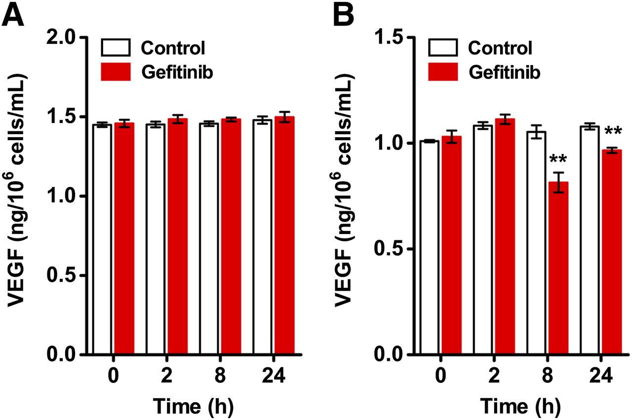
    Quantification of VEGF expression in tumor cells by ELISA. VEGF levels in cell culture supernatants of 22B (A) and A549 (B) cells were evaluated at 0, 2, 8, and 24 h with or without 1 μM gefitinib treatment (n = 6). **P < 0.01.

  • FIGURE 2.
    • Download figure
    • Open in new tab
    • Download powerpoint
    FIGURE 2.

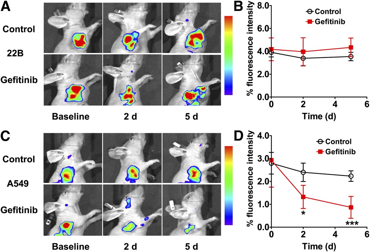
    (A and C) In vivo optical imaging of 22B (A) and A549 (C) tumor–bearing mice at 4 h after intravenous injection of 0.5 nmol of Dye-BevF(ab′)2 on days 0, 2, and 5 after initiation of gefitinib treatment (80 mg/kg). (B and D) Quantified 22B (B) and A549 (D) tumor uptake from A and C (n = 5). *P < 0.05. ***P < 0.001.

  • FIGURE 3.
    • Download figure
    • Open in new tab
    • Download powerpoint
    FIGURE 3.

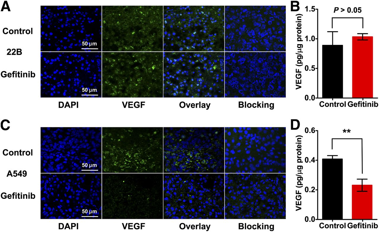
    VEGF expression in vehicle (control) or gefitinib-treated tumors as determined by immunofluorescence staining and ELISA. (A and C) Immunofluorescence staining of VEGF in 22B (A) and A549 (C) tumor tissues using FITC-bevacizumab (with or without blocking using unlabeled bevacizumab). (B and D) VEGF expression levels in 22B (B) and A549 (D) tumor lysates as determined by ELISA (n = 5). **P < 0.01.

  • FIGURE 4.
    • Download figure
    • Open in new tab
    • Download powerpoint
    FIGURE 4.

    Antitumorigenic effects of gefitinib treatment. Growth curves of 22B (A) and A549 (B) tumors in nude mice after intraperitoneal administration of 6 doses of gefitinib (80 mg/kg in 50 μL daily) or DMSO (control; 50 μL daily) (n = 5–7). **P < 0.01. ***P < 0.001.

  • FIGURE 5.
    • Download figure
    • Open in new tab
    • Download powerpoint
    FIGURE 5.

    CD31 and Ki67 staining of vehicle (control) or gefitinib-treated 22B (A) and A549 (B) tumor tissues.

Additional Files

  • Figures
  • Supplemental Data

    Files in this Data Supplement:

    • Supplemental Data
PreviousNext
Back to top

In this issue

Journal of Nuclear Medicine: 55 (5)
Journal of Nuclear Medicine
Vol. 55, Issue 5
May 1, 2014
  • Table of Contents
  • Table of Contents (PDF)
  • About the Cover
  • Index by author
Print
Download PDF
Article Alerts
Sign In to Email Alerts with your Email Address
Email Article

Thank you for your interest in spreading the word on Journal of Nuclear Medicine.

NOTE: We only request your email address so that the person you are recommending the page to knows that you wanted them to see it, and that it is not junk mail. We do not capture any email address.

Enter multiple addresses on separate lines or separate them with commas.
Early Assessment of Tumor Response to Gefitinib Treatment by Noninvasive Optical Imaging of Tumor Vascular Endothelial Growth Factor Expression in Animal Models
(Your Name) has sent you a message from Journal of Nuclear Medicine
(Your Name) thought you would like to see the Journal of Nuclear Medicine web site.
Citation Tools
Early Assessment of Tumor Response to Gefitinib Treatment by Noninvasive Optical Imaging of Tumor Vascular Endothelial Growth Factor Expression in Animal Models
Zhaofei Liu, Xianlei Sun, Hao Liu, Teng Ma, Jiyun Shi, Bing Jia, Huiyun Zhao, Fan Wang
Journal of Nuclear Medicine May 2014, 55 (5) 818-823; DOI: 10.2967/jnumed.113.133660

Citation Manager Formats

  • BibTeX
  • Bookends
  • EasyBib
  • EndNote (tagged)
  • EndNote 8 (xml)
  • Medlars
  • Mendeley
  • Papers
  • RefWorks Tagged
  • Ref Manager
  • RIS
  • Zotero
Share
Early Assessment of Tumor Response to Gefitinib Treatment by Noninvasive Optical Imaging of Tumor Vascular Endothelial Growth Factor Expression in Animal Models
Zhaofei Liu, Xianlei Sun, Hao Liu, Teng Ma, Jiyun Shi, Bing Jia, Huiyun Zhao, Fan Wang
Journal of Nuclear Medicine May 2014, 55 (5) 818-823; DOI: 10.2967/jnumed.113.133660
Twitter logo Facebook logo LinkedIn logo Mendeley logo
  • Tweet Widget
  • Facebook Like
  • Google Plus One
Bookmark this article

Jump to section

  • Article
    • Abstract
    • MATERIALS AND METHODS
    • RESULTS
    • DISCUSSION
    • CONCLUSION
    • DISCLOSURE
    • Acknowledgments
    • Footnotes
    • REFERENCES
  • Figures & Data
  • Supplemental
  • Info & Metrics
  • PDF

Related Articles

  • This Month in JNM
  • PubMed
  • Google Scholar

Cited By...

  • Molecular Imaging of Post-Src Inhibition Tumor Signatures for Guiding Dasatinib Combination Therapy
  • Google Scholar

More in this TOC Section

  • PET/MRI of Hypoxic Atherosclerosis Using 64Cu-ATSM in a Rabbit Model
  • Tumor Uptake of Anti-CD20 Fabs Depends on Tumor Perfusion
  • How Sensitive Is the Upper Gastrointestinal Tract to 90Y Radioembolization? A Histologic and Dosimetric Analysis in a Porcine Model
Show more Basic Science Investigations

Similar Articles

Keywords

  • EGFR
  • angiogenesis
  • VEGF
  • Molecular imaging
  • therapy response
SNMMI

© 2025 SNMMI

Powered by HighWire