


Article Figures & Data
Figures
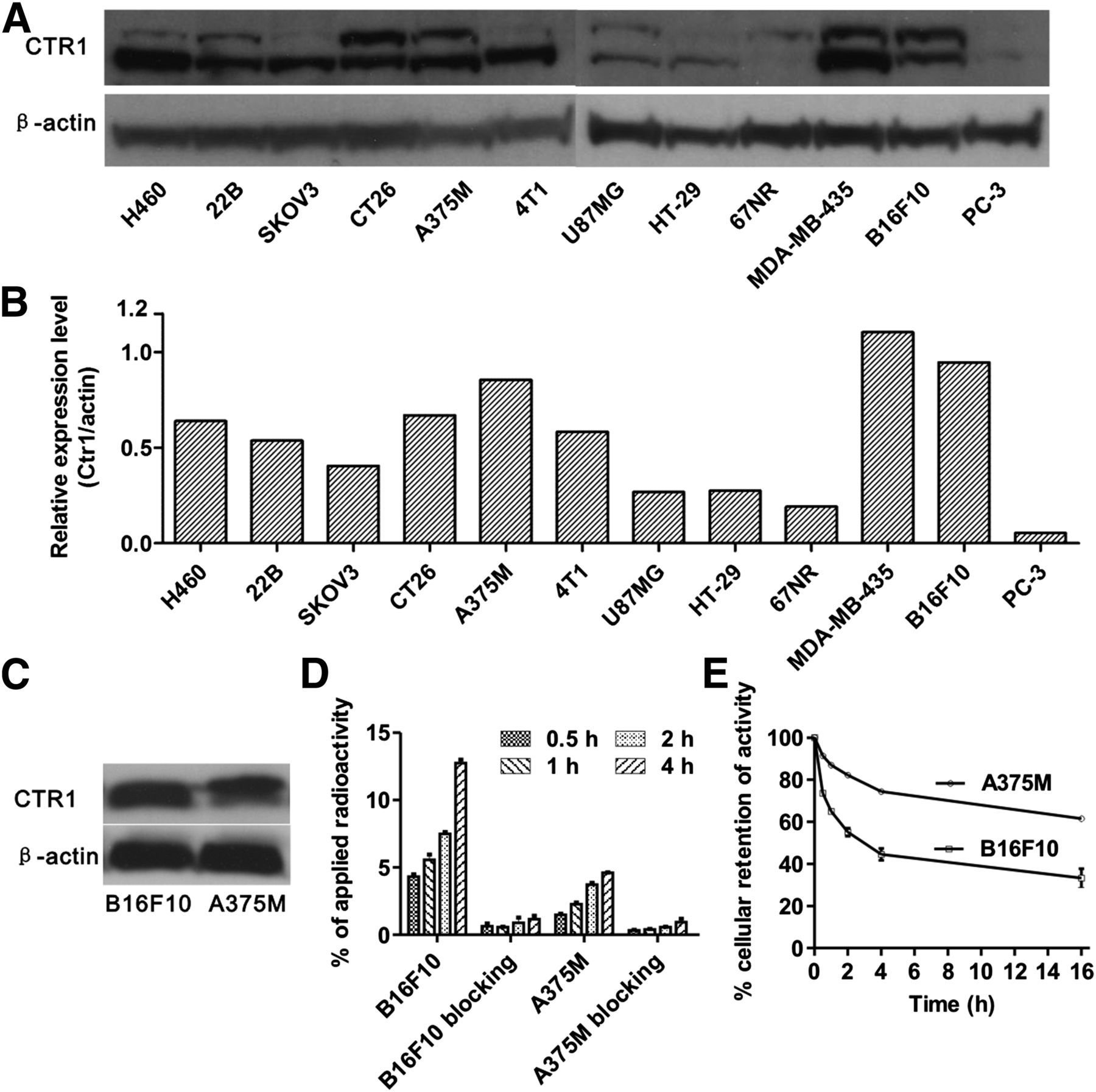
- FIGURE 1.
(A) Western blots of CTR1 in 12 cancer cell lines. (B) Quantitative analysis of Western blot results. (C) Western blots of CTR1 in B16F10 and A375M tumor tissues. (D) B16F10 and A375M’s cellular uptake of 64CuCl2 at 0.5, 1, 2, and 4 h time points and blocking studies (excess cold CuCl2 was added) at each time point. (E) Efflux of 64CuCl2 from A375M and B16F10 cells at 0.5, 1, 2, 4, and 16 h after 2 h incubation with 64CuCl2.
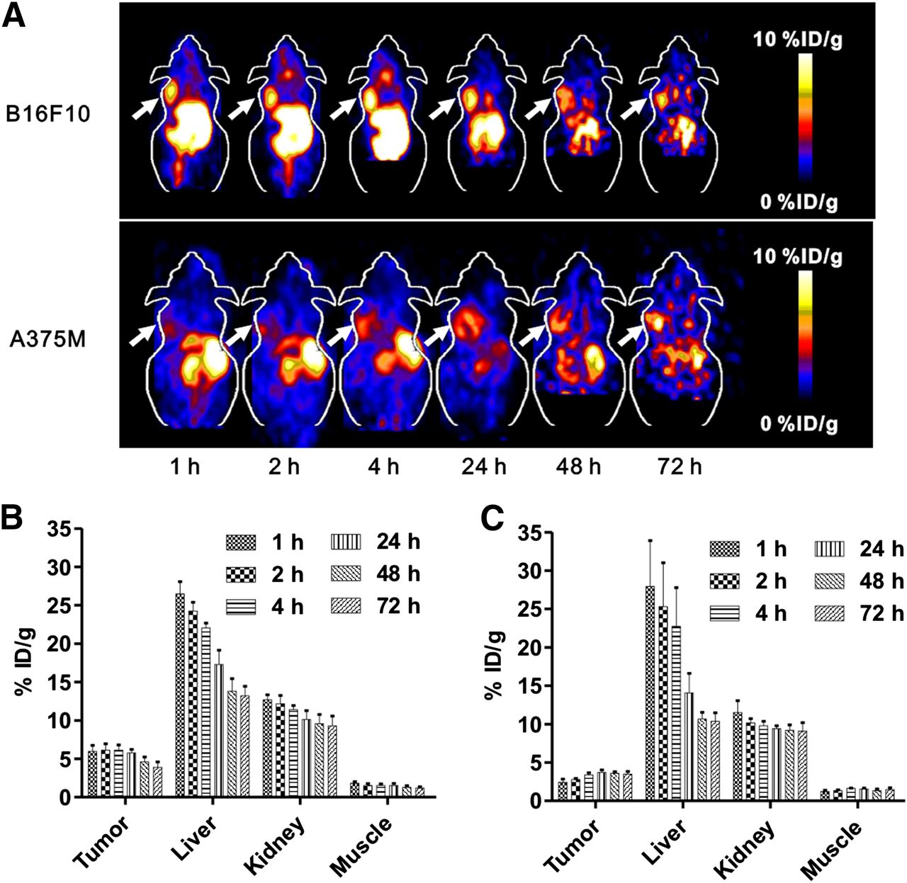
- FIGURE 2.
(A) Decay-corrected whole-body coronal small-animal PET images of C57BL/6 mice bearing B16F10 murine melanoma tumors (upper) and athymic nude mice bearing A375M human melanoma (lower) from 5-min static scans at 1, 2, 4, 24, 48, and 72 h after intravenous injection of 64CuCl2. Tumors are indicated by arrows. (B and C) Small-animal PET quantification of tumors and major organs, including liver, kidney, and muscle, at 1, 2, 4, 24, 48, and 72 h after intravenous injection of 64CuCl2 in B16F10 (B) and A375M (C) tumor–bearing mice, respectively (n = 4).
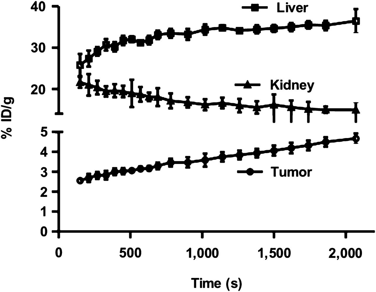
- FIGURE 3.
Time–activity curves of tumor and major organs of C57BL/6 mice bearing B16F10 murine melanoma tumors from 35-min dynamic scans after intravenous injection of 64CuCl2 (3 MBq [∼80 μCi]/mouse, n = 4).
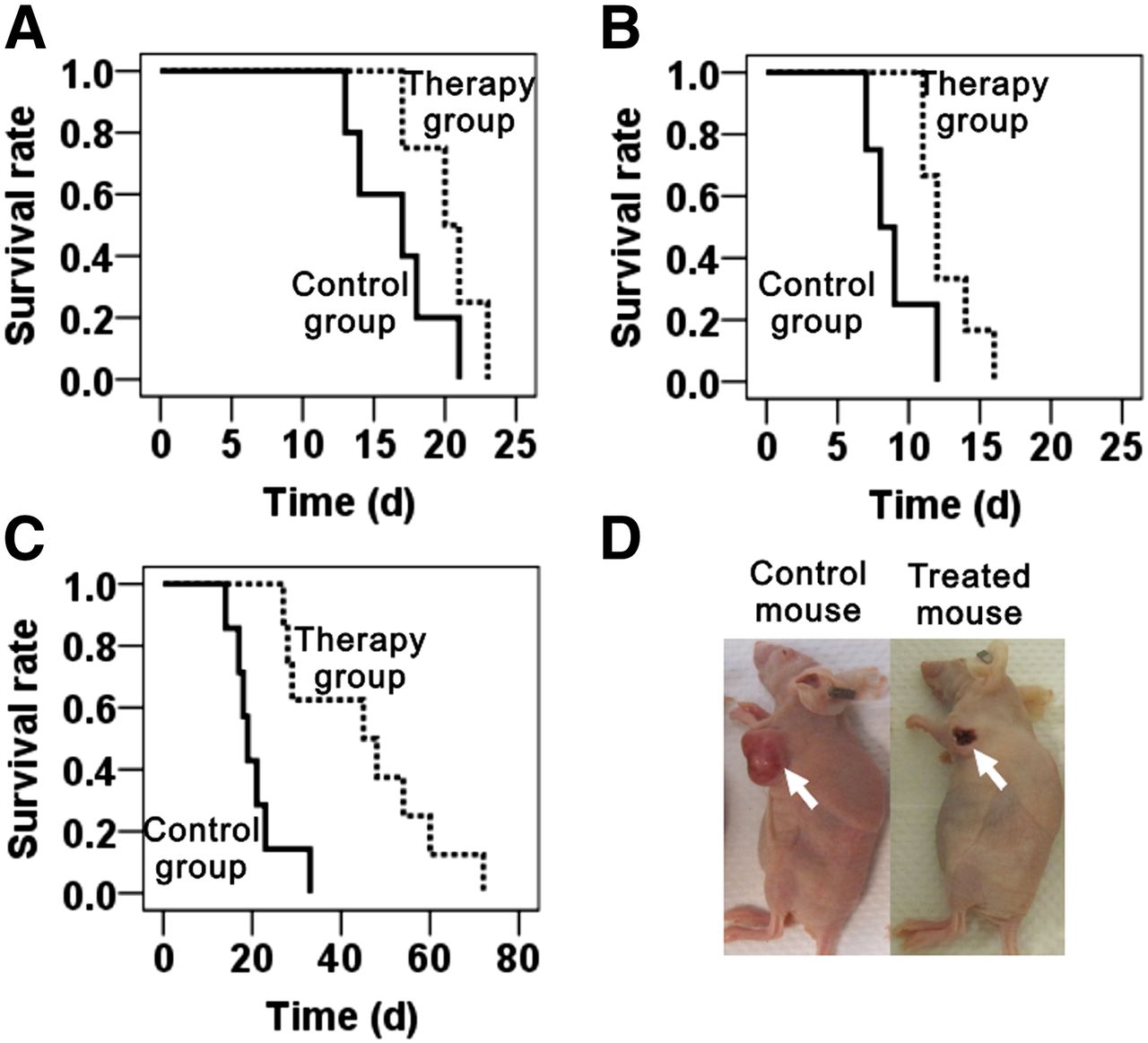
- FIGURE 4.
(A–C) Kaplan–Meier plot of time-to-sacrifice for therapy group (dotted line) and control group (solid line). (A) Group of mice inoculated with 1 million B16F10 cells. (B) Group of mice inoculated with 2 million B16F10 cells. (C) Group of A375M tumor models. Time is expressed in days from therapy. (D) Representative images of A375M tumor necrosis after 64CuCl2 treatment (left, control mouse; right, treated mouse).
- FIGURE 5.
H&E staining of livers and kidneys from therapy group and control group.


Tables
Organ B16F10 A375M Blood 1.51 ± 0.19 1.94 ± 0.56 Heart 5.16 ± 0.73 8.85 ± 0.65 Lungs 6.64 ± 0.48 10.17 ± 1.58 Liver 14.06 ± 2.33 13.37 ± 1.32 Spleen 3.46 ± 0.42 4.59 ± 0.10 Pancreas 2.74 ± 0.36 3.48 ± 0.61 Stomach 3.92 ± 0.82 6.47 ± 0.86 Brain 0.88 ± 0.09 1.00 ± 0.07 Intestine 4.35 ± 0.99 6.82 ± 0.40 Kidneys 11.86 ± 0.56 10.34 ± 0.53 Skin 1.18 ± 0.51 1.95 ± 0.36 Muscle 1.03 ± 0.18 1.13 ± 0.36 Bone 1.66 ± 0.24 1.82 ± 0.24 Tumor 4.14 ± 0.24 3.59 ± 0.36 Uptake ratio Tumor to blood 2.79 ± 0.42 1.94 ± 0.44 Tumor to lung 0.63 ± 0.07 0.36 ± 0.04 Tumor to liver 0.30 ± 0.04 0.26 ± 0.04 Tumor to muscle 4.11 ± 0.07 3.46 ± 1.25 Data are presented as percentage injected dose per gram (%ID/g) ± SD.
Supplemental Data
Files in this Data Supplement:


Jump to section
Related Articles
Cited By...
- Insights into Trace Metal Metabolism in Health and Disease from PET: "PET Metallomics"
- Monte Carlo N-Particle (MCNP) Modeling of the Cellular Dosimetry of 64Cu: Comparison with MIRDcell S Values and Implications for Studies of Its Cytotoxic Effects
- Detection of Increased 64Cu Uptake by Human Copper Transporter 1 Gene Overexpression Using PET with 64CuCl2 in Human Breast Cancer Xenograft Model