


Article Figures & Data
Figures
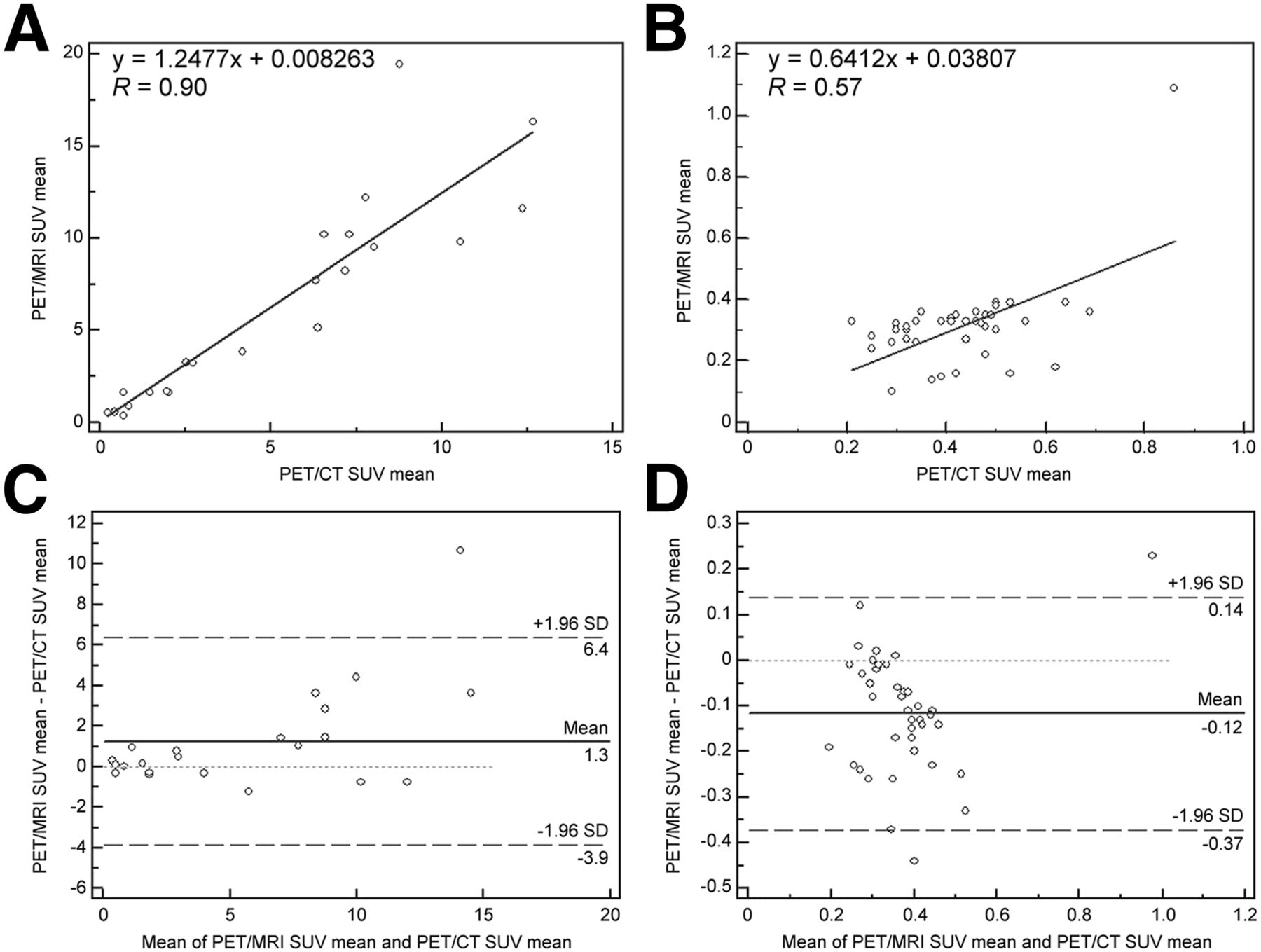
- FIGURE 1.
Correlation analysis of tracer uptake between PET/CT and subsequent PET/MR imaging as assessed by SUVmean in lung lesions (A) and normal lung parenchyma (B). x-axis displays quantitative values as obtained by PET/CT, and y-axis displays corresponding values by PET/MR imaging. High correlations as expressed by Spearman correlation coefficient (R) are found for SUVmean (0.9 for normal lung parenchyma, 0.57 for lung lesions) between findings from both modalities. Difference between 2 SUV measurements is shown by Bland–Altman (for SUVmean in normal lung parenchyma [C] and for SUVmean in lung lesions [D]) on which difference between 2 SUV measurements is plotted against their average. For SUVmean-healthy lung (mean SUV in healthy lung), mean difference is 1.3 SUV; 95% CI is +6.4 and −3.9 SUV; for SUVmean_lung lesion (mean SUV of lung lesion), mean difference is −0.12 SUV; 95% CI for SUVmax is +0.14 and −0.37 SUV.
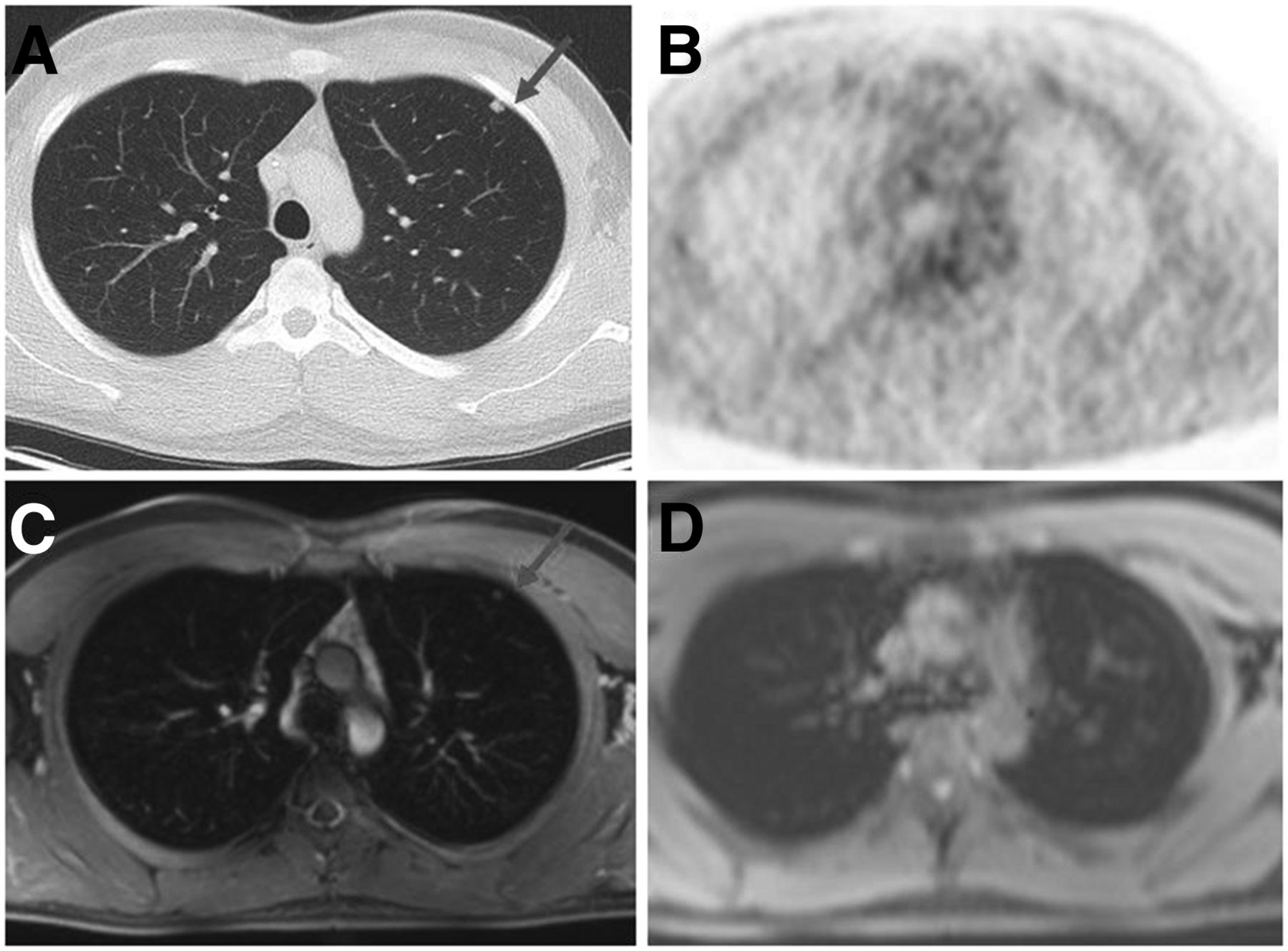
- FIGURE 2.
A 26-y-old patient with testicular cancer. (A, C, and D) Morphologic datasets (axial CT [A], axial VIBE [C], and axial Dixon sequence [D] [in phase]). (B) Corresponding PET of PET/CT. Lung lesion (arrow) was seen in CT and VIBE scan but not in Dixon sequence. Additionally, there was no suspected 18F-FDG uptake.


Tables
Sequence T1-weighted VIBE Dixon T1-weighted VIBE fat saturated Repetition time/echo time (ms)* 3.61/1.23 and 2.46 3.29/1.16 Slice thickness (mm) 3.1 5 Gap (%) 0 0 Matrix 79 × 192 320 × 240 Field of view (mm) 500 460 Bandwidth (kHz) 960 540 Voxel size (mm3) 4.1 × 2.6 × 3.1 1.8 × 1.4 × 5 Flip angle (°) 10 9 % phase field of view 65.5 75.0 Acquisition time (min:sec) 0:19 0:16 No. of excitations 1 1 Integrated parallel acquisition technique factor 2 2 ↵* Attenuation-correction technique with T1 VIBE Dixon requires 2 echo times.
No. of lung lesions Size of lung lesions (mm ± SD) Group CT (standard of reference) Dixon VIBE PET MR imaging PET CT CT (standard of reference) Dixon VIBE PET MR imaging PET CT All 47 15 32 22 22 10.0 ± 11.4 (2–60) 12.2 ± 5.0 (4–21) 11.9 ± 11.7 (2–59) 13.5 ± 10.2 (4–49) 13.5 ± 10.2 (4–49) <1 cm diameter 33 9 15 10 10 >1 cm diameter 14 6 17 12 12 Data in parentheses are ranges.
Modality 1—most likely unspecific/benign 2—indeterminate 3—suggestive of malignancy CT (n = 47) 21 2 24 VIBE (n = 32) 9 7 16 Dixon (n = 15) 2 4 9
Supplemental Data
Files in this Data Supplement:


Jump to section
Related Articles
Cited By...
- Evaluation of Prostate Cancer with PET/MRI
- Comparative Performance of 18F-FDG PET/MRI and 18F-FDG PET/CT in Detection and Characterization of Pulmonary Lesions in 121 Oncologic Patients
- 18F-FDG PET/CT and PET/MRI Perform Equally Well in Cancer: Evidence from Studies on More Than 2,300 Patients
- Evaluation of the Outcome of Lung Nodules Missed on 18F-FDG PET/MRI Compared with 18F-FDG PET/CT in Patients with Known Malignancies