


Article Figures & Data
Figures
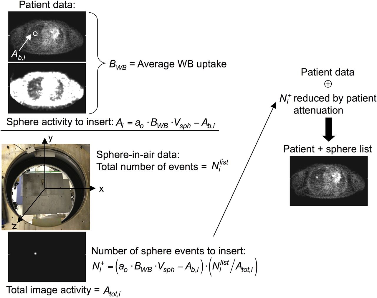
- FIGURE 1.
Schematic of sphere insertion process for list-mode data. Sphere activity to insert (Ai) depends on desired activity ratio (ao) with respect to average whole-body (WB) uptake per unit volume (BWB), reduced by activity already present in patient image at location of sphere (Ab,i). Sphere-in-air data were acquired at known locations on grid (photograph). Sphere data were reconstructed, and ratio of sphere-in-air list-mode events to total sphere image activity (
 ) was used to scale Ai to determine number of list-mode events () that would generate that activity. These list-mode events were reduced by sampling of probability of attenuation by body for line of response of given event and then were merged with subject’s list-mode data (⊕). This procedure was adapted from that used in earlier lesion detectability studies (16,17).
 - FIGURE 2.
Sphere NUV [
] for 1 replicate in 1 subject as function of image noise for each of 6 spheres inserted in lung (A) and liver (B), demonstrating typical variations seen across locations and organs. Solid symbols indicate TOF reconstruction; open symbols indicate non-TOF reconstruction. Each curve represents 1 sphere; data points correspond to each of 20 iterations used. - FIGURE 3.
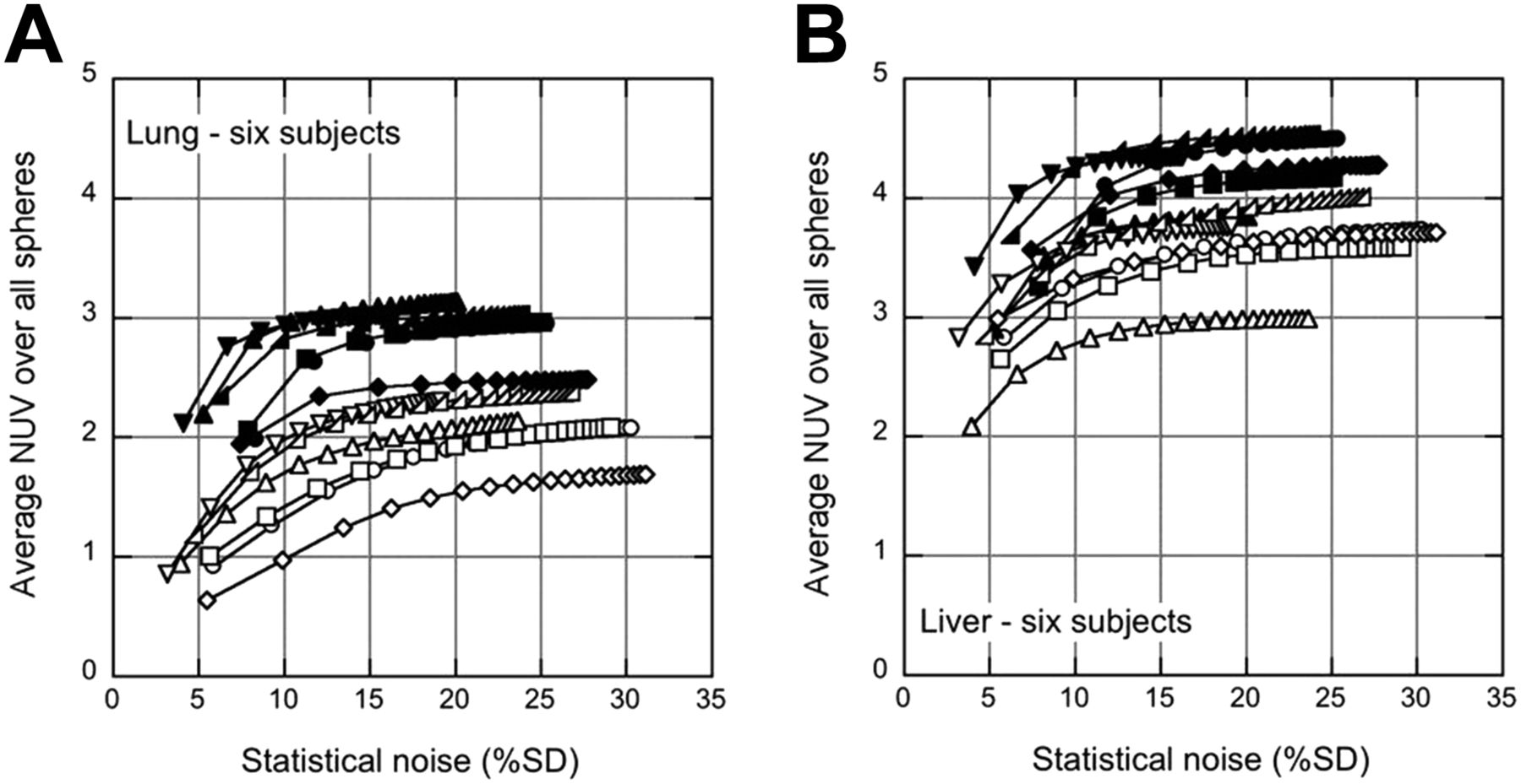
Average NUV across all 6 spheres [
] in given organ for each of 6 subjects as function of image noise in lung (A) and liver (B). Solid symbols indicate TOF reconstruction; open symbols indicate non-TOF reconstruction. Each curve represents 1 subject; data points correspond to each of 20 iterations used. - FIGURE 4.
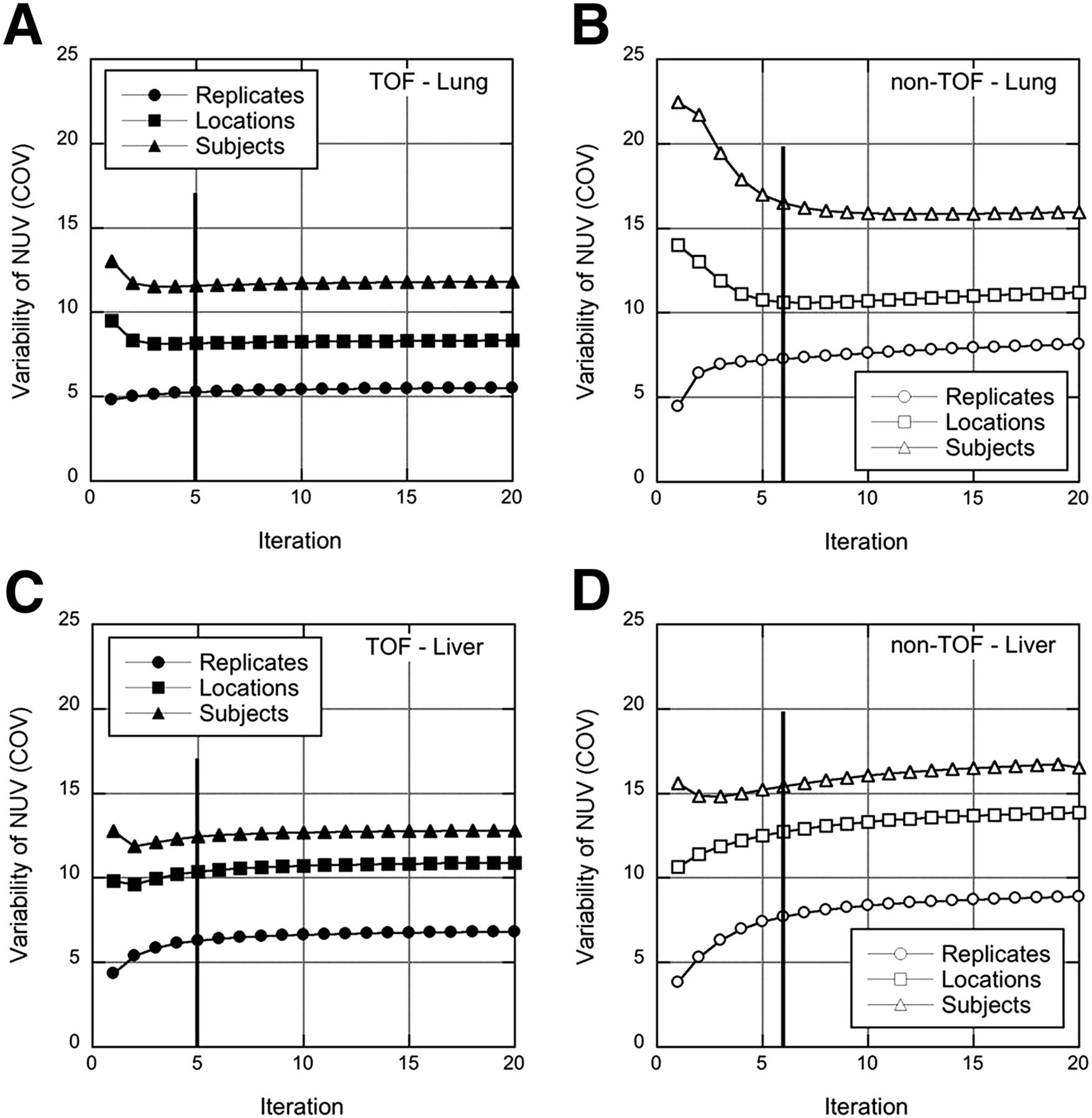
Variability measures for TOF (left) and non-TOF (right) reconstructions of spheres in lung (A and B) and liver (C and D). Variabilities across replicates (
), across sphere locations within organ (), and across spheres and subjects () are shown as function of iteration. Vertical lines at 5 TOF iterations (6 non-TOF iterations) indicate points (with similar image noise) at which reconstructions were stopped for subsequent analysis. - FIGURE 5.
Average uptake and variability at fixed number of iterations and corresponding image noise (5 TOF iterations; 6 non-TOF iterations). (A) Sphere uptake (
) in lung and liver, averaged across all spheres in all subjects. (B) Variability of NUV measurements across replicates (Repl.) (), across sphere locations (Loc.) (), and across subjects (Subj.) () for spheres inserted in lung and liver.


Jump to section
Related Articles
Cited By...
- Total-Body PET System Designs with Axial and Transverse Gaps: A Study of Lesion Quantification and Detectability
- Performance Characteristics of a New Generation 148-cm Axial Field-of-View uMI Panorama GS PET/CT System with Extended NEMA NU 2-2018 and EARL Standards
- Benefit of Improved Performance with State-of-the Art Digital PET/CT for Lesion Detection in Oncology
- Display of 3D Multimodality Cardiac Images With 2D Polar Maps: Simplicity Can Be a Virtue
- Techniques, Benefits, and Challenges of PET-MR
- Improving the Detection of Small Lesions Using a State-of-the-Art Time-of-Flight PET/CT System and Small-Voxel Reconstructions
- Update on Time-of-Flight PET Imaging