


Article Figures & Data
Figures
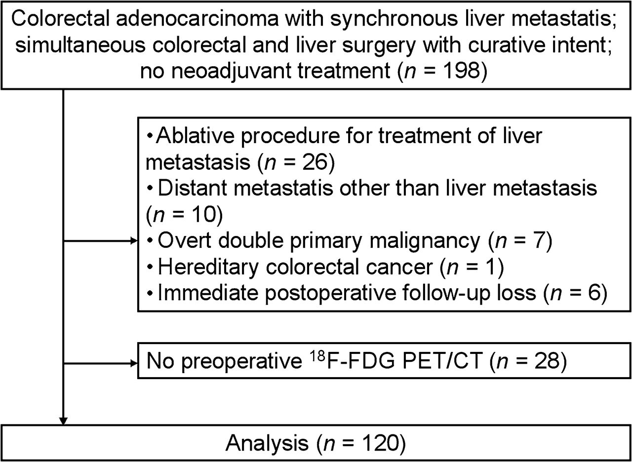
- FIGURE 1.
Flow diagram outlining criteria used for patient inclusion and exclusion.
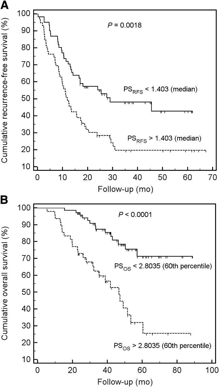
- FIGURE 2.
Kaplan–Meier curves for RFS dichotomized using median cutoff value of PSRFS (A) and for OS dichotomized using 60th-percentile cutoff value of PSOS (B).

Tables
- TABLE 1
Demographics and Clinicopathologic Features of Primary Colorectal Cancers and Liver Metastases
Characteristic n or mean ± SD Age (y) 59.9 ± 10.1 (range, 35−80) Sex Male 82 (68%) Female 38 (32%) Preoperative carcinoembryonic antigen (ng/mL) 35.2 ± 99.8 (range, 0.32−812.8) Pathologic stage T T2 1 (1%) T3 107 (89%) T4 12 (10%) N N0 29 (24%) N1 47 (39%) N2 44 (37%) Location of primary tumor Colon 74 (62%) Rectum 46 (38%) Bilobar hepatic metastasis Yes 21 (17%) No 99 (83%) Hepatic resection margin Positive 13 (11%) Negative 107 (89%) Differentiation grade Well differentiated 4 (3%) Moderately differentiated 110 (92%) Poorly differentiated 6 (5%) Number of hepatic metastasis 1 70 (59%) 2 29 (24%) 3 10 (8%) >3 11 (9%) Size of primary tumor (cm) 5.5 ± 1.8 (range, 1.7−11.2) Size of hepatic metastasis (cm) 3.5 ± 2.4 (range, 0.4−14.0) Adjuvant chemotherapy No 4 (3%) Yes 116 (97%) Total 120 (100%) Metabolic parameter Mean ± SD SUVmean of normal liver 1.8 ± 0.3 (1.2−2.9) SUVpeak of primary tumor 8.7 ± 4.0 (2.1−29.9) nSUVpeak of primary tumor 5.0 ± 2.3 (1.4−16.9) SUVpeak of hepatic metastasis 5.4 ± 2.8 (1.8−20.6) nSUVpeak of hepatic metastasis 3.1 ± 1.6 (0.9−11.6) MTV of primary tumor (cm3) 20.6 ± 15.3 (3.0−124.7) MTV of hepatic metastasis (cm3) 32.2 ± 61.9 (0.0−450.4) nTLG of primary tumor (cm3) 79.5 ± 75.4 (6.0−555.0) nTLG of hepatic metastasis (cm3) 91.6 ± 182.7 (0.0−1215.6) Data in parentheses are ranges.
- TABLE 3
Univariate Cox Regression Analysis for Clinicopathologic Risk Factors Associated with Survival
RFS OS Variable n Hazard ratio 95% CI P Hazard ratio 95% CI P Age (y) 120 1.009 0.987−1.032 0.428 1.034 1.000−1.069 0.052 Sex Female 38 — — — — — — Male 82 1.462 0.885−2.414 0.138 1.308 0.639−2.677 0.462 Size of primary tumor (cm) 120 1.034 0.908−1.176 0.617 0.982 0.826−1.168 0.838 Size of hepatic metastasis (cm) 120 1.088 0.998−1.187 0.055 1.131 1.016−1.259 0.025 Preoperative serum carcinoembryonic antigen (ng/mL) 120 1.001 0.999−1.003 0.298 1.002 0.999−1.004 0.179 T stage T2, T3 108 — — — — — — T4 12 1.194 0.595−2.396 0.617 1.019 0.361−2.876 0.972 N stage N0 29 — — 0.822 — — 0.776 N1 47 0.881 0.494−1.571 0.667 1.281 0.557−2.947 0.560 N2 44 1.031 0.578−1.839 0.919 1.035 0.434−2.471 0.937 Positive hepatic resection margin No 107 — — — — — — Yes 13 1.315 0.656−2.639 0.440 1.518 0.637−3.620 0.346 No. of hepatic metastasis 120 1.200 1.009−1.427 0.039 1.058 0.858−1.304 0.596 Bilobar hepatic metastasis No 99 — — — — — — Yes 21 0.886 0.488−1.609 0.691 0.735 0.288−1.880 0.521 Differentiation grade Well or moderately differentiated 114 — — — — — — Poorly differentiated 6 1.934 0.703−5.324 0.202 5.234 1.803−15.196 0.002 Location of primary tumor Colon 74 — — — — — — Rectum 46 1.265 0.803−1.992 0.310 0.986 0.524−1.857 0.966 Adjuvant chemotherapy No 4 — — — — — — Yes 116 0.585 0.214−1.605 0.298 0.414 0.099−1.738 0.228 Fong’s clinical risk score 120 1.225 0.891−1.683 0.214 1.310 0.850−2.021 0.224 - TABLE 4
Univariate Cox Regression Analysis for Metabolic Risk Factors Associated with Survival
RFS OS Variable n Hazard ratio 95% CI P Hazard ratio 95% CI P nSUVpeak of primary tumor 120 0.932 0.837−1.038 0.202 0.984 0.860−1.127 0.820 nSUVpeak of hepatic metastasis 120 1.106 0.982−1.245 0.097 1.209 1.055−1.386 0.006 M/P ratio of nSUVpeak 120 2.351 1.308−4.226 0.004 1.849 0.891−3.837 0.099 MTV of primary tumor (cm3) 120 1.012 0.996−1.028 0.139 1.005 0.988−1.021 0.589 MTV of hepatic metastasis (cm3) 120 1.003 1.000−1.006 0.068 1.003 1.000−1.006 0.058 M/P ratio of MTV 120 1.036 0.983−1.092 0.184 1.051 0.989−1.117 0.108 nTLG of primary tumor (cm3) 120 1.001 0.997−1.005 0.702 1.000 0.996−1.004 0.949 nTLG of hepatic metastasis (cm3) 120 1.001 1.000−1.002 0.065 1.001 1.000−1.002 0.023 M/P ratio of nTLG 120 1.040 0.989−1.093 0.129 1.049 0.989−1.112 0.108 Model Variable β Hazard ratio 95% CI P 1 Sex (male) 0.491 1.634 0.979−2.726 0.060 nSUVpeak of primary tumor −0.136 0.872 0.763−0.998 0.047 nSUVpeak of hepatic metastasis 0.218 1.244 1.002−1.543 0.048 Size of hepatic metastasis (cm) 0.050 1.051 0.937−1.180 0.393 No. of hepatic metastasis 0.169 1.184 0.994−1.410 0.058 2 Sex (male) 0.508 1.663 0.997−2.774 0.051 M/P ratio of SUVpeak 0.735 2.086 1.041−4.180 0.038 Size of hepatic metastasis (cm) 0.070 1.073 0.967−1.190 0.183 No. of hepatic metastasis 0.185 1.204 1.011−1.433 0.037 3 Sex (male) 0.492 1.635 0.979−2.730 0.060 MTV of hepatic metastasis (cm3) 0.000 1.000 0.994−1.005 0.887 Size of hepatic metastasis (cm) 0.127 1.136 0.964−1.338 0.129 No. of hepatic metastasis 0.213 1.237 1.040−1.472 0.016 4 Sex (male) 0.490 1.633 0.978−2.726 0.061 nTLG of hepatic metastasis (cm3) 0.000 1.000 0.998−1.002 0.937 Size of hepatic metastasis (cm) 0.123 1.131 0.955−1.338 0.153 No. of hepatic metastasis 0.212 1.237 1.039−1.472 0.017 Model Variable β Hazard ratio 95% CI P 5 Age (y) 0.035 1.036 1.001−1.073 0.047 nSUVpeak of primary tumor −0.152 0.859 0.717−1.030 0.101 nSUVpeak of hepatic metastasis 0.363 1.438 1.080−1.916 0.013 Size of hepatic metastasis (cm) 0.032 1.033 0.895−1.191 0.660 Differentiation grade (poor) 1.344 3.836 1.255−11.724 0.018 6 Age (y) 0.032 1.032 0.997−1.069 0.070 M/P ratio of SUVpeak 0.233 1.263 0.497−3.207 0.624 Size of hepatic metastasis (cm) 0.112 1.118 0.986−1.269 0.082 Differentiation grade (poor) 1.546 4.694 1.515−14.515 0.007 7 Age (y) 0.031 1.031 0.997−1.067 0.077 MTV of hepatic metastasis (cm3) 0.000 1.000 0.993−1.007 0.914 Size of hepatic metastasis (cm) 0.138 1.148 0.915−1.440 0.234 Differentiation grade (poor) 1.635 5.127 1.756−14.975 0.003 8 Age (y) 0.031 1.031 0.997−1.067 0.077 nTLG of hepatic metastasis (cm3) 0.001 1.001 0.998−1.003 0.643 Size of hepatic metastasis (cm) 0.078 1.081 0.854−1.368 0.516 Differentiation grade (poor) 1.651 5.214 1.783−15.248 0.003
Supplemental Data
Files in this Data Supplement:


Jump to section
Related Articles
Cited By...
- No citing articles found.