


Article Figures & Data
Figures
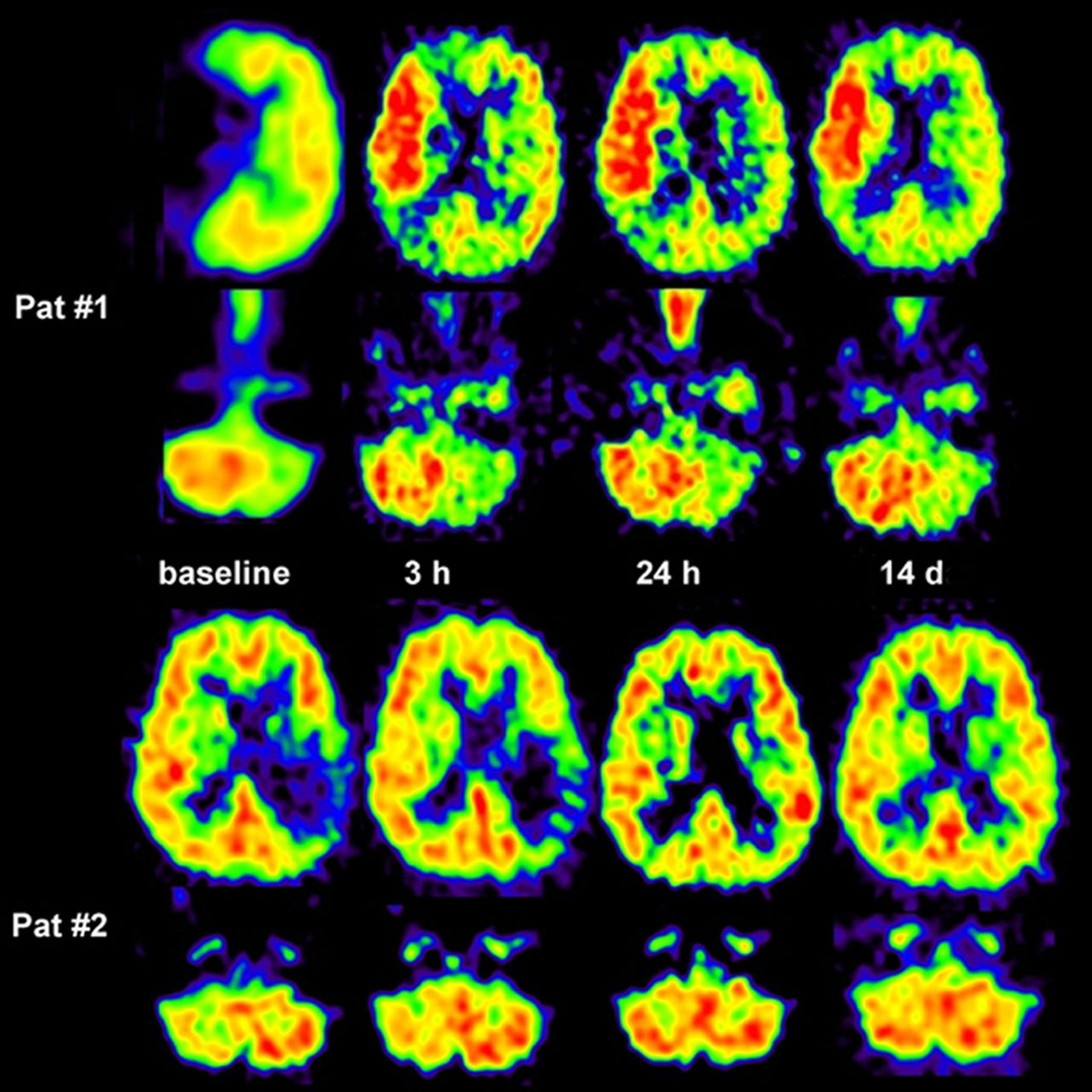
- FIGURE 1.
Four SPECT studies of 99mTc-HMPAO distribution in 47-y-old man. First study was approximately 20 h after right-sided hemiplegia and sensory aphasia had occurred. Profound decrease in uptake is seen in left parietal and temporal region. This area is surrounded in central and frontal directions by slightly increased tracer deposition. Seven and 14 d later, high uptake is seen in previously low-perfusion region. Last control study 29 d after insult shows decreased uptake in left parietal and temporal lobe and in left central and laterofrontal regions. Clinically, hemiplegia improved to hemiparesis and speech was still disturbed but improved. Late CT scan showed hypodense lesion in left parietal lobe.
- FIGURE 2.
PET images of CBF, CMRO2, and OEF in patients with acute ischemic stroke. (Left 2 columns) Areas with preserved OEF are not infarcted and can survive in spontaneous course (posterior part of ischemic cortex in patient 1, anterior part in patient 2 as indicated on CMRGlc and on late MR imaging and CT). (Right 2 columns) In patients receiving rtPA treatment, measurements of CMRO2 and OEF are not feasible, but flow determinations show effect. If reperfusion occurs early enough and before tissue damage, tissue can be salvaged (patient 3). If reperfusion is achieved too late, tissue cannot be salvaged despite hyperperfusion in some parts (patient 4).
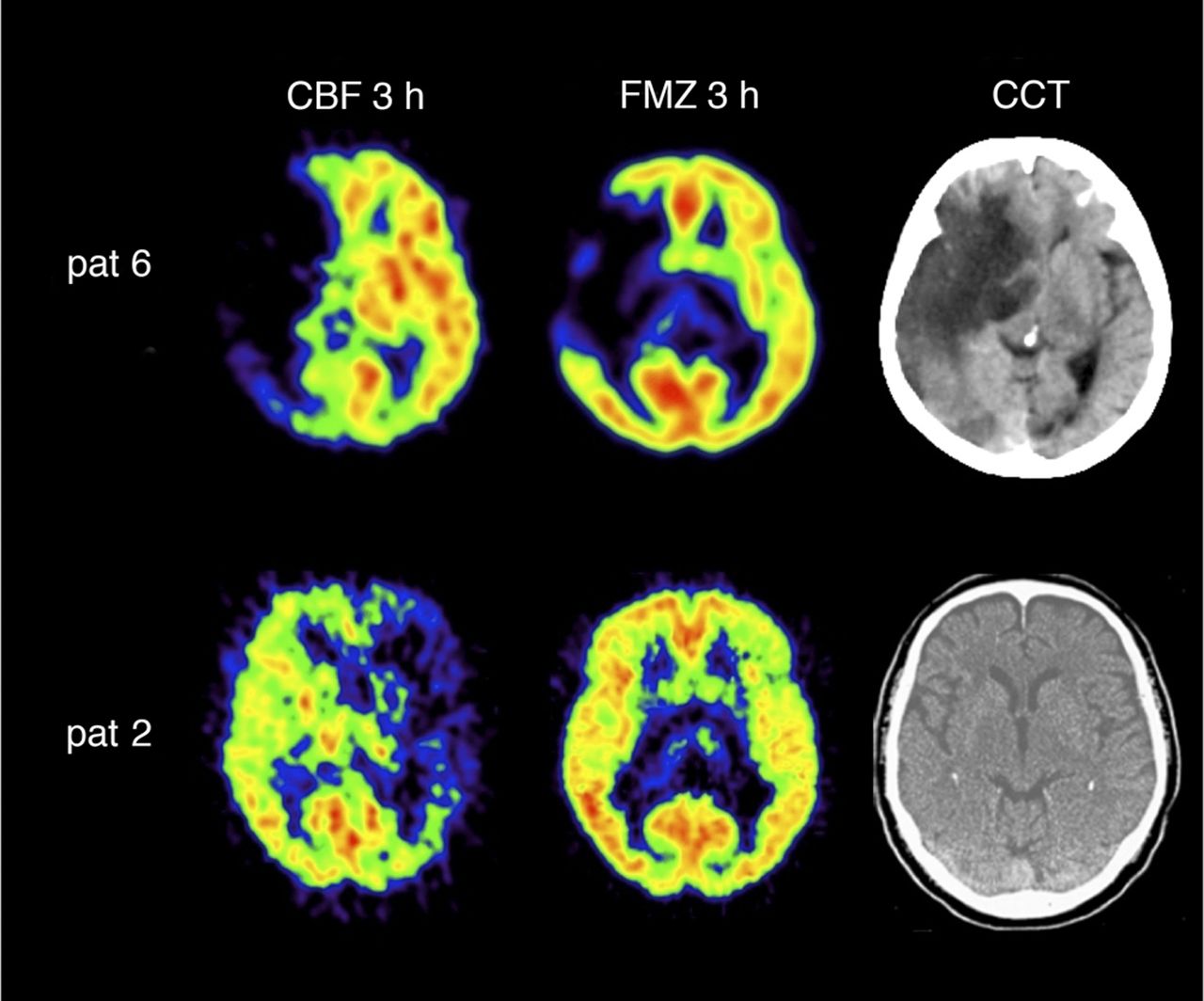
- FIGURE 3.
11C-flumazenil distribution and 11C-flumazenil binding in patients with large ischemic areas and effect of rtPA treatment. In patient 6, with area of decreased 11C-flumazenil (FMZ) binding indicating irreversible damage, rtPA was not effective and large malignant infarction developed (late cranial CT [CCT]). In patient 2, with no significant decrease of 11C-flumazenil binding in area of low blood flow, reperfusion by rtPA was successful and no infarction is seen on late CT.
- FIGURE 4.
Coregistered images of multitracer PET (top row) and MR PWI and DWI (middle row) in patient with severe right-sided hemiparesis and aphasia 4 h after onset of left middle cerebral artery stroke. Penumbra is characterized by hypoperfusion (CBF), preserved CMRO2, and elevated OEF (top row). These findings correspond grossly to mismatch between DWI hyperintensity in anterior and deep parts of middle cerebral artery territory and PWI delay of 4 s in posterior parts compared with unaffected hemisphere. After rtPA treatment, demarcation of small infarct on DWI on day 2 persisted on T2-weighted MR imaging (bottom row).
- FIGURE 5.
Crossed cerebellar diaschisis in acute human stroke and response to supratentorial reperfusion. In patient 1, crossed cerebellar diaschisis persisted despite marked supratentorial reperfusion or hyperperfusion. Infarct volume was 60 cm3, and clinical outcome was poor (National Institutes of Health Stroke Scale, 9 points). In patient 2, supratentorial reperfusion was accompanied by crossed cerebellar diaschisis decrease. Follow-up CT showed no infarct; outcome score on National Institutes of Health Stroke Scale was 0 points. (Reprinted with permission of (85).)
- FIGURE 6.
Representative case with carotid stenosis before and after stenting. (A) Preoperative left common carotid artery angiogram shows severe internal carotid artery stenosis (anteroposterior view). (B) Left carotid arterial stenting was performed, and postoperative angiogram reveals no significant residual stenosis (anteroposterior view). (C and D) Preoperative 123I-isopropyl iodoamphetamine SPECT shows hypoperfusion (C) and low cerebrovascular reactivity after acetazolamide (D) in both hemispheres. In right hemisphere, steal phenomenon is detected (D). (E and F) Postoperative SPECT obtained 3 mo after carotid arterial stenting demonstrates increased resting CBF (E) and cerebrovascular reactivity (F) on both ipsilateral and contralateral carotid occluded sides. (Reprinted with permission of (80).)
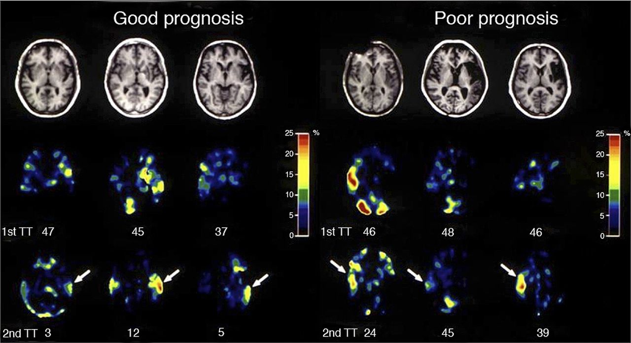
- FIGURE 7.
T1-weighted transaxial MR images (top row) and images of relative metabolic increase in PET (middle and bottom rows) in 6 patients with poststroke aphasia. Middle row shows metabolic increase during single-word repetition in subacute state after stroke, and bottom row shows metabolic increase more than 1 y after stroke. Numbers below images of metabolic increase give number of token test (TT) errors at time of PET. The 3 patients on the right had poor outcome of aphasia and showed maximum metabolic activation of right superior temporal cortex, whereas the 3 patients on the left, with better outcome, showed recovery of metabolic activation in left superior temporal cortex (arrows).


Jump to section
- Article
- Abstract
- PHYSIOLOGIC VARIABLES AFFECTED IN ISCHEMIC STROKE
- RADIOACTIVE TRACERS USED IN STROKE
- APPLICATIONS OF RADIOISOTOPE STUDIES IN STROKE
- RADIOISOTOPE IMAGING AS A SURROGATE MARKER FOR TREATMENT EFFICIENCY AND FOR SELECTION OF PATIENTS FOR SPECIAL THERAPEUTIC STRATEGIES
- MICROGLIAL ACTIVATION AS AN INDICATOR OF INFLAMMATION
- HEMODYNAMIC AND METABOLIC RESERVE IN ARTERIAL OCCLUSIVE DISEASE
- DEACTIVATION OF REMOTE TISSUE (DIASCHISIS)
- ACTIVATION STUDIES IN STROKE PATIENTS
- CONCLUSION AND FUTURE PERSPECTIVES
- Footnotes
- REFERENCES
- Figures & Data
- Info & Metrics
Related Articles
Cited By...
- Erythropoietin Pretreatment of Transplanted Endothelial Colony-Forming Cells Enhances Recovery in a Cerebral Ischemia Model by Increasing Their Homing Ability: A SPECT/CT Study
- Spatiotemporal PET Imaging of Dynamic Metabolic Changes After Therapeutic Approaches of Induced Pluripotent Stem Cells, Neuronal Stem Cells, and a Chinese Patent Medicine in Stroke
- Imaging Inflammation in Cerebrovascular Disease