


Article Figures & Data
Figures
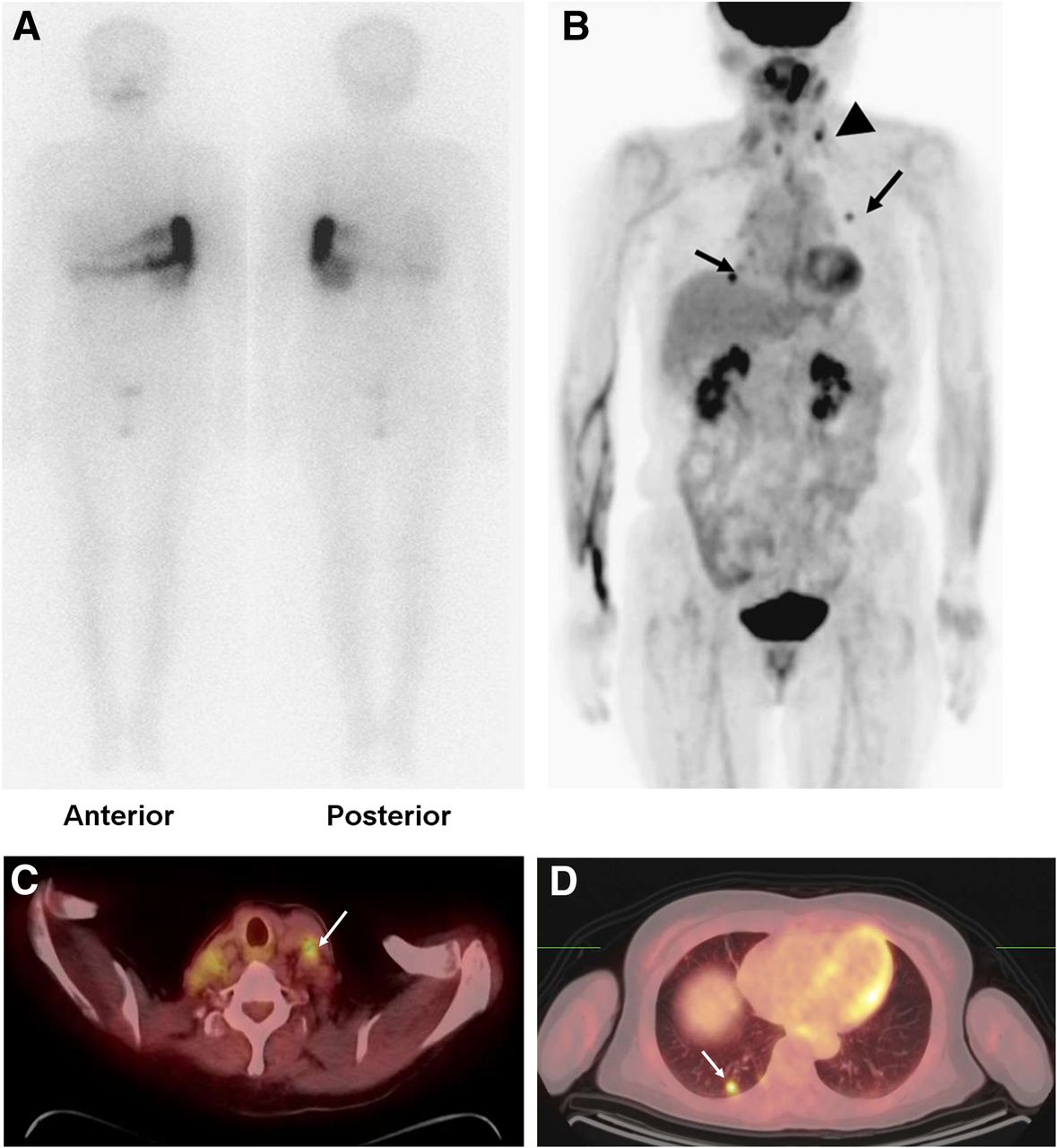
- FIGURE 1.
A 72-y-old woman with papillary thyroid cancer underwent 131I ablation (3.7 GBq) 4 mo after total thyroidectomy (histopathologic stage, T3N1). Posttherapy 131I scan (A) showed no abnormal 131I uptake, suggesting remnant thyroid uptake or metastatic lesion. However, maximal-intensity-projection image (B) and transaxial images (C and D) of 18F-FDG PET/CT showed areas of increased focal uptake in left neck region (arrowhead) and in lung (arrow), suggesting multiple metastatic lesions. Neck lesion was histopathologically diagnosed as metastatic lesion.
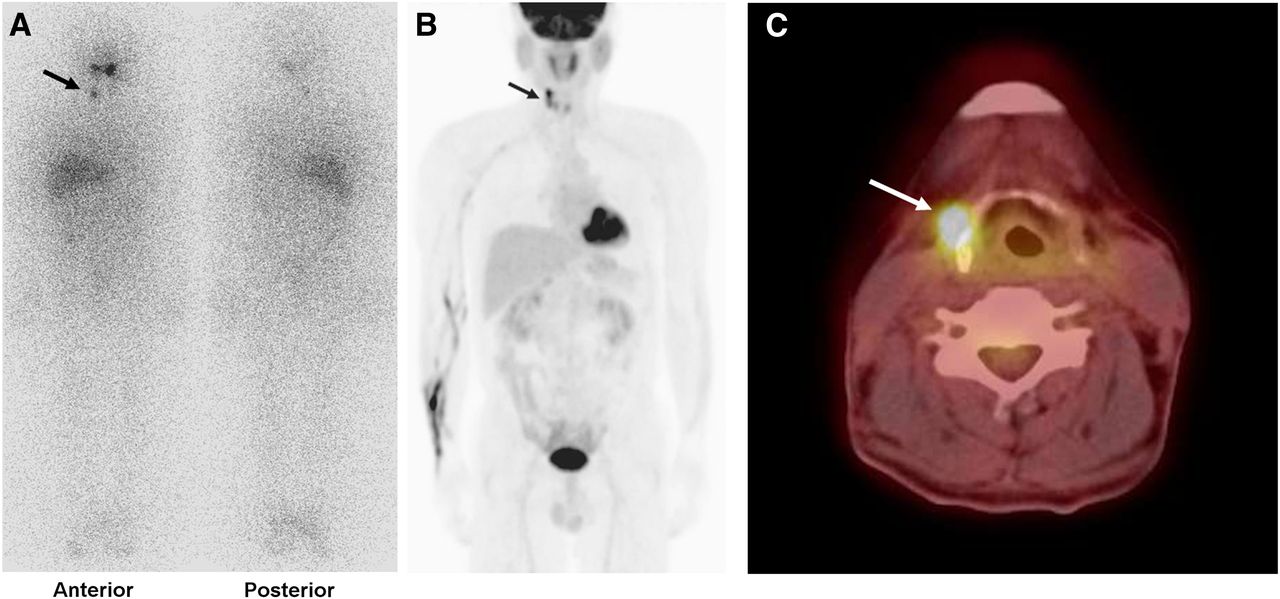
- FIGURE 2.
A 61-y-old man with papillary thyroid cancer underwent adjuvant 131I treatment (5.6 GBq) 3 mo after neck lymph node dissection for recurrent cancer. Posttherapy 131I scan (A) showed focus of increased 131I uptake in right neck area (arrow), which also showed increased focal uptake (arrow) on maximal-intensity-projection (B) and transaxial 18F-FDG PET/CT (C) images. Lesion was histopathologically confirmed as metastatic lesion.
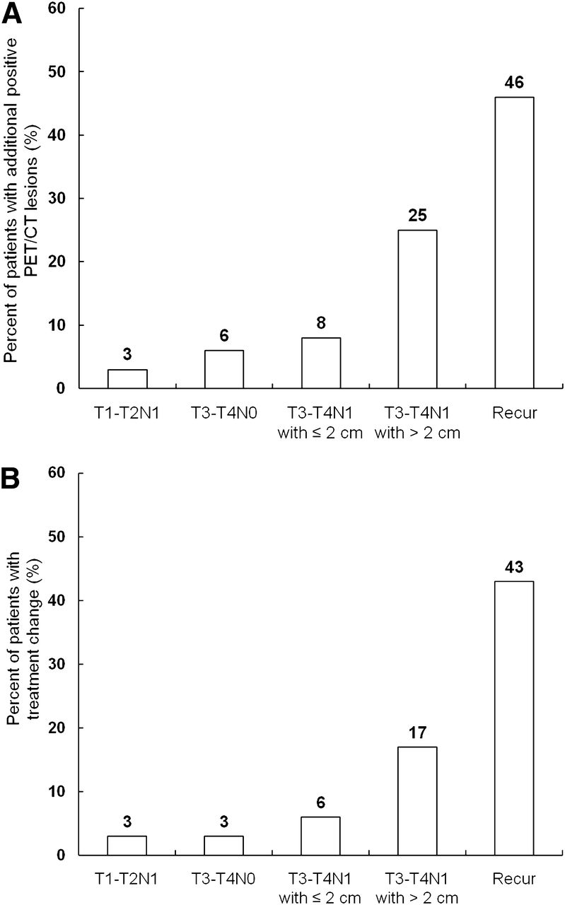
- FIGURE 3.
Ratios of patients with additional positive lesions on 18F-FDG PET/CT (A) and ratios of patients with treatment change due to PET/CT findings (B). Stage T3–T4N1 patients with tumor size > 2.0 cm and patients who underwent adjuvant 131I treatment after operation for recurrence had higher frequency of positive PET/CT findings and treatment change than stage T3–T4N1 patients with tumor size ≤ 2.0 cm, stage T3–T4N0 patients, or stage T1–T2N1 patients (P < 0.05). T3–T4N1 with ≤ 2 cm = stage T3–T4N1 patients with tumor size ≤ 2 cm; T3–T4N1 with > 2 cm = stage T3–T4N1 patients with tumor size > 2 cm; Recur = patients who underwent adjuvant 131I treatment after operation for recurrent tumor.
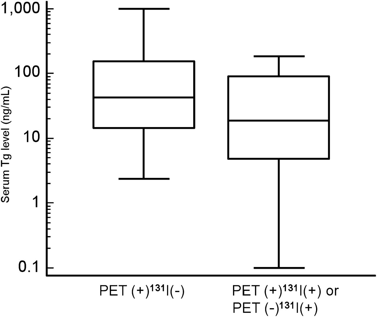
- FIGURE 4.
Serum thyroglobulin values in patients with additional positive lesions on 18F-FDG PET/CT (PET(+)131I(−) group) and in patients with positive lesions on both PET/CT and 131I scan or with positive lesions on only 131I scan (PET(+)131I(+) or PET(−)131I(+) group). There was no significant difference in serum thyroglobulin level between the 2 groups (P > 0.05).


Tables
Characteristic n or mean ± SD % or range Age (y) 51 ± 13 18–87 Sex Male 57 20 Female 229 80 Histopathology Papillary 280 98 Follicular 6 2 Stage* T1–T2N0 2 1 T1–T2N1 66 23 T3–T4N0 44 15 T3–T4N1 174 61 Dose of 131I 3.7 GBq (100 mCi) 41 14 5.6 GBq (150 mCi) 235 82 7.4 GBq (200 mCi) 10 4 Size of primary tumor (cm)† 1.5 ± 1.1 0.3–6.8 Serum TSH level (IU/mL) 88.1 ± 22.1 30.7–100.0 Serum thyroglobulin level (ng/mL)‡ 25.8 ± 91.8 0.1–1000.0 - TABLE 2
Positive 18F-FDG PET/CT Finding and Subsequent Treatment Change According to Stage
Stage Total patients Positive PET/CT lesions Additional positive PET/CT lesions Treatment change Recurrence* 28 16 (57) 13 (46) 12 (43) T3–T4N1 159 28 (18) 21 (13) 15 (9) T3–T4N0 64 4 (6) 4 (6) 2 (3) T1–T2N1 35 2 (6) 1 (3) 1 (3) P <0.0001† <0.0001† <0.0001†


