


Article Figures & Data
Figures
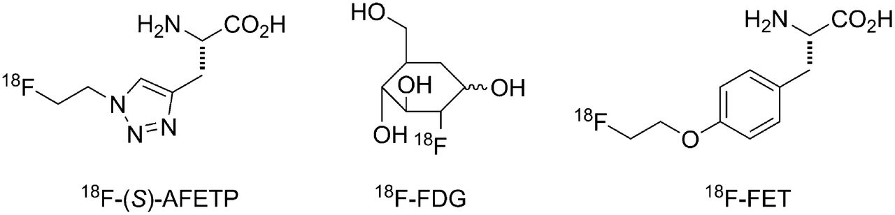
- FIGURE 1.
Structures of 18F-labeled PET tracers evaluated in the mouse DBT glioma model.
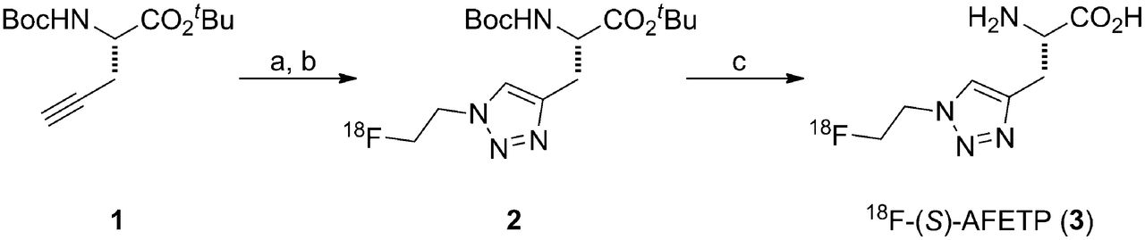
- FIGURE 2.
Radiosynthesis of 18F-AFETP. (A) CuSO4, sodium ascorbate, N,N-dimethylformamide/water 2-18F-fluoroethyl azide. (B) HPLC purification. (C) 1 M aqueous HCl, 60W microwave.
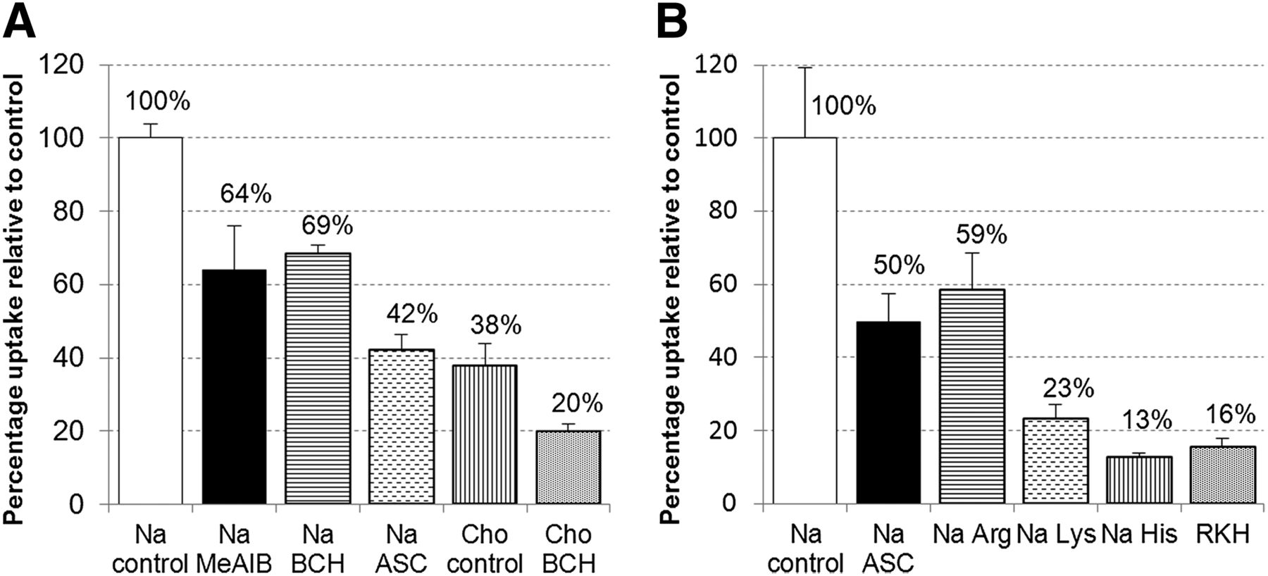
- FIGURE 3.
In vitro uptake of 18F-AFETP by DBT glioma cells in presence of neutral (A) and cationic (B) competitive inhibitors of the amino acid transport. Uptake data are normalized based on amount of activity added to each well and total amount of protein in each well. Data are expressed as percentage uptake relative to sodium control condition, and values for each condition are noted in appropriate bars. To provide consistent osmolarity, compared with inhibitory conditions, Na control and Cho controls contain 10 mM sucrose. ASC = 3.3 mM each of l-Ala, l-Ser, l-Cys; BCH = 10 mM 2-aminobicyclo(2,2,1)-heptane-2-carboxylic acid (system L inhibitor); Cho = assay buffer containing choline ions; MeAIB = 10 mM N-methyl,α-aminoisobutryic acid (system A inhibitor); Na = assay buffer containing sodium ions; RKH = 3.3 mM each of l-arginine, l-lysine, and l-histidine.
- FIGURE 4.
Representative small-animal PET and MR images from mice with intracranial DBTs. Small-animal PET images in A and B and MR image in C were obtained from same animal, whereas small-animal PET image in D was from mouse in different cohort. Tumors are indicated in each panel with arrows. (A, B, and D) Summed images from 50 to 60 min after injection of 18F-AFETP (A), 18F-FDG (B), or 18F-FET (D). MR image (C) was obtained usingT2-weighted sequence, as described in “Materials and Methods” section. Tumors were confirmed histologically (data not shown).
- FIGURE 5.
Representative time–activity curves and tumor-to-brain ratio curves observed with dynamic small-animal PET studies in mice with intracranial DBTs. Time–activity curves for 18F-AFETP (A), 18F-FDG (B), and 18F-FET (C) are displayed as average SUVs. 18F-AFETP and 18F-FDG data in A and B were obtained in same animal, whereas 18F-FET data in C was from animal from different cohort. Data from time–activity curves are expressed as tumor-to-brain ratios over time for all 3 tracers in D. Figure 6 shows average data from these small-animal PET studies from all animals (n = 3 or 4) for each tracer.
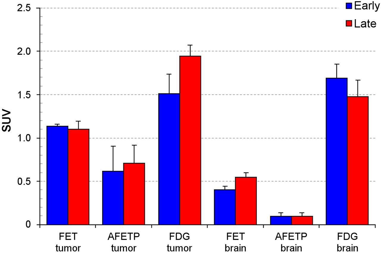
- FIGURE 6.
Average SUVs in intracranial DBTs and contralateral normal brain measured through small-animal PET studies performed with 18F-FET (n = 3), 18F-AFETP (n = 4), and 18F-FDG (n = 4). Early (7–12.5 min) and late (47.5–57.5 min) time points are depicted. Small-animal PET data for 18F-AFETP and 18F-FDG were obtained in same animals, whereas 18F-FET data were obtained in second cohort of animals. Error bars show SD.
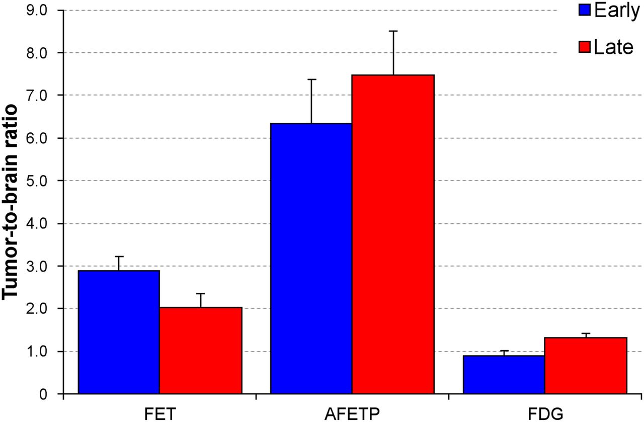
- FIGURE 7.
Tumor-to-brain ratios between intracranial DBTs and contralateral normal brain measured through small-animal PET studies performed with 18F-FET (n = 3), 18F-AFETP (n = 4), and 18F-FDG (n = 4). Data for early (7–12.5 min) and late (47.5–57.5 min) time points are depicted. Small-animal PET data for 18F-AFETP and 18F-FDG were obtained in same animals, whereas 18F-FET data were obtained in second cohort of animals. Error bars show SD.


Tables
Organ or tissue 5 min 30 min 60 min Blood 3.99 ± 0.38 1.66 ± 0.20 0.69 ± 0.18 Bone 0.91 ± 0.17 1.46 ± 0.14 0.99 ± 0.47 Brain 0.25 ± 0.03 0.34 ± 0.40 0.23 ± 0.03 Fat 0.90 ± 0.08 0.61 ± 0.20 0.39 ± 0.17 Heart 2.19 ± 0.19 2.10 ± 0.33 1.64 ± 0.27 Kidney 65.6 ± 10.2 51.4 ± 6.2 17.8 ± 3.7 Large intestine 1.81 ± 0.39 1.79 ± 0.24 1.08 ± 0.29 Liver 3.19 ± 0.50 1.56 ± 0.15 0.65 ± 0.12 Lung 6.06 ± 0.95 5.53 ± 1.87 2.40 ± 0.67 Muscle 1.34 ± 0.19 1.22 ± 0.12 1.02 ± 0.12 Pancreas 36.1 ± 5.1 21.9 ± 4.1 8.75 ± 3.0 Salivary glands 5.23 ± 0.41 2.96 ± 0.46 1.28 ± 0.22 Small intestine 3.97 ± 0.90 2.56 ± 0.38 1.42 ± 0.27 Spleen 3.40 ± 0.42 2.67 ± 0.20 1.67 ± 0.27 Tumor 4.39 ± 1.10 4.91 ± 1.15 2.46 ± 1.02 Data are expressed as %ID/g ± SD; n = 3 or 4 for each value.


