


Article Figures & Data
Figures
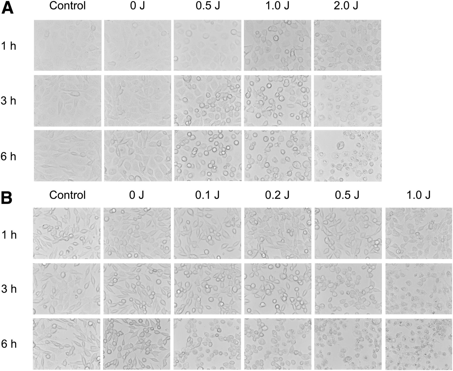
- FIGURE 1.
In vitro microscopy studies on A431 cells (A) and 3T3/HER2 cells (B) after photoimmunotherapy. In both cell lines, excitation light induced cellular swelling, bleb formation, and rupture of vesicles representing necrotic cell death. Change of cell morphology correlated with dose of light and also increased over time after photoimmunotherapy.
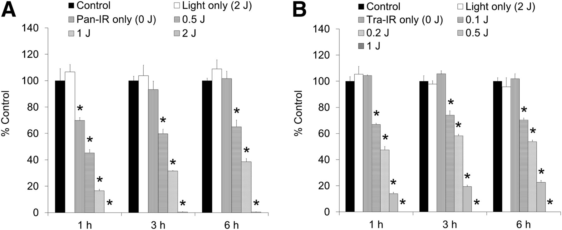
- FIGURE 2.
In vitro 18F-FDG cell uptake studies on A431 cells (A) and 3T3/HER2 cells (B) after photoimmunotherapy. In both cell lines, the uptake of 18F-FDG was reduced in NIR light dose-dependent manner. High light doses completely (>99%) shut down glucose metabolism. Data are represented as mean ± SEM (n = 4 wells). *P < 0.01, compared with control group, Dunnett test.
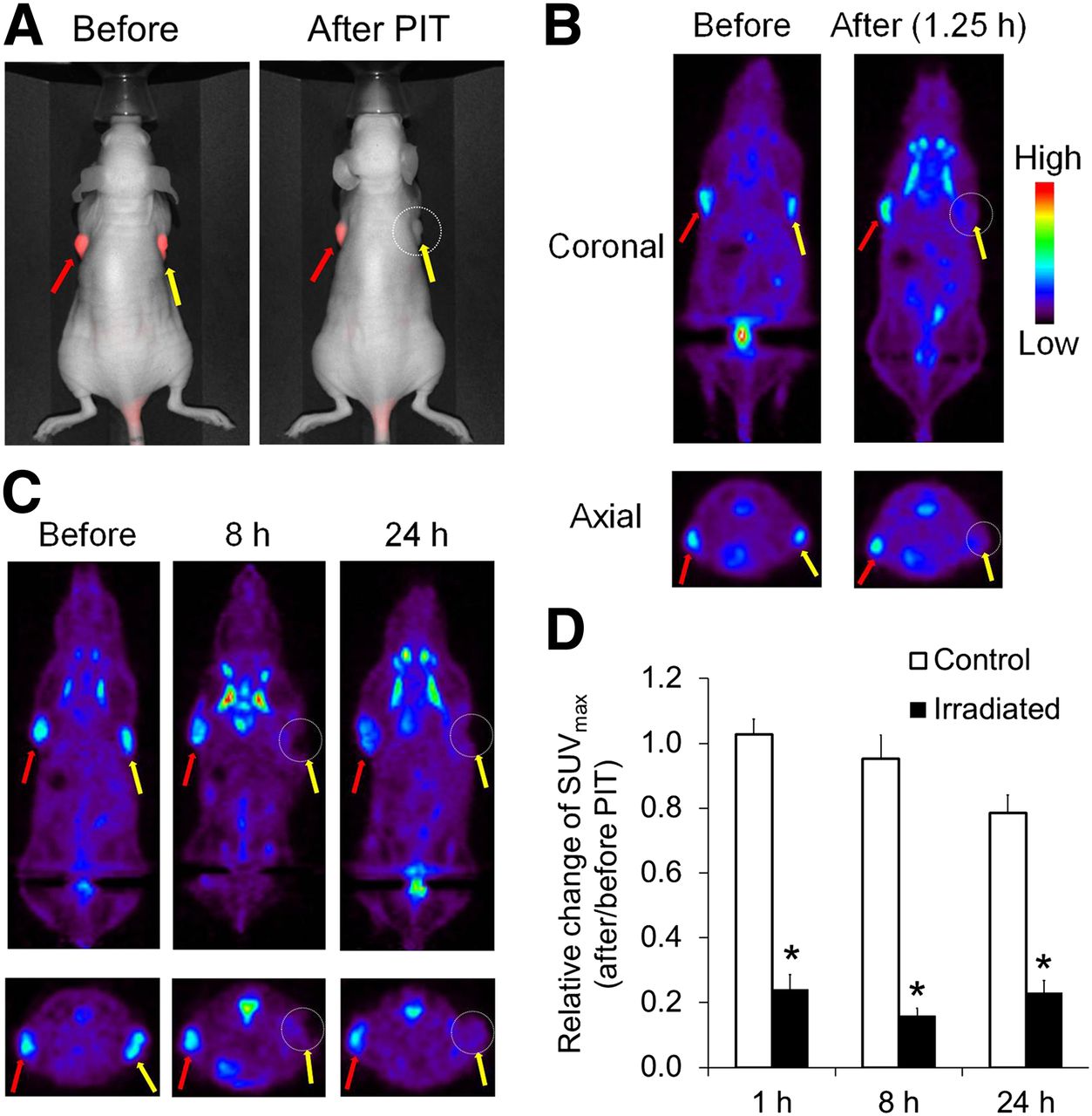
- FIGURE 3.
In vivo 18F-FDG PET imaging before and after photoimmunotherapy. Panitumumab-IR700 was injected intravenously into mice bearing A431 (HER1-positive) tumors on both shoulders. Photoimmunotherapy was performed only in tumors on right shoulders 24 h after injection of panitumumab-IR700. (A) In vivo fluorescence images of IR700 in mice bearing A431 tumors on both shoulders. Fluorescence signals of treated tumors (right side: circled; yellow arrow) were almost completely absent after photoimmunotherapy, whereas those on covered side (left side: red arrow) were unchanged. (B and C) 18F-FDG PET images were acquired 1.25, 8, and 24 h after photoimmunotherapy in mice bearing A431 tumors. Photoimmunotherapy resulted in significant decrease of 18F-FDG uptake within only irradiated tumors (right side: circled; yellow arrow) beginning 1.25 h after treatment. This effect was sustained for at least 24 h. (D) Quantitative data analysis on relative change of SUVmax (after and before photoimmunotherapy). Data are represented as mean ± SEM (n = 4–6 mice). *P < 0.01, compared with control group, Tukey–Kramer test. PIT = photoimmunotherapy.
- FIGURE 4.
In vivo 18F-FDG PET imaging before and after photoimmunotherapy in target-negative animal model. (A) 18F-FDG PET images were acquired 1.25 h after photoimmunotherapy with panitumumab-IR700 in Balb3T3/DsRed tumor (HER1-negative)–bearing mice. (B) Quantitative data analysis on relative change of SUVmax (after and before photoimmunotherapy). Data are represented as mean ± SEM (n = 4–6 mice). Uptake of 18F-FDG was not changed before and after photoimmunotherapy. PIT = photoimmunotherapy.
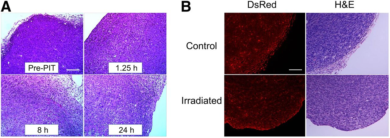
- FIGURE 5.
Histologic findings immediately after photoimmunotherapy. (A) Histologic specimens of A431 tumors before and 1.25, 8, and 24 h after photoimmunotherapy with 100 J/cm2 are shown. All specimens are stained with hematoxylin and eosin. A few scattered clusters of damaged tumor cells are seen within background of diffuse cellular necrosis and microhemorrhage at all time points after photoimmunotherapy. (B) Histologic specimens of Balb3T3/DsRed tumors and control tumors at 1.25 h after photoimmunotherapy. No obvious damage was observed. Scale indicates 50 μm. H&E = hematoxylin and eosin; PIT = photoimmunotherapy.


Jump to section
Related Articles
Cited By...
- No citing articles found.