


Article Figures & Data
Figures
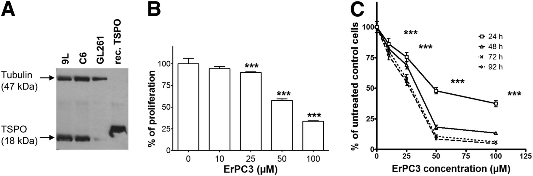
- FIGURE 1.
Expression levels of TSPO and effect of ErPC3 treatment on proliferation and viability of glioma cells in culture. (A) TSPO expression levels assessed by Western blot analysis of 9L, C6, and GL261 cells. β-tubulin was used as protein-loading index. TSPO recombinant protein has a molecular weight of 20.4 kDa, slightly larger than native TSPO. (B) Effect of ErPC3 on 9L cell proliferation as measured by bromodeoxyuridine incorporation. ***P < 0.001 for 25, 50, and 100 μM ErPC3 versus control. (C) Effect of ErPC3 on 9L cell viability using MTT assay. ***P < 0.001, ErPC3- versus control-treated cells, 24 h. Results are expressed as mean ± SD percentage of untreated controls.
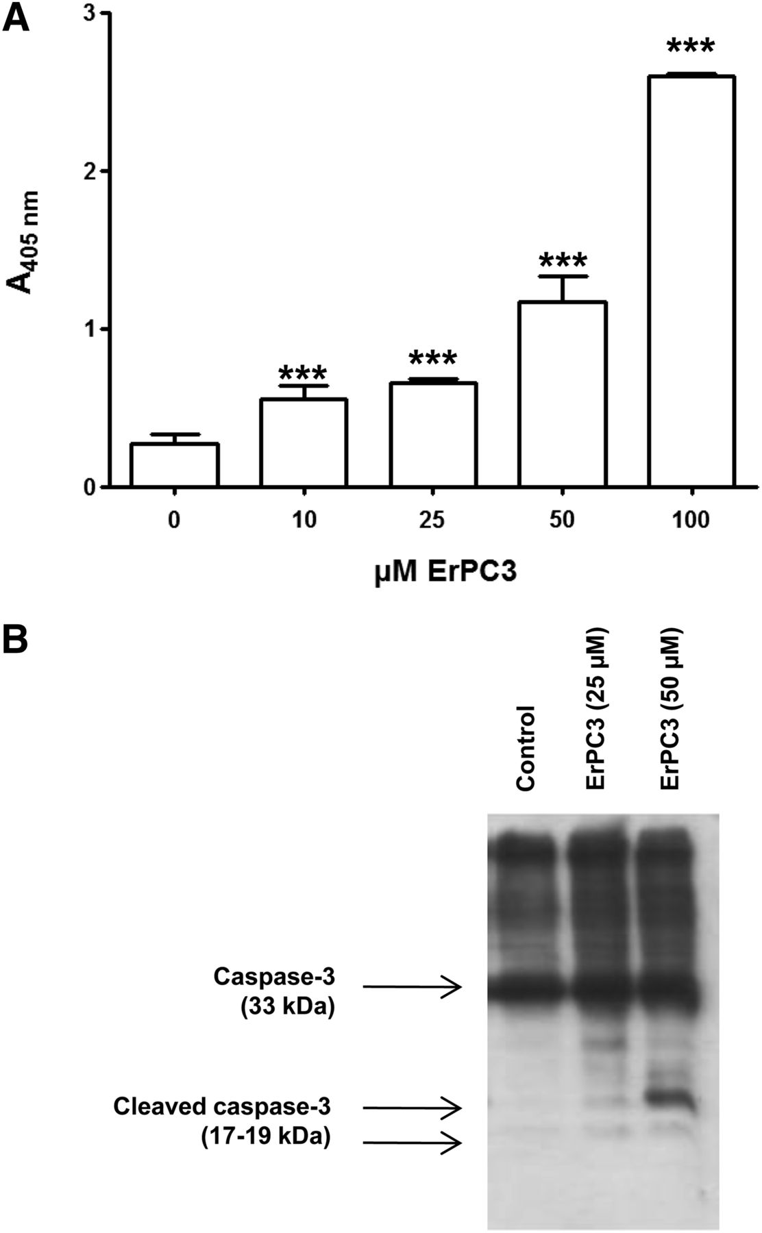
- FIGURE 2.
Effect of ErPC3 on apoptosis. (A) Absorbance at wavelength of 405 nm indicates level of apoptosis. Results are mean ± SD. ***P < 0.001, ErPC3 versus control treatment. (B) Western blot analysis of caspase-3 processing after 0, 25, and 50 μM ErPC3 for 24 h.
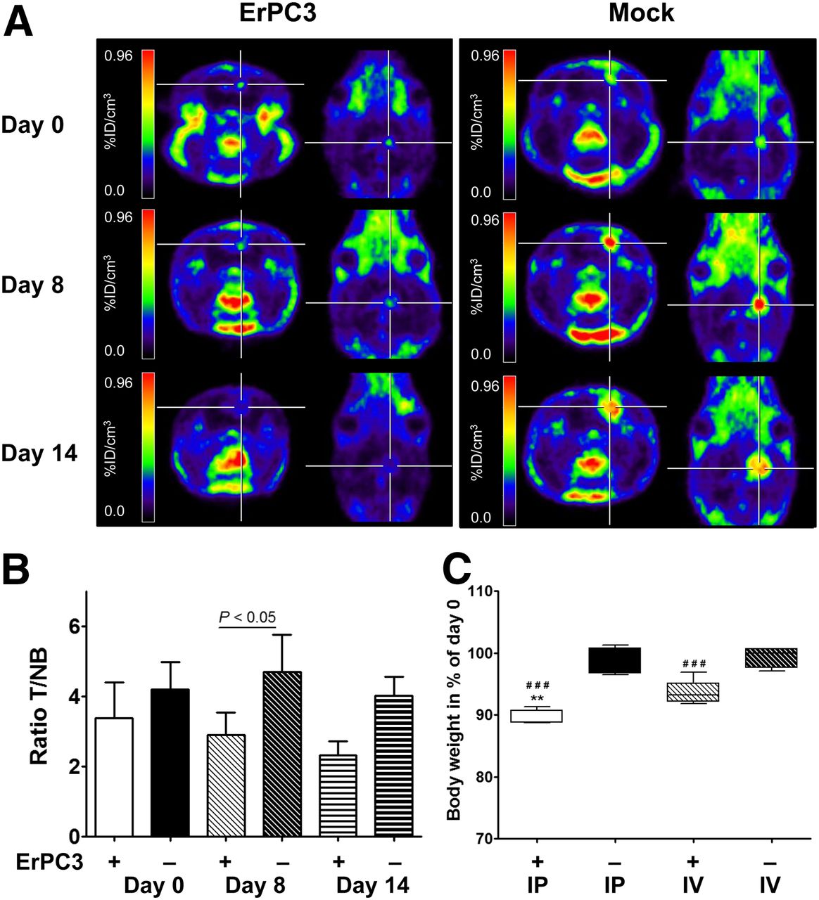
- FIGURE 3.
Effect of ErPC3 in vivo. (A) ErPC3 treatment reduces 18F-DPA-714 tumor uptake: representative coregistered brain 18F-DPA-714 PET images in ErPC3- and mock-treated animals before and during ErPC3 treatment. (B) Graphs of mean ± SD of tumor-to-contralateral 18F-DPA-714 uptake ratios for ErPC3- and control-treated animals at day 0, 8, and 14 after intraperitoneal (IP) administration. T/NB ratios show significant difference (P < 0.05) between 2 groups at day 8 of ErPC3 administration. (C) One week of ErPC3 treatment reduces body weight significantly (###P < 0.001, compared with control group). Intraperitoneal injections of ErPC3 induce more severe loss in body weight than intravenous (IV) injections (**P < 0.01). %ID/cm3 = percentage injected dose per cubic centimeter.
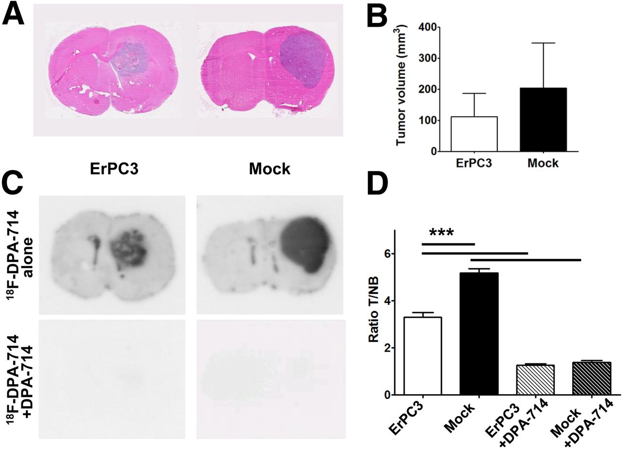
- FIGURE 4.
ErPC3 decreases tumor volume. (A) Hematoxylin and eosin staining of brain sections from ErPC3- and mock-treated animals. (B) Quantitative analysis using CellProfiler. Tumor volumes are smaller in ErPC3-treated than in control animals at day 14 after treatment, but this difference is not statistically significant (P = 0.37). (C) Autoradiography of tumor-bearing brain sections. 18F-DPA-714 binding is significantly reduced in ErPC3-treated versus control animals and in presence of nonlabeled DPA-714 (P < 0.0001).
- FIGURE 5.
TUNEL staining in experimental gliomas. (A) Increased levels of TUNEL staining (green) in tumors of ErPC3-treated animals, compared with controls, is indicative of increased apoptosis. TSPO staining (red) shows lack of TSPO expression in some TUNEL-positive cells (combined image). (B) Quantitative evaluation of tumor area demonstrates increase in percentage of TUNEL-positive cells in ErPC3-treated animals, compared with mock-treated animals.
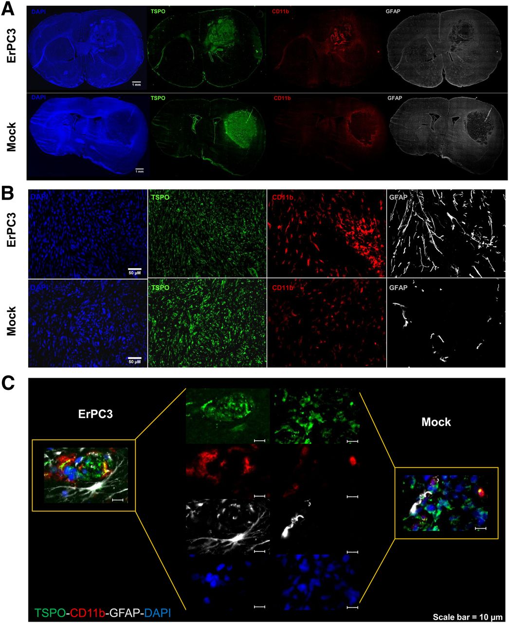
- FIGURE 6.
Increased infiltration of microglia and astrocytes in ErPC3-treated tumors. (A) Immunohistochemical staining of TSPO (green), CD11b (red), and GFAP (white) of whole coronal brain sections from animals bearing intracranial 9L glioma. (B) More CD11b-positive and GFAP-positive cells are found within tumor core of ErPC3-treated animals than controls. (C) TSPO expression in CD11b-positive and GFAP-positive cells in tumor core of ErPC3-treated animals is hardly detectable in control animals. DAPI = 4′,6-diamidino-2-phenylindole.


Additional Files
Supplemental Data
Files in this Data Supplement:


