
Main menu

  • Home
  • Content
    • Current
    • Ahead of print
    • Past Issues
    • JNM Supplement
    • SNMMI Annual Meeting Abstracts
    • Continuing Education
    • JNM Podcasts
  • Subscriptions
    • Subscribers
    • Institutional and Non-member
    • Rates
    • Journal Claims
    • Corporate & Special Sales
  • Authors
    • Submit to JNM
    • Information for Authors
    • Assignment of Copyright
    • AQARA requirements
  • Info
    • Reviewers
    • Permissions
    • Advertisers
  • About
    • About Us
    • Editorial Board
    • Contact Information
  • More
    • Alerts
    • Feedback
    • Help
    • SNMMI Journals
  • SNMMI
    • JNM
    • JNMT
    • SNMMI Journals
    • SNMMI

User menu

  • Subscribe
  • My alerts
  • Log in
  • My Cart

Search

  • Advanced search
Journal of Nuclear Medicine
  • SNMMI
    • JNM
    • JNMT
    • SNMMI Journals
    • SNMMI
  • Subscribe
  • My alerts
  • Log in
  • My Cart
Journal of Nuclear Medicine

Advanced Search

  • Home
  • Content
    • Current
    • Ahead of print
    • Past Issues
    • JNM Supplement
    • SNMMI Annual Meeting Abstracts
    • Continuing Education
    • JNM Podcasts
  • Subscriptions
    • Subscribers
    • Institutional and Non-member
    • Rates
    • Journal Claims
    • Corporate & Special Sales
  • Authors
    • Submit to JNM
    • Information for Authors
    • Assignment of Copyright
    • AQARA requirements
  • Info
    • Reviewers
    • Permissions
    • Advertisers
  • About
    • About Us
    • Editorial Board
    • Contact Information
  • More
    • Alerts
    • Feedback
    • Help
    • SNMMI Journals
  • View or Listen to JNM Podcast
  • Visit JNM on Facebook
  • Join JNM on LinkedIn
  • Follow JNM on Twitter
  • Subscribe to our RSS feeds
Research ArticleBasic Science Investigations

Preclinical Evaluation of 18F-LMI1195 for In Vivo Imaging of Pheochromocytoma in the MENX Tumor Model

Florian C. Gaertner, Tobias Wiedemann, Behrooz H. Yousefi, Misu Lee, Ines Repokis, Takahiro Higuchi, Stephan G. Nekolla, Ming Yu, Simon Robinson, Markus Schwaiger and Natalia S. Pellegata
Journal of Nuclear Medicine December 2013, 54 (12) 2111-2117; DOI: https://doi.org/10.2967/jnumed.113.119966
Florian C. Gaertner
1Klinikum Rechts der Isar der Technischen Universität München, Nuklearmedizinische Klinik, München, Germany
  • Find this author on Google Scholar
  • Find this author on PubMed
  • Search for this author on this site
Tobias Wiedemann
2Helmholtz Zentrum München, Deutsches Forschungszentrum für Gesundheit und Umwelt (GmbH), Institut für Pathologie, Neuherberg, Germany
  • Find this author on Google Scholar
  • Find this author on PubMed
  • Search for this author on this site
Behrooz H. Yousefi
1Klinikum Rechts der Isar der Technischen Universität München, Nuklearmedizinische Klinik, München, Germany
3Pharmaceutical Radiochemistry, Faculties of Chemistry and Medicine, Technische Universität München, Garching, Germany
  • Find this author on Google Scholar
  • Find this author on PubMed
  • Search for this author on this site
Misu Lee
2Helmholtz Zentrum München, Deutsches Forschungszentrum für Gesundheit und Umwelt (GmbH), Institut für Pathologie, Neuherberg, Germany
  • Find this author on Google Scholar
  • Find this author on PubMed
  • Search for this author on this site
Ines Repokis
2Helmholtz Zentrum München, Deutsches Forschungszentrum für Gesundheit und Umwelt (GmbH), Institut für Pathologie, Neuherberg, Germany
  • Find this author on Google Scholar
  • Find this author on PubMed
  • Search for this author on this site
Takahiro Higuchi
4Universitätsklinikum Würzburg, Comprehensive Heart Failure Center, Nuklearmedizinische Klinik und Poliklinik, Würzburg, Germany; and
  • Find this author on Google Scholar
  • Find this author on PubMed
  • Search for this author on this site
Stephan G. Nekolla
1Klinikum Rechts der Isar der Technischen Universität München, Nuklearmedizinische Klinik, München, Germany
  • Find this author on Google Scholar
  • Find this author on PubMed
  • Search for this author on this site
Ming Yu
5Discovery Research, Lantheus Medical Imaging, North Billerica, Massachusetts
  • Find this author on Google Scholar
  • Find this author on PubMed
  • Search for this author on this site
Simon Robinson
5Discovery Research, Lantheus Medical Imaging, North Billerica, Massachusetts
  • Find this author on Google Scholar
  • Find this author on PubMed
  • Search for this author on this site
Markus Schwaiger
1Klinikum Rechts der Isar der Technischen Universität München, Nuklearmedizinische Klinik, München, Germany
  • Find this author on Google Scholar
  • Find this author on PubMed
  • Search for this author on this site
Natalia S. Pellegata
2Helmholtz Zentrum München, Deutsches Forschungszentrum für Gesundheit und Umwelt (GmbH), Institut für Pathologie, Neuherberg, Germany
  • Find this author on Google Scholar
  • Find this author on PubMed
  • Search for this author on this site
  • Article
  • Figures & Data
  • Supplemental
  • Info & Metrics
  • PDF
Loading

Article Figures & Data

Figures

  • Tables
  • Additional Files
  • FIGURE 1.
    • Download figure
    • Open in new tab
    • Download powerpoint
    FIGURE 1.

    (A and B) Pharmacokinetics of 18F-LMI1195 in healthy Wistar control rats (n = 6), evaluated by dynamic PET imaging. Maximal adrenal uptake was reached 1 min after injection. Background organs reached low activity after approximately 45 min. (C and D) Maximum-intensity projection of PET images 5 and 45 min after injection. Scale bars represent SUV. Adrenal glands (arrows) can already be delineated well in early PET images (C). Late images show lower background activity; however, bone uptake is increased (D). p.i. = after injection.
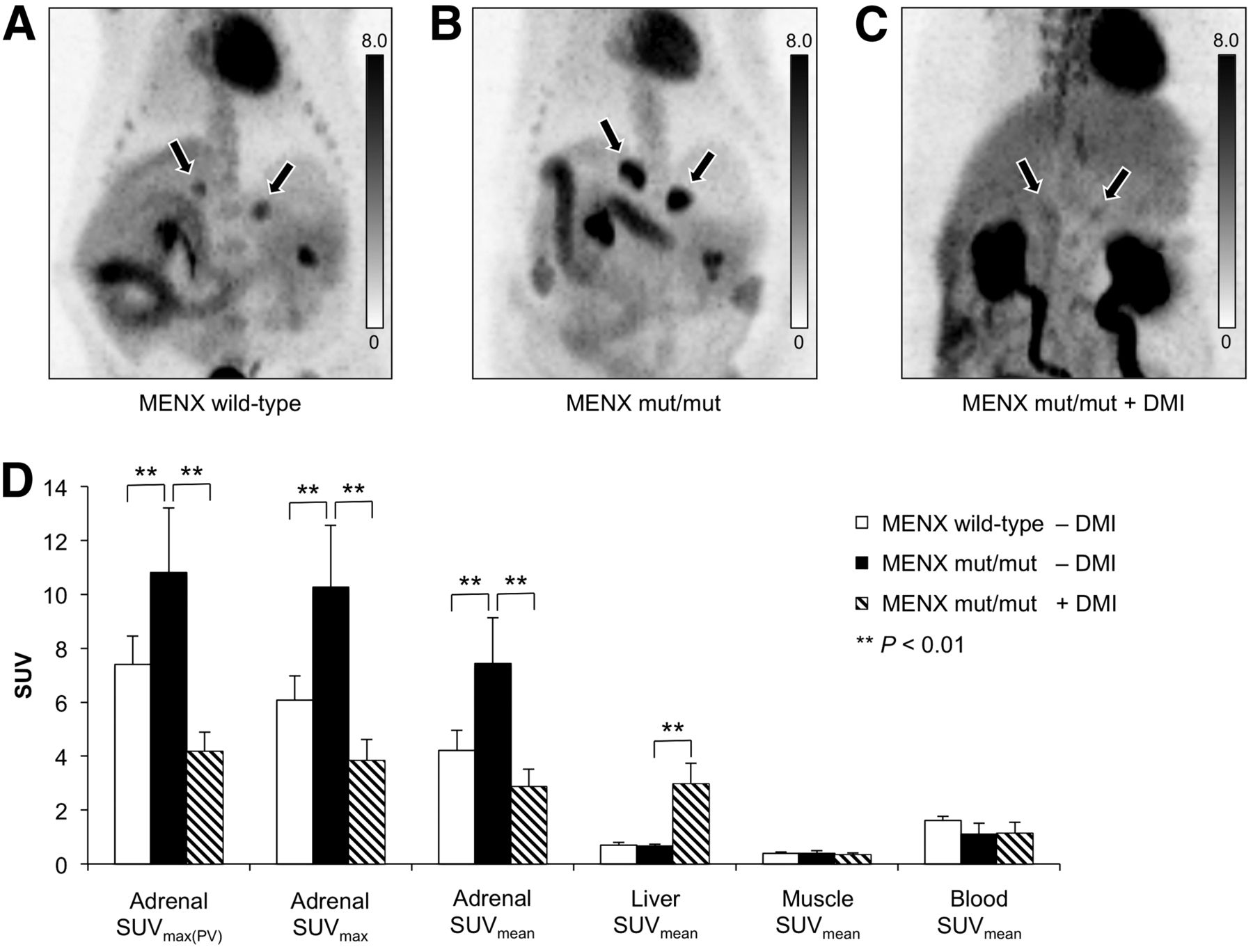
  • FIGURE 2.
    • Download figure
    • Open in new tab
    • Download powerpoint
    FIGURE 2.

    (A–C) 18F-LMI1195 PET images (maximum-intensity projection) 45 min after injection. Scale bars represent SUV. Normal adrenal glands of MENX wild-type rat show moderate tracer accumulation (A), whereas intense tracer accumulation is observed in adrenal glands of tumor-bearing mut/mut rat (B). After inhibition of NET with desipramine (DMI), tracer uptake in adrenal glands is significantly reduced (C). (D) Quantitative 18F-LMI1195 PET analysis of MENX wild-type control rats (n = 4), mut/mut rats (n = 10), and mut/mut rats after inhibition of NET with desipramine (n = 6). Bars represent mean SUVmax corrected for partial volume (SUVmax(PV)), mean SUVmax, or mean SUVmean as indicated in legend. Error bars represent SD. SUV of adrenal glands of mut/mut animals were significantly higher than those of wild-type controls (P < 0.01). After desipramine treatment, SUV of adrenal glands decreased significantly (P < 0.01), whereas significantly higher SUV was observed in liver (P < 0.01).
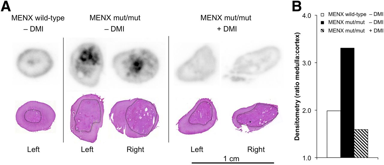
  • FIGURE 3.
    • Download figure
    • Open in new tab
    • Download powerpoint
    FIGURE 3.

    (A) Autoradiography of adrenal glands 60 min after injection of 18F-LMI1195 (top) and corresponding H&E stained slices (bottom). Low tracer uptake is observed in adrenal medulla (black outline in H&E staining) and in outer margin of adrenal cortex of wild-type control animal (left). Inhomogeneous tracer uptake is observed in enlarged adrenal medulla of tumor-bearing mut/mut rat, with focal areas of high tracer accumulation (middle). After desipramine (DMI) injection, virtually no tracer uptake is observed in area of pheochromocytoma in mut/mut animals (right). (B) Densitometry of autoradiography slices shown in A. Bars represent ratio of medulla to cortex of wild-type rat (n = 1), mean ratio of mut/mut animal (n = 2), and mean ratio of desipramine-blocked mut/mut rat (n = 2).
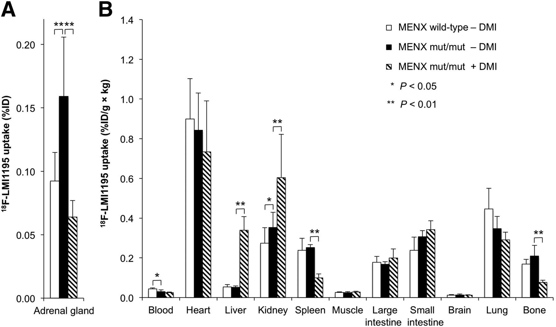
  • FIGURE 4.
    • Download figure
    • Open in new tab
    • Download powerpoint
    FIGURE 4.

    Biodistribution 60 min after injection of 18F-LMI1195 in MENX wild-type control rats (n = 4), mut/mut rats (n = 10), and mut/mut rats after desipramine (DMI) treatment (n = 6). (A) Bars represent total tracer uptake in adrenal glands (%ID), and error bars represent SD. (B) Bars represent tracer uptake in remaining organs normalized to organ weight and body weight, and error bars represent SD. When wild-type and nonblocked mut/mut rats were compared, total activity of 18F-LMI1195 in adrenal glands of mut/mut rats was significantly higher (A, P < 0.01). Furthermore, significant differences were observed for blood and kidneys (B). When nonblocked and blocked mut/mut rats were compared, tracer accumulation in adrenal glands was significantly lower in blocked animals (A). Tracer uptake in liver was significantly higher after desipramine treatment (B). Further significant differences were observed for kidneys, spleen, and bone.

  • FIGURE 5.
    • Download figure
    • Open in new tab
    • Download powerpoint
    FIGURE 5.

    Biodistribution of 18F-LMI1195 (n = 10) and 123I-MIBG (n = 6) in tumor-bearing mut/mut rats (60 min after injection). (A) Bars represent total tracer uptake in adrenal glands, and error bars represent SD. No significant differences are observed in adrenal tracer uptake between 18F-LMI1195 and 123I-MIBG (P > 0.05). (B) Bars represent tracer uptake in remaining organs normalized to organ weight and body weight, and error bars represent SD. Liver uptake of 18F-LMI1195 was significantly lower than 123I-MIBG (P < 0.01), whereas bone uptake was significantly higher (P < 0.01). Furthermore, significant differences were observed regarding kidneys, spleen, muscle, small intestine, brain, and lung.

Tables

  • Figures
  • Additional Files
    • View popup
    TABLE 1

    Biodistribution of 18F-LMI1195 and 123I-MIBG in MENX Wild-Type Rats and MENX mut/mut Rats With and Without Previous Blocking of NET With Desipramine

    18F-LMI1195
    ParameterMENX wild-typeMENX mut/mutMENX mut/mut123I-MIBG, MENX mut/mut
    Time after injection60 min60 min60 min60 min
    BlockingNoneNoneDesipramine, 10 mg/kgNone
    No. of rats41066
    Animal weight358 ± 101 g445 ± 95 g450 ± 115 g443 ± 99 g
    Organ (%ID/g × kg)
     Blood0.044 ± 0.0050.030 ± 0.008 (P = 0.024*)0.024 ± 0.003 (P = 0.18)0.035 ± 0.008 (P = 0.23)
     Heart0.90 ± 0.200.84 ± 0.19 (P = 0.57)0.73 ± 0.26 (P = 0.42)0.83 ± 0.11 (P = 0.59)
     Liver0.053 ± 0.0120.051 ± 0.007 (P = 0.78)0.34 ± 0.068 (P < 0.001*)0.300 ± 0.025 (P = 0.001*)
     Kidney0.27 ± 0.0790.35 ± 0.076 (P = 0.025*)0.60 ± 0.22 (P = 0.002*)0.29 ± 0.12 (P = 0.038*)
     Spleen0.24 ± 0.0600.25 ± 0.015 (P = 0.83)0.10 ± 0.020 (P = 0.002*)0.28 ± 0.019 (P = 0.009*)
     Muscle0.026 ± 0.0030.025 ± 0.005 (P = 0.48)0.027 ± 0.004 (P = 0.26)0.020 ± 0.002 (P = 0.009*)
     Large intestine0.18 ± 0.0300.17 ± 0.012 (P = 0.39)0.20 ± 0.046 (P = 0.39)0.17 ± 0.014 (P = 0.52)
     Small intestine0.24 ± 0.0670.31 ± 0.030 (P = 0.088)0.34 ± 0.046 (P = 0.18)0.43 ± 0.084 (P = 0.006*)
     Brain0.012 ± 0.0030.012 ± 0.005 (P = 0.67)0.012 ± 0.003 (P = 1.0)0.006 ± 0.003 (P = 0.037*)
     Lung0.45 ± 0.110.35 ± 0.062 (P = 0.20)0.29 ± 0.039 (P = 0.18)0.56 ± 0.10 (P = 0.006*)
     Bone0.17 ± 0.0240.21 ± 0.054 (P = 0.20)0.077 ± 0.010 (P = 0.002*)0.034 ± 0.006 (P = 0.004*)
    Adrenal gland (%ID)0.092 ± 0.0230.16 ± 0.047 (P < 0.001*)0.064 ± 0.013 (P < 0.001*)0.15 ± 0.028 (P = 0.60)
    • ↵* Significant P values.

    • Biodistribution values are %ID ± SD for adrenal glands and %ID/g × kg ± SD for other organs. P values for nonblocked 18F-LMI1195 in MENX mut/mut rats are with respect to 18F-LMI1195 in wild-type rats. P values for other 2 categories are with respect to nonblocked 18F-LMI1195 in mut/mut rats.

Additional Files

  • Figures
  • Tables
  • Supplemental Data

    Files in this Data Supplement:

    • Supplemental Data
PreviousNext
Back to top

In this issue

Journal of Nuclear Medicine: 54 (12)
Journal of Nuclear Medicine
Vol. 54, Issue 12
December 1, 2013
  • Table of Contents
  • Table of Contents (PDF)
  • About the Cover
  • Index by author
Print
Download PDF
Article Alerts
Sign In to Email Alerts with your Email Address
Email Article

Thank you for your interest in spreading the word on Journal of Nuclear Medicine.

NOTE: We only request your email address so that the person you are recommending the page to knows that you wanted them to see it, and that it is not junk mail. We do not capture any email address.

Enter multiple addresses on separate lines or separate them with commas.
Preclinical Evaluation of 18F-LMI1195 for In Vivo Imaging of Pheochromocytoma in the MENX Tumor Model
(Your Name) has sent you a message from Journal of Nuclear Medicine
(Your Name) thought you would like to see the Journal of Nuclear Medicine web site.
Citation Tools
Preclinical Evaluation of 18F-LMI1195 for In Vivo Imaging of Pheochromocytoma in the MENX Tumor Model
Florian C. Gaertner, Tobias Wiedemann, Behrooz H. Yousefi, Misu Lee, Ines Repokis, Takahiro Higuchi, Stephan G. Nekolla, Ming Yu, Simon Robinson, Markus Schwaiger, Natalia S. Pellegata
Journal of Nuclear Medicine Dec 2013, 54 (12) 2111-2117; DOI: 10.2967/jnumed.113.119966

Citation Manager Formats

  • BibTeX
  • Bookends
  • EasyBib
  • EndNote (tagged)
  • EndNote 8 (xml)
  • Medlars
  • Mendeley
  • Papers
  • RefWorks Tagged
  • Ref Manager
  • RIS
  • Zotero
Share
Preclinical Evaluation of 18F-LMI1195 for In Vivo Imaging of Pheochromocytoma in the MENX Tumor Model
Florian C. Gaertner, Tobias Wiedemann, Behrooz H. Yousefi, Misu Lee, Ines Repokis, Takahiro Higuchi, Stephan G. Nekolla, Ming Yu, Simon Robinson, Markus Schwaiger, Natalia S. Pellegata
Journal of Nuclear Medicine Dec 2013, 54 (12) 2111-2117; DOI: 10.2967/jnumed.113.119966
Twitter logo Facebook logo LinkedIn logo Mendeley logo
  • Tweet Widget
  • Facebook Like
  • Google Plus One
Bookmark this article

Jump to section

  • Article
    • Abstract
    • MATERIALS AND METHODS
    • RESULTS
    • DISCUSSION
    • CONCLUSION
    • DISCLOSURE
    • Acknowledgments
    • Footnotes
    • REFERENCES
  • Figures & Data
  • Supplemental
  • Info & Metrics
  • PDF

Related Articles

  • This Month in JNM
  • PubMed
  • Google Scholar

Cited By...

  • First Experience Using 18F-Flubrobenguane PET Imaging in Patients with Suspected Pheochromocytoma or Paraganglioma
  • Radiolabeled (4-Fluoro-3-Iodobenzyl)Guanidine Improves Imaging and Targeted Radionuclide Therapy of Norepinephrine Transporter-Expressing Tumors
  • Animal models of MEN1
  • Targeting PI3K/mTOR signaling exerts potent antitumor activity in pheochromocytoma in vivo
  • Google Scholar

More in this TOC Section

  • 11C-Methionine PET of Myocardial Inflammation in a Rat Model of Experimental Autoimmune Myocarditis
  • Counting Rate Characteristics and Image Distortion in Preclinical PET Imaging During Radiopharmaceutical Therapy
  • Design and Fabrication of Kidney Phantoms for Internal Radiation Dosimetry Using 3D Printing Technology
Show more Basic Science Investigations

Similar Articles

Keywords

  • oncology
  • pheochromocytoma
  • MIBG
  • 18F-LMI1195
  • MENX
SNMMI

© 2025 SNMMI

Powered by HighWire