
Main menu

  • Home
  • Content
    • Current
    • Ahead of print
    • Past Issues
    • JNM Supplement
    • SNMMI Annual Meeting Abstracts
    • Continuing Education
    • JNM Podcasts
  • Subscriptions
    • Subscribers
    • Institutional and Non-member
    • Rates
    • Journal Claims
    • Corporate & Special Sales
  • Authors
    • Submit to JNM
    • Information for Authors
    • Assignment of Copyright
    • AQARA requirements
  • Info
    • Reviewers
    • Permissions
    • Advertisers
  • About
    • About Us
    • Editorial Board
    • Contact Information
  • More
    • Alerts
    • Feedback
    • Help
    • SNMMI Journals
  • SNMMI
    • JNM
    • JNMT
    • SNMMI Journals
    • SNMMI

User menu

  • Subscribe
  • My alerts
  • Log in
  • My Cart

Search

  • Advanced search
Journal of Nuclear Medicine
  • SNMMI
    • JNM
    • JNMT
    • SNMMI Journals
    • SNMMI
  • Subscribe
  • My alerts
  • Log in
  • My Cart
Journal of Nuclear Medicine

Advanced Search

  • Home
  • Content
    • Current
    • Ahead of print
    • Past Issues
    • JNM Supplement
    • SNMMI Annual Meeting Abstracts
    • Continuing Education
    • JNM Podcasts
  • Subscriptions
    • Subscribers
    • Institutional and Non-member
    • Rates
    • Journal Claims
    • Corporate & Special Sales
  • Authors
    • Submit to JNM
    • Information for Authors
    • Assignment of Copyright
    • AQARA requirements
  • Info
    • Reviewers
    • Permissions
    • Advertisers
  • About
    • About Us
    • Editorial Board
    • Contact Information
  • More
    • Alerts
    • Feedback
    • Help
    • SNMMI Journals
  • View or Listen to JNM Podcast
  • Visit JNM on Facebook
  • Join JNM on LinkedIn
  • Follow JNM on Twitter
  • Subscribe to our RSS feeds
Research ArticleClinical Investigations

18F-FDG PET/CT in Staging Patients with Locally Advanced or Inflammatory Breast Cancer: Comparison to Conventional Staging

David Groheux, Sylvie Giacchetti, Marc Delord, Elif Hindié, Laetitia Vercellino, Caroline Cuvier, Marie-Elisabeth Toubert, Pascal Merlet, Christophe Hennequin and Marc Espié
Journal of Nuclear Medicine January 2013, 54 (1) 5-11; DOI: https://doi.org/10.2967/jnumed.112.106864
David Groheux
1Department of Nuclear Medicine, Saint-Louis Hospital, Paris, France
2B2T, Doctoral School, IUH, University of Paris VII, France
  • Find this author on Google Scholar
  • Find this author on PubMed
  • Search for this author on this site
Sylvie Giacchetti
3Breast Diseases Unit, Department of Medical Oncology, Saint-Louis Hospital, Paris, France
  • Find this author on Google Scholar
  • Find this author on PubMed
  • Search for this author on this site
Marc Delord
4Department of Biostatistics and Bioinformatics, Institut Universitaire d’Hématologie, Paris, France
  • Find this author on Google Scholar
  • Find this author on PubMed
  • Search for this author on this site
Elif Hindié
2B2T, Doctoral School, IUH, University of Paris VII, France
5Department of Nuclear Medicine, Haut-Lévêque Hospital, CHU Bordeaux, University Bordeaux-Segalen, Bordeaux, France
  • Find this author on Google Scholar
  • Find this author on PubMed
  • Search for this author on this site
Laetitia Vercellino
1Department of Nuclear Medicine, Saint-Louis Hospital, Paris, France
  • Find this author on Google Scholar
  • Find this author on PubMed
  • Search for this author on this site
Caroline Cuvier
3Breast Diseases Unit, Department of Medical Oncology, Saint-Louis Hospital, Paris, France
  • Find this author on Google Scholar
  • Find this author on PubMed
  • Search for this author on this site
Marie-Elisabeth Toubert
1Department of Nuclear Medicine, Saint-Louis Hospital, Paris, France
  • Find this author on Google Scholar
  • Find this author on PubMed
  • Search for this author on this site
Pascal Merlet
1Department of Nuclear Medicine, Saint-Louis Hospital, Paris, France
6Service Hospitalier Frédéric Joliot, SHFJ/I2BM/DSVCEA, Orsay, France; and
  • Find this author on Google Scholar
  • Find this author on PubMed
  • Search for this author on this site
Christophe Hennequin
7Department of Radiation Oncology, Saint-Louis Hospital, Paris, France
  • Find this author on Google Scholar
  • Find this author on PubMed
  • Search for this author on this site
Marc Espié
3Breast Diseases Unit, Department of Medical Oncology, Saint-Louis Hospital, Paris, France
  • Find this author on Google Scholar
  • Find this author on PubMed
  • Search for this author on this site
  • Article
  • Figures & Data
  • Info & Metrics
  • PDF
Loading

Article Figures & Data

Figures

  • Tables
  • FIGURE 1.
    • Download figure
    • Open in new tab
    • Download powerpoint
    FIGURE 1.

    Study flow diagram. US = ultrasonography.
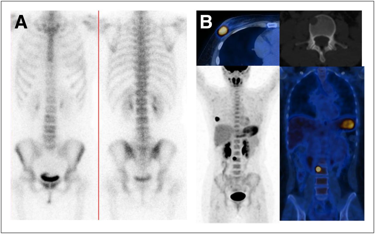
  • FIGURE 2.
    • Download figure
    • Open in new tab
    • Download powerpoint
    FIGURE 2.

    Bone scan (A) and PET/CT (B) findings in patient with clinical T4bN0 (stage IIIB), estrogen receptor–positive, progesterone receptor–positive, HER2-negative, grade 3 invasive ductal carcinoma of right breast. PET/CT showed primary tumor and depicted lytic metastasis of vertebral body of L3. MR imaging confirmed bone metastasis (not shown). Bone scanning was falsely negative (slight heterogeneity of upper border of L3 was considered suggestive of arthrosis).
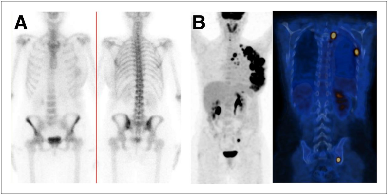
  • FIGURE 3.
    • Download figure
    • Open in new tab
    • Download powerpoint
    FIGURE 3.

    Bone scan (A) and PET/CT (B) findings in patient with clinical T4bN3 (stage IIIC) estrogen receptor–positive, progesterone receptor–negative, HER2-negative, grade 3 invasive ductal carcinoma of left breast. PET/CT showed locally advanced primary breast cancer with axillary and supraclavicular lymph nodes, as well as numerous distant metastases to bones and pleura. Bone scanning was true-positive, showing faint uptake at lower part of left sacroiliac joint.
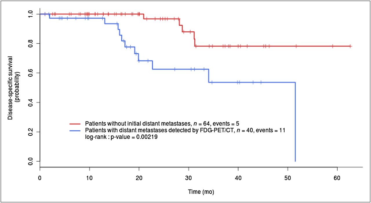
  • FIGURE 4.
    • Download figure
    • Open in new tab
    • Download powerpoint
    FIGURE 4.

    Kaplan–Meier disease-specific survival for 104 patients with adequate follow-up.

Tables

  • Figures
    • View popup
    TABLE 1

    Patients and Tumor Characteristics

    CharacteristicNo. of patients (of 117 total)
    Noninflammatory LABC*82 (70)
     T1 N2 M01 (1)
     T1 N3 M02 (2)
     T2 N2 M011 (9)
     T2 N3 M01 (1)
     T3 N2 M014 (12)
     T3 N3 M05 (4)
     T4a/b/c N0 M013 (11)
     T4a/b/c N1 M020 (18)
     T4a/b/c N2 M012 (10)
     T4a/b/c N3 M03 (3)
    Inflammatory breast cancer*35 (30)
     T4d N0 M01 (1)
     T4d N1 M016 (14)
     T4d N2 M012 (10)
     T4d N3 M06 (5)
    Tumor type
     Invasive ductal carcinoma98 (84)
     Invasive lobular carcinoma12 (10)
     Other7 (6)
    Grade
     Grade 12 (2)
     Grade 249 (42)
     Grade 358 (49)
     Unspecified8 (7)
    Estrogen receptor status†
     Positive59 (50)
     Negative57 (49)
     Unspecified1 (1)
    Progesterone receptor status†
     Positive28 (24)
     Negative87 (75)
     Unspecified1 (1)
    HER2 status‡
     Positive22 (19)
     Negative94 (80)
     Unspecified1 (1)
    • ↵* Clinical classification before PET/CT and conventional distant work-up.

    • ↵† Tumors were considered positive for ER or for PR if >10% of cells showed staining by immunohistochemistry.

    • ↵‡ Tumors were considered to overexpress c-erbB-2 oncoprotein (HER2-positive) if >30% of invasive tumor cells showed definite membrane staining resulting in so-called fishnet appearance.

    • Data in parentheses are percentages.

    • View popup
    TABLE 2

    Findings with 18F-FDG PET/CT in 3 Different Groups

    Results expressed on per-patient basisNoninflammatory LABCInflammatory breast cancerWhole population
    Patients82 (70)35 (30)117 (100)
    Overall stage modifications*39 (48)22 (63)61 (52)
    Lymph nodes† outside level I and level II axillary levels27 (33)22 (63)49 (42)
     Internal mammary node involvement12 (15)10 (28)22 (19)
     Infraclavicular19 (23)15 (43)34 (29)
     Supraclavicular13 (16)13 (37)26 (22)
    Distant metastases‡27 (33)16 (46)43 (37)
     Bone20 (24)10 (29)30 (26)
     Lung3 (4)3 (9)6 (5)
     Pleura2 (2)02 (2)
     Distant lymph nodes§11 (13)8 (23)19 (16)
     Liver6 (7)4 (11)10 (8)
    Second cancer02 (6)2 (2)
    • ↵* Some women had extraaxillary lymph nodes as well as distant metastases.

    • ↵† Some women had lymph node metastases in different areas.

    • ↵‡ Some women had distant metastases in different viscera.

    • ↵§ Distant lymph nodes were cervical, mediastinal, hilar, contralateral axillary, or abdominopelvic.

    • Data are n, with percentages in parentheses.

    • View popup
    TABLE 3

    Performance of PET/CT Versus Conventional Imaging Work-up to Depict Distant Metastases in Overall Series

    SitePET/CTBone scanningChest imaging (radiography or dedicated CT)Abdominal imaging (sonography or enhanced CT)Total*
    Bone metastases3023†——30
    Lung metastases6‡—7—7
    Pleura2—1—2
    Distant lymph node metastases19—10§119
    Liver metastases10——9║10
    • ↵† 19 positive bone scans and 4 suggestive nonequivocal bone scans.

    • ↵* In total, 43 patients had distant metastases. Some women had metastases in different viscera.

    • ↵‡ Two women with lung metastases had no 18F-FDG uptake; metastases were detected only on CT part of PET/CT imaging.

    • ↵§ Among 19 patients with lymph node metastases detected by PET/CT, 18 were positive in supradiaphragmatic area. All these patients had chest radiography, and 13 had dedicated chest CT.

    • ↵║ Among 10 patients with liver metastases, 7 had liver sonography and 8 had abdominal enhanced CT.

    • Data are expressed per patient (not per lesion).

PreviousNext
Back to top

In this issue

Journal of Nuclear Medicine: 54 (1)
Journal of Nuclear Medicine
Vol. 54, Issue 1
January 1, 2013
  • Table of Contents
  • Table of Contents (PDF)
  • About the Cover
  • Index by author
Print
Download PDF
Article Alerts
Sign In to Email Alerts with your Email Address
Email Article

Thank you for your interest in spreading the word on Journal of Nuclear Medicine.

NOTE: We only request your email address so that the person you are recommending the page to knows that you wanted them to see it, and that it is not junk mail. We do not capture any email address.

Enter multiple addresses on separate lines or separate them with commas.
18F-FDG PET/CT in Staging Patients with Locally Advanced or Inflammatory Breast Cancer: Comparison to Conventional Staging
(Your Name) has sent you a message from Journal of Nuclear Medicine
(Your Name) thought you would like to see the Journal of Nuclear Medicine web site.
Citation Tools
18F-FDG PET/CT in Staging Patients with Locally Advanced or Inflammatory Breast Cancer: Comparison to Conventional Staging
David Groheux, Sylvie Giacchetti, Marc Delord, Elif Hindié, Laetitia Vercellino, Caroline Cuvier, Marie-Elisabeth Toubert, Pascal Merlet, Christophe Hennequin, Marc Espié
Journal of Nuclear Medicine Jan 2013, 54 (1) 5-11; DOI: 10.2967/jnumed.112.106864

Citation Manager Formats

  • BibTeX
  • Bookends
  • EasyBib
  • EndNote (tagged)
  • EndNote 8 (xml)
  • Medlars
  • Mendeley
  • Papers
  • RefWorks Tagged
  • Ref Manager
  • RIS
  • Zotero
Share
18F-FDG PET/CT in Staging Patients with Locally Advanced or Inflammatory Breast Cancer: Comparison to Conventional Staging
David Groheux, Sylvie Giacchetti, Marc Delord, Elif Hindié, Laetitia Vercellino, Caroline Cuvier, Marie-Elisabeth Toubert, Pascal Merlet, Christophe Hennequin, Marc Espié
Journal of Nuclear Medicine Jan 2013, 54 (1) 5-11; DOI: 10.2967/jnumed.112.106864
Twitter logo Facebook logo LinkedIn logo Mendeley logo
  • Tweet Widget
  • Facebook Like
  • Google Plus One
Bookmark this article

Jump to section

  • Article
    • Abstract
    • MATERIALS AND METHODS
    • RESULTS
    • DISCUSSION
    • CONCLUSION
    • DISCLOSURE
    • Footnotes
    • REFERENCES
  • Figures & Data
  • Info & Metrics
  • PDF

Related Articles

  • This Month in JNM
  • PubMed
  • Google Scholar

Cited By...

  • Assessment of Bone Lesions with 18F-FDG PET Compared with 99mTc Bone Scintigraphy Leads to Clinically Relevant Differences in Metastatic Breast Cancer Management
  • Assessing 18F-FDG Uptake in the Sentinel Lymph Node in Breast Cancer
  • 18F-FDG PET/CT for Systemic Staging of Newly Diagnosed Breast Cancer in Men
  • 18F-FDG PET/CT for Staging and Restaging of Breast Cancer
  • Translation of New Molecular Imaging Approaches to the Clinical Setting: Bridging the Gap to Implementation
  • Google Scholar

More in this TOC Section

  • Cardiac Presynaptic Sympathetic Nervous Function Evaluated by Cardiac PET in Patients with Chronotropic Incompetence Without Heart Failure
  • Validation and Evaluation of a Vendor-Provided Head Motion Correction Algorithm on the uMI Panorama PET/CT System
  • Prognostic Role of 68Ga-PSMA11 PET–Based Response in Patients with Prostate Cancer Undergoing Taxane-Based Chemotherapy
Show more Clinical Investigations

Similar Articles

Keywords

  • 18F-FDG-PET/CT
  • locally advanced breast cancer
  • Inflammatory breast cancer
  • work-up
  • initial staging
SNMMI

© 2025 SNMMI

Powered by HighWire