


Article Figures & Data
Figures
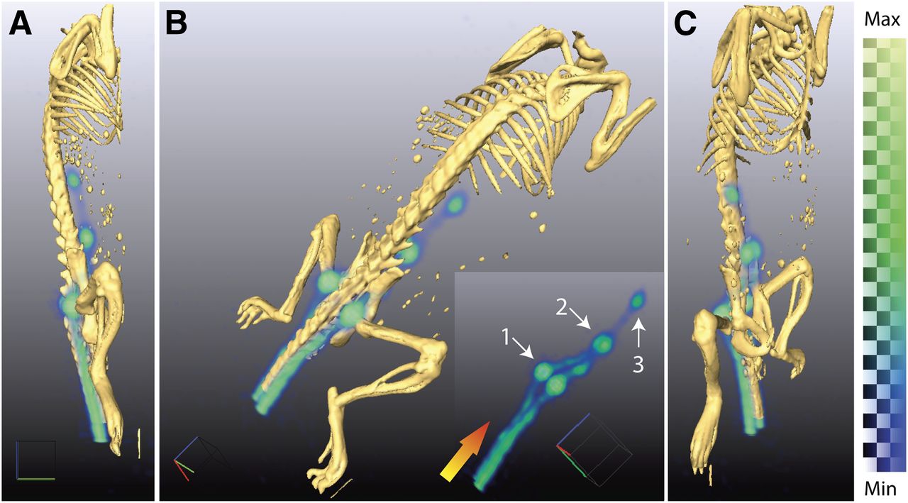
- FIGURE 1.
Three-dimensionally rendered positron lymphography images demonstrate high-resolution mapping using intradermal administration of 18F-FDG to a male nu/nu mouse at multiple angles (A–C). Radiotracer is transported from site of injection through lymphatic system, enabling visualization of lymph flow. Delineation of LN draining injection site is clearly distinguished (B; insert is without fused CT). At 10 min after injection, we can identify lymphatic vessels in tail leading first to sacral nodes (1). Tracer then moves proximally to caudal (2) and then mesenteric nodes (3). Gradient arrow indicates direction of flow. Three-dimensionally rendered PET data are generated from weighted average of intensities from all 2-dimensional slices. As such, it is strictly semiquantitative; color bar indicates range of intensities. Max = maximum; Min = minimum.
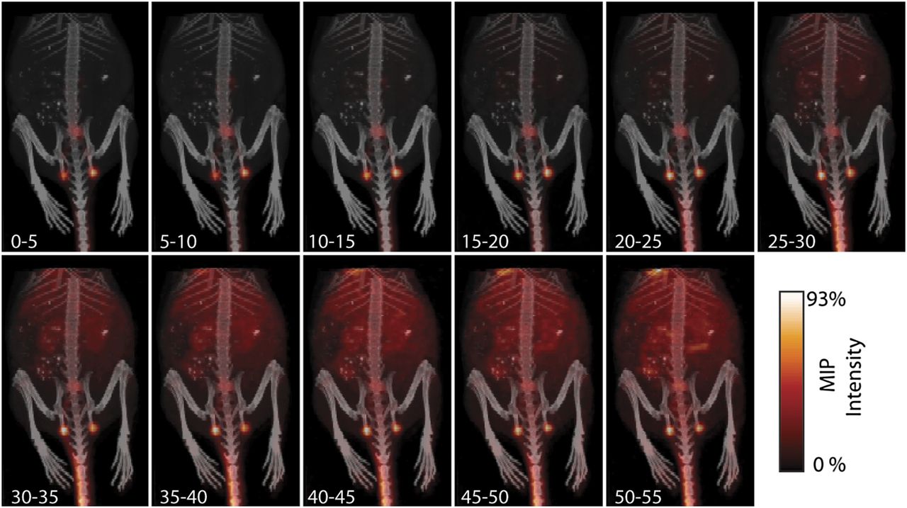
- FIGURE 2.
Dynamic imaging of progress of lymphatic transport of small-molecule radiotracer (5-min intervals). Within minutes, 18F-FDG has begun to be cleared from injection site through lymphatic system. Transport through and uptake at LN makes these features obviously apparent. Background tissue uptake of tracer builds slowly over approximately 30–35 min, via lymphatic clearance and diffusion of the small molecule. However, clearly identifiable presence of nodes even at late time points is significant, enabling ready identification of these nodes at 1 h after injection. Detailed imaging of transport immediately after injection is shown in Figure 3. MIP = maximum-intensity projection.
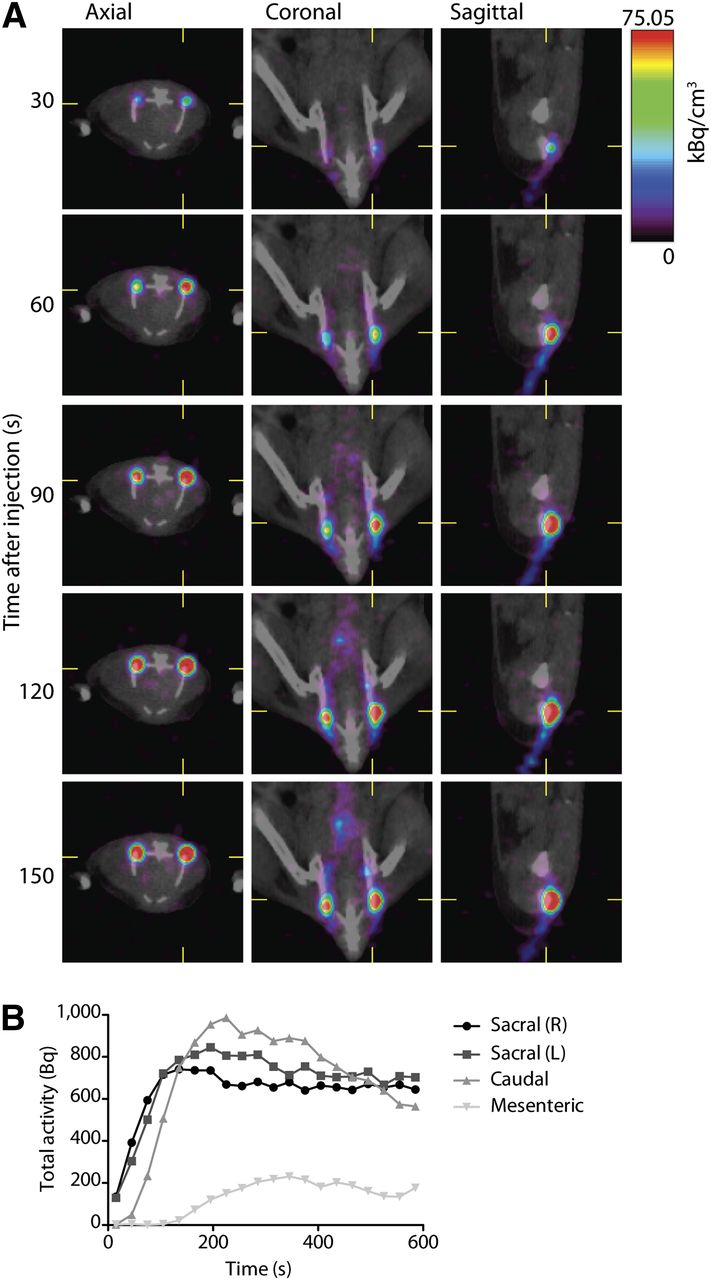
- FIGURE 3.
Quantitative and dynamic imaging of lymphatic 18F-FDG transport. (A) Axial, coronal, and sagittal views of dynamic imaging (30-s frames starting at time of injection) of 18F-FDG transport in lymphatics. Acquisition of rapidity of flow through vessels and uptake into nodes, with both high spatial and longitudinal resolution, is an advantage of this technique. (B) PET information is inherently quantitative, providing activity concentration information. Here, regions of interest were drawn around LN to assess lymphatic function by computing uptake over time (over linear regions of curve). For right sacral, left sacral, caudal, and mesenteric nodes, this yields 6.47, 6.57, 5.67, and 1.13 Bq/s, respectively.
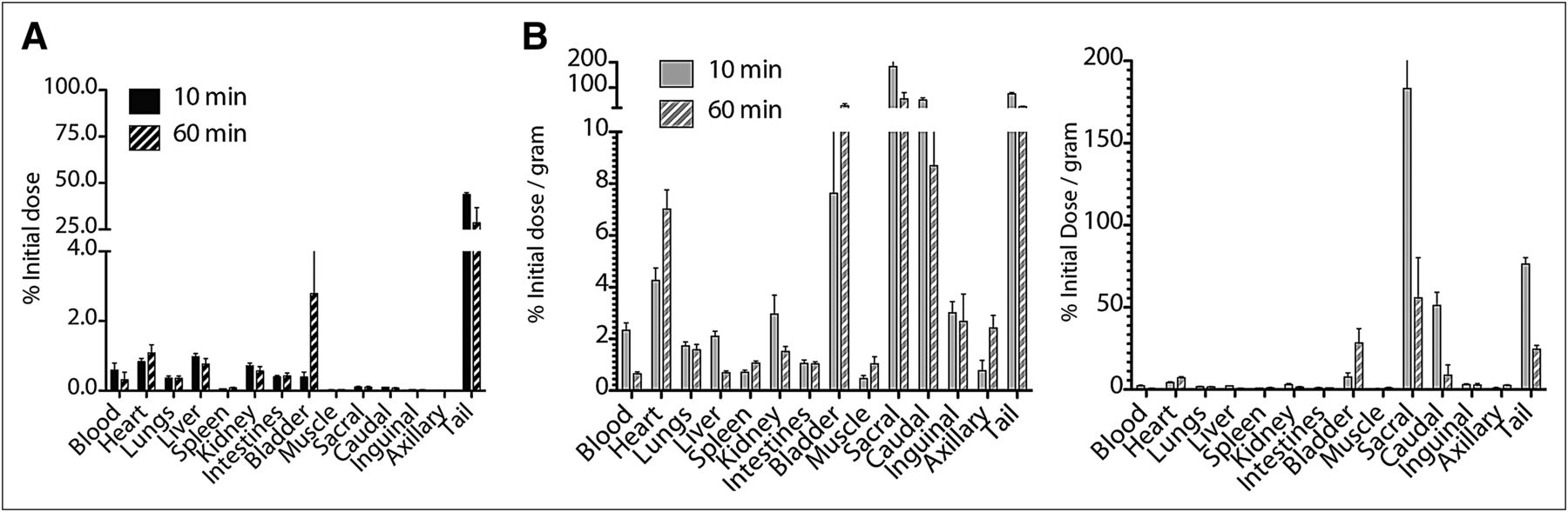
- FIGURE 4.
Biodistribution of intradermally injected small-molecule tracer. (A) Bar chart showing %ID distribution of locally administered 18F-FDG in major organs and LNs at 10 and 60 min after injection. Radiotracer slowly leaves dermal site of tail to circulation, renal excretion, and accumulation in bladder. (B) Normalizing dose distribution for mass of tissue (%ID/g) demonstrates draining-lymph–specific transit of tracer. Retained radiotracer at injection site continues to be carried through lymphatics from injection site to draining nodes, enabling continued visualization. Left and right plots have different scales to show organ and nodal %ID/g, respectively.
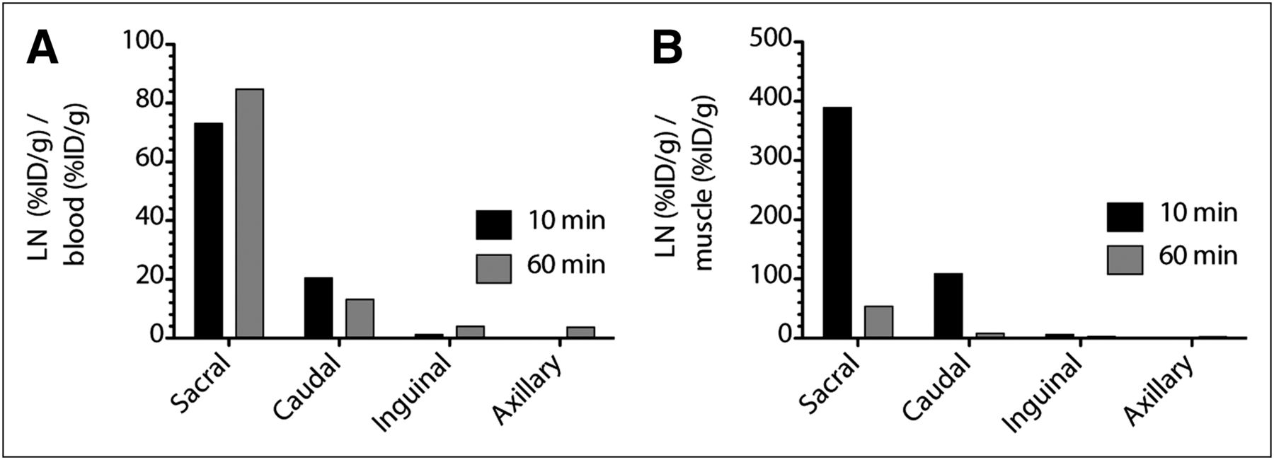
- FIGURE 5.
Contrast ratio of 18F-FDG to blood and muscle. (A) Ratios of %ID/g from draining LN and blood are high, affording clear distinction between lymphatics and surrounding tissues. (B) Contrast derived from %ID/g between LN and muscle enables clear delineation of nodes. LNs not in the draining path from the injection site (inguinal and axillary) do not produce significant ratios with respect to background. From 10 to 60 min, ratio of LN to muscle drops as conscious and ambulatory mice increase muscle uptake. Inguinal (and axillary) uptake is low because these nodes are not within drainage of injection site, which drains to deep pelvic nodes
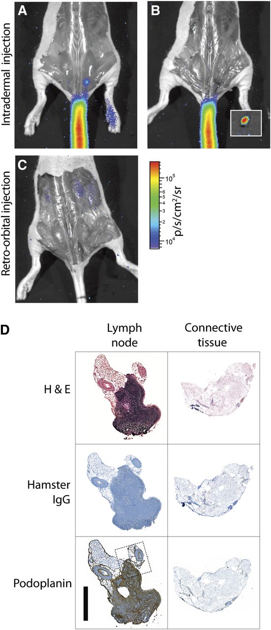
- FIGURE 6.
Cerenkov-guided surgical resection of 18F-FDG–bearing LN, 10 min after injection, with surgical validation. (A) Lateral-tail intradermal injection yields greater uptake in 1 sacral node, seen with just skin removed. PET image is included as Supplemental Figure 2. (B) CR guides resection of node, magnified in inset. (C) Systemic administration of 18F-FDG (retroorbital injection) does not enable identification of nodes, even after surgical exposure. Signal from renal clearance can be seen. (D) Immunohistologic verification of Cerenkov-guided excised tissues. Resected node as indicated by CR, along with surrounding connective tissue, was stained with hematoxylin and eosin, control hamster IgG, and podoplanin. The stromal region of node is indicated by dense hematoxylin and eosin and positive podoplanin staining. Dotted line indicates region of magnification (included as Supplemental Fig. 4). Scale bar, 1 mm. H&E = hematoxylin and eosin.


Additional Files
Supplemental Data
Files in this Data Supplement:


Jump to section
Related Articles
Cited By...
- Intestinal Lymphatic Biology, Drug Delivery, and Therapeutics: Current Status and Future Directions
- Optical Imaging Modalities: Principles and Applications in Preclinical Research and Clinical Settings
- Positron Lymphography via Intracervical 18F-FDG Injection for Presurgical Lymphatic Mapping in Cervical and Endometrial Malignancies
- Dynamic 18F-FDG PET Lymphography for In Vivo Identification of Lymph Node Metastases in Murine Melanoma
- Cerenkov-Specific Contrast Agents for Detection of pH In Vivo
- Clinical Cerenkov Luminescence Imaging of 18F-FDG
- Sentinel Lymph Node Biopsy for Prostate Cancer: A Hybrid Approach