
Main menu

  • Home
  • Content
    • Current
    • Ahead of print
    • Past Issues
    • JNM Supplement
    • SNMMI Annual Meeting Abstracts
    • Continuing Education
    • JNM Podcasts
  • Subscriptions
    • Subscribers
    • Institutional and Non-member
    • Rates
    • Journal Claims
    • Corporate & Special Sales
  • Authors
    • Submit to JNM
    • Information for Authors
    • Assignment of Copyright
    • AQARA requirements
  • Info
    • Reviewers
    • Permissions
    • Advertisers
  • About
    • About Us
    • Editorial Board
    • Contact Information
  • More
    • Alerts
    • Feedback
    • Help
    • SNMMI Journals
  • SNMMI
    • JNM
    • JNMT
    • SNMMI Journals
    • SNMMI

User menu

  • Subscribe
  • My alerts
  • Log in
  • My Cart

Search

  • Advanced search
Journal of Nuclear Medicine
  • SNMMI
    • JNM
    • JNMT
    • SNMMI Journals
    • SNMMI
  • Subscribe
  • My alerts
  • Log in
  • My Cart
Journal of Nuclear Medicine

Advanced Search

  • Home
  • Content
    • Current
    • Ahead of print
    • Past Issues
    • JNM Supplement
    • SNMMI Annual Meeting Abstracts
    • Continuing Education
    • JNM Podcasts
  • Subscriptions
    • Subscribers
    • Institutional and Non-member
    • Rates
    • Journal Claims
    • Corporate & Special Sales
  • Authors
    • Submit to JNM
    • Information for Authors
    • Assignment of Copyright
    • AQARA requirements
  • Info
    • Reviewers
    • Permissions
    • Advertisers
  • About
    • About Us
    • Editorial Board
    • Contact Information
  • More
    • Alerts
    • Feedback
    • Help
    • SNMMI Journals
  • View or Listen to JNM Podcast
  • Visit JNM on Facebook
  • Join JNM on LinkedIn
  • Follow JNM on Twitter
  • Subscribe to our RSS feeds
Research ArticleClinical Investigations

Added Value of 99mTc-HMPAO–Labeled Leukocyte SPECT/CT in the Characterization and Management of Patients with Infectious Endocarditis

Paola A. Erba, Umberto Conti, Elena Lazzeri, Martina Sollini, Roberta Doria, Salvatore M. De Tommasi, Francesco Bandera, Carlo Tascini, Francesco Menichetti, Rudi A.J.O. Dierckx, Alberto Signore and Giuliano Mariani
Journal of Nuclear Medicine August 2012, 53 (8) 1235-1243; DOI: https://doi.org/10.2967/jnumed.111.099424
Paola A. Erba
  • Find this author on Google Scholar
  • Find this author on PubMed
  • Search for this author on this site
Umberto Conti
  • Find this author on Google Scholar
  • Find this author on PubMed
  • Search for this author on this site
Elena Lazzeri
  • Find this author on Google Scholar
  • Find this author on PubMed
  • Search for this author on this site
Martina Sollini
  • Find this author on Google Scholar
  • Find this author on PubMed
  • Search for this author on this site
Roberta Doria
  • Find this author on Google Scholar
  • Find this author on PubMed
  • Search for this author on this site
Salvatore M. De Tommasi
  • Find this author on Google Scholar
  • Find this author on PubMed
  • Search for this author on this site
Francesco Bandera
  • Find this author on Google Scholar
  • Find this author on PubMed
  • Search for this author on this site
Carlo Tascini
  • Find this author on Google Scholar
  • Find this author on PubMed
  • Search for this author on this site
Francesco Menichetti
  • Find this author on Google Scholar
  • Find this author on PubMed
  • Search for this author on this site
Rudi A.J.O. Dierckx
  • Find this author on Google Scholar
  • Find this author on PubMed
  • Search for this author on this site
Alberto Signore
  • Find this author on Google Scholar
  • Find this author on PubMed
  • Search for this author on this site
Giuliano Mariani
  • Find this author on Google Scholar
  • Find this author on PubMed
  • Search for this author on this site
  • Article
  • Figures & Data
  • Info & Metrics
  • PDF
Loading

Article Figures & Data

Figures

  • Tables
  • FIGURE 1.
    • Download figure
    • Open in new tab
    • Download powerpoint
    FIGURE 1.

    99mTc-HMPAO-WBC scintigraphic images for patient with aortic endocarditis. Maximum-intensity-projection image (A) demonstrates focal increase of radiolabeled WBC in heart region. Transaxial SPECT/CT images (B) show that such focal uptake is localized at mechanical prosthesis of aortic valve (CT [left], fused SPECT/CT [center], and SPECT [right]).
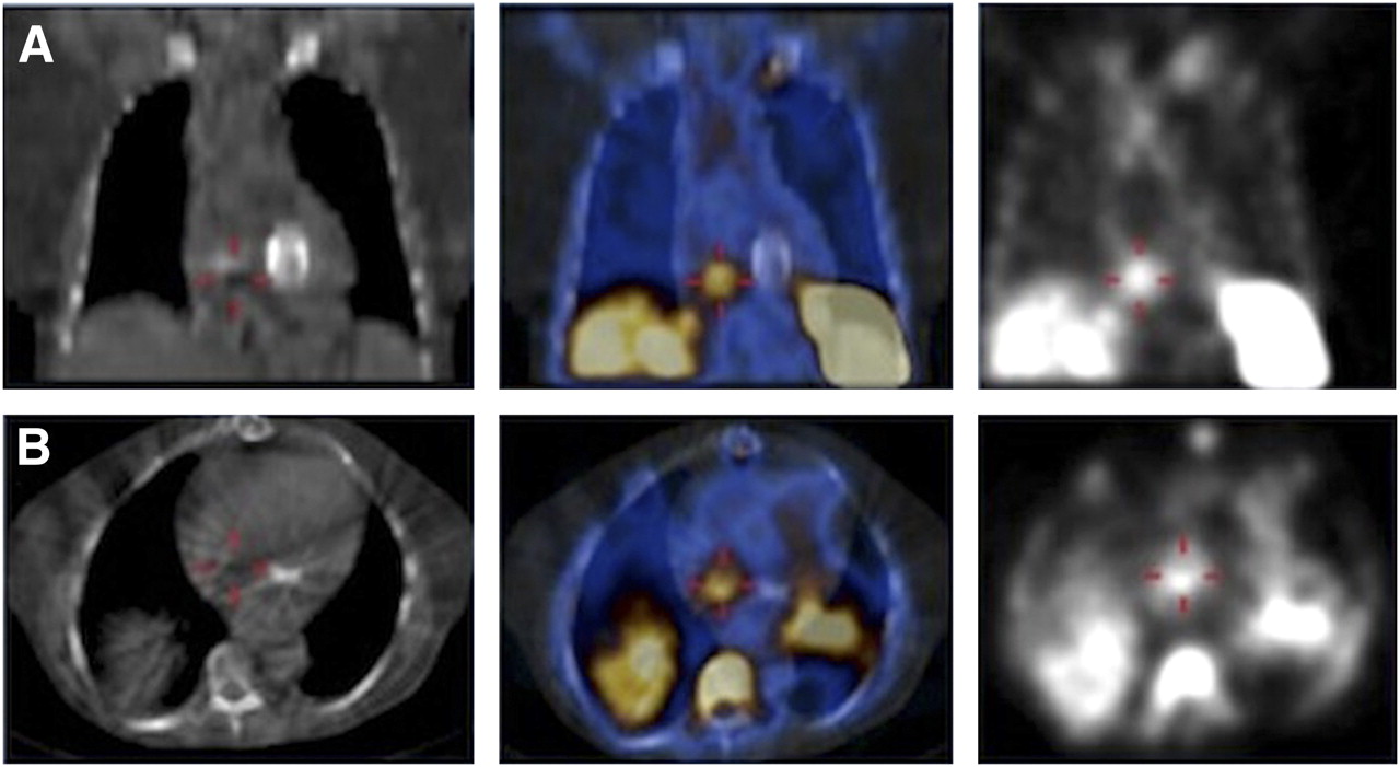
  • FIGURE 2.
    • Download figure
    • Open in new tab
    • Download powerpoint
    FIGURE 2.

    99mTc-HMPAO-WBC SPECT/CT images for patient with positive blood cultures and fever that arose a few months after substitution of mitral valve with mechanical prosthesis (coronal views [top], transaxial views [bottom]; CT [left], fused SPECT/CT [center], and SPECT [right]). SPECT images demonstrate clear focus of uptake in right heart, identified as endocarditis of native tricuspid valve by superimposed SPECT/CT images. Endocarditis of mechanical prosthesis, expected site of infection before 99mTc-HMPAO-WBC was performed, was therefore excluded.
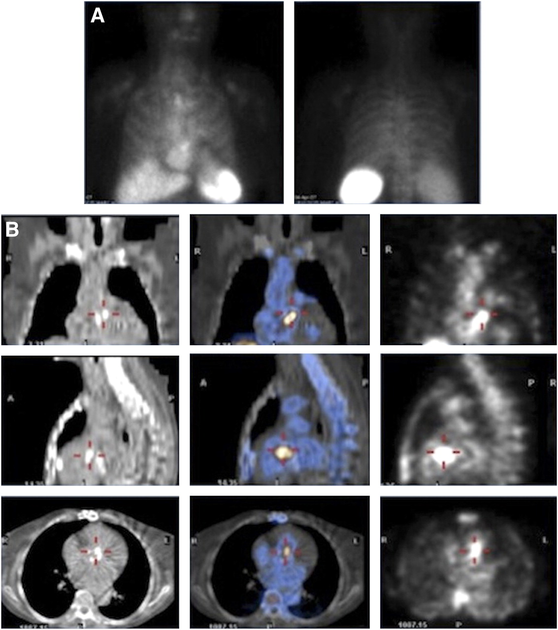
  • FIGURE 3.
    • Download figure
    • Open in new tab
    • Download powerpoint
    FIGURE 3.

    99mTc-HMPAO-WBC scintigraphy demonstrating value of SPECT/CT for precisely localizing site of infection. (A) Planar anterior (left) and posterior (right) views, where focal uptake of radiolabeled WBC mimics sternal osteomyelitis. (B) Coronal, sagittal, and transaxial CT (left); fused SPECT/CT (middle); and SPECT (right). Tomographic images correctly localize uptake of 99mTc-HMPAO-WBC at mitral valve prosthesis.
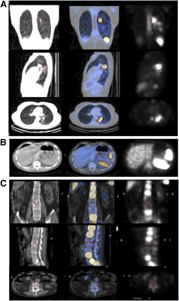
  • FIGURE 4.
    • Download figure
    • Open in new tab
    • Download powerpoint
    FIGURE 4.

    Examples of septic embolism at different sites as detected by 99mTc-HMPAO-WBC SPECT/CT. (A) Patient with septic embolism in left lung (coronal, sagittal, and transaxial CT [left]; fused SPECT/CT [middle]; and SPECT [right]). (B) Patient with septic embolism in spleen, where infection shows as photopenic area in splenic parenchyma (transaxial CT [left], fused SPECT/CT [center], and SPECT [right]). (C) Patient with septic embolism in spine (coronal, sagittal, and transaxial CT [left]; fused SPECT/CT [middle]; and SPECT [right]). Similarly, as in case of spleen, infection shows as photopenic area, which in this patient involves 2 vertebral bodies.
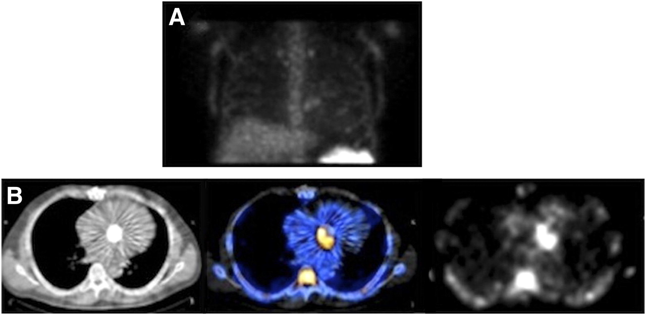
  • FIGURE 5.
    • Download figure
    • Open in new tab
    • Download powerpoint
    FIGURE 5.

    False-positive 18F-FDG PET/CT result in patient with fever. Area of increased 18F-FDG uptake suspected of being endocarditis at mitral valve mechanical prosthesis (A; fused transaxial PET/CT [left] and PET alone [right]) turned out to be negative with 99mTc-HMPAO-WBC SPECT/CT (B; fused transaxial SPECT/CT [left] and SPECT [right]). Clinical follow-up confirmed absence of infection. (C) CT transaxial image.

Tables

  • Figures
    • View popup
    TABLE 1

    Patient Characteristics (n = 131)

    Characteristicn
    Sex
     Female45 (34)
     Male86 (66)
    Risk factors
     Diabetes20 (15)
     Renal failure24 (18)
     Cutaneous lesions10 (8)
    Blood tests
     Erythrocyte sedimentation rate110 (84)
     C-reactive protein78 (60)
     Leukocytosis55 (42)
    Blood culture
     Positive67 (51)
     Negative64 (49)
    Duke criteria
     Definite28 (21)
     Possible55 (42)
     Rejected48 (37)
    • Median age was 66 y (mean age ± SD, 62.8 ± 16.6 y; age range, 19–89 y). Data in parentheses are percentages.

    • View popup
    TABLE 2

    Type of Valve, Site of IE, Type of Infection, and Time of Infection Onset

    Parametern
    Type of valve (n = 51)
     Native16 (31)
     Biologic prosthesis19 (38)
     Mechanical prosthesis16 (31)
    Site of IE
     Native
      Aortic (n = 30)9 (30)
      Mitral (n = 19)6 (32)
      Tricuspid (n = 1)1/1 (100)
     Biologic prosthesis
      Aortic (n = 30)10 (33)
      Mitral (n = 19)8 (42)
      Aortic + mitral (n = 1)1/1 (100)
     Mechanical prosthesis
      Aortic (n = 30)11 (37)
      Mitral (n = 19)5 (26)*
    Type of infection (n = 35)†
     Early IE9 (26)
     Somewhat-late IE11 (31)
     Late IE15 (43)
    Time of infection onset (months after valve replacement)
     Native
      Mean1.39
      Range0.5–2
     Biologic prosthesis
      Mean6
      Range3–10
     Mechanical prosthesis
      Mean51.4
      Range6–204
    • ↵* Including 2 patients with anuloplasty.

    • ↵† Only for patients with valve prosthesis either biologic or mechanical.

    • Data in parentheses are percentages.

    • View popup
    TABLE 3

    Results of 99mTc-HMPAO-WBC Scintigraphy in the 51 Patients with Final Diagnosis of IE, Stratified According to Duke Criteria

    Positive results
    Duke criterionCardiac onlyCardiac and extracardiacExtracardiac onlyNegative results
    Definite IE (n = 24)911*04
    Possible IE (n = 25)1311†1*0
    Rejected IE (n = 2)11*00
    • ↵* Septic embolism consequent to IE.

    • ↵† Eight patients with septic embolism, 1 with vasculitis, and 2 false-positive scans due to vertebral crush and metastasis from prostate cancer.

    • View popup
    TABLE 4

    Results of 99mTc-HMPAO-WBC Scintigraphy in the 51 Patients with Final Diagnosis of IE, Stratified According to Echocardiography

    EchocardiographyPositive resultsNegative results
    Positive (n = 40)355
    Negative (n = 11)110
    • View popup
    TABLE 5

    Results of 99mTc-HMPAO-WBC Scintigraphy in the 51 Patients with Final Diagnosis of IE, Stratified According to Blood Culture

    Positive results
    Blood cultureCardiac onlyCardiac and extracardiacExtracardiac onlyNegative results
    Positive (n = 32)1514*03
    Negative (n = 19)†89‡11
    • ↵* Patients (n = 11/14) with septic embolism consequent to IE and 2 false-positive scans due to vertebral crush.

    • ↵† During antibiotic therapy in 44 of 64 patients.

    • ↵‡ Patients (n = 8/9) with septic embolism consequent to IE and 1 false-positive scan due to vasculitis.

    • View popup
    TABLE 6

    Results of All Diagnostic Procedures in the 80 Patients Without IE

    99mTc-HMPAO-WBC scintigraphy
    ProcedureResultsPositive results*Negative results
    EchocardiographyPositive (n = 3)03
    Negative (n = 77)077
    Blood culturePositive (n = 35)269
    Negative (n = 45)2421
    Duke criteriaDefinitive IE (n = 4)40
    Possible IE (n = 30)1911
    Rejected IE (n = 46)2719
    • ↵* All patients presented with only extracardiac site(s) of radiopharmaceutical uptake.

PreviousNext
Back to top

In this issue

Journal of Nuclear Medicine: 53 (8)
Journal of Nuclear Medicine
Vol. 53, Issue 8
August 1, 2012
  • Table of Contents
  • Table of Contents (PDF)
  • About the Cover
  • Index by author
Print
Download PDF
Article Alerts
Sign In to Email Alerts with your Email Address
Email Article

Thank you for your interest in spreading the word on Journal of Nuclear Medicine.

NOTE: We only request your email address so that the person you are recommending the page to knows that you wanted them to see it, and that it is not junk mail. We do not capture any email address.

Enter multiple addresses on separate lines or separate them with commas.
Added Value of 99mTc-HMPAO–Labeled Leukocyte SPECT/CT in the Characterization and Management of Patients with Infectious Endocarditis
(Your Name) has sent you a message from Journal of Nuclear Medicine
(Your Name) thought you would like to see the Journal of Nuclear Medicine web site.
Citation Tools
Added Value of 99mTc-HMPAO–Labeled Leukocyte SPECT/CT in the Characterization and Management of Patients with Infectious Endocarditis
Paola A. Erba, Umberto Conti, Elena Lazzeri, Martina Sollini, Roberta Doria, Salvatore M. De Tommasi, Francesco Bandera, Carlo Tascini, Francesco Menichetti, Rudi A.J.O. Dierckx, Alberto Signore, Giuliano Mariani
Journal of Nuclear Medicine Aug 2012, 53 (8) 1235-1243; DOI: 10.2967/jnumed.111.099424

Citation Manager Formats

  • BibTeX
  • Bookends
  • EasyBib
  • EndNote (tagged)
  • EndNote 8 (xml)
  • Medlars
  • Mendeley
  • Papers
  • RefWorks Tagged
  • Ref Manager
  • RIS
  • Zotero
Share
Added Value of 99mTc-HMPAO–Labeled Leukocyte SPECT/CT in the Characterization and Management of Patients with Infectious Endocarditis
Paola A. Erba, Umberto Conti, Elena Lazzeri, Martina Sollini, Roberta Doria, Salvatore M. De Tommasi, Francesco Bandera, Carlo Tascini, Francesco Menichetti, Rudi A.J.O. Dierckx, Alberto Signore, Giuliano Mariani
Journal of Nuclear Medicine Aug 2012, 53 (8) 1235-1243; DOI: 10.2967/jnumed.111.099424
Twitter logo Facebook logo LinkedIn logo Mendeley logo
  • Tweet Widget
  • Facebook Like
  • Google Plus One
Bookmark this article

Jump to section

  • Article
    • Abstract
    • MATERIALS AND METHODS
    • RESULTS
    • DISCUSSION
    • CONCLUSION
    • DISCLOSURE STATEMENT
    • Acknowledgments
    • Footnotes
    • REFERENCES
  • Figures & Data
  • Info & Metrics
  • PDF

Related Articles

  • This Month in JNM
  • PubMed
  • Google Scholar

Cited By...

  • Infection Imaging: Focus on New Tracers?
  • Radiolabeled-White Blood Cell Imaging in Cardiac Device-Related Infective Endocarditis: Worth All the Effort?
  • The Prognostic Value of 99mTc-HMPAO-Labeled Leucocyte SPECT/CT in Cardiac Device-Related Infective Endocarditis
  • Infective endocarditis complicating transcatheter aortic valve implantation
  • Methicillin-Resistant Staphylococcus aureus Prosthetic Valve Endocarditis: Pathophysiology, Epidemiology, Clinical Presentation, Diagnosis, and Management
  • Approach to Diagnosis of Cardiovascular Implantable-Electronic-Device Infection
  • Targeting Cardiovascular Implant Infection: Multimodality and Molecular Imaging
  • Clinical Trial Principles and Endpoint Definitions for Paravalvular Leaks in Surgical Prosthesis: An Expert Statement
  • 18F-Fluorodeoxyglucose Positron Emission Tomography-Computed Tomography in Cardiac Implantable Electronic Devices Infection: Ready for Routine Care!
  • 18F-Fluorodeoxyglucose Imaging of Inflammation: Ready to Represent a Standard in Diagnosing Endocarditis?
  • Challenges in Infective Endocarditis
  • Diagnostic Accuracy of 18F-FDG PET/CT in Infective Endocarditis and Implantable Cardiac Electronic Device Infection: A Cross-Sectional Study
  • Improving the Diagnosis of Infective Endocarditis in Prosthetic Valves and Intracardiac Devices With 18F-Fluordeoxyglucose Positron Emission Tomography/Computed Tomography Angiography: Initial Results at an Infective Endocarditis Referral Center
  • Respective Performance of 18F-FDG PET and Radiolabeled Leukocyte Scintigraphy for the Diagnosis of Prosthetic Valve Endocarditis
  • Radiolabeled WBC Scintigraphy in the Diagnostic Workup of Patients With Suspected Device-Related Infections
  • Positron Emission Tomography/Computed Tomography for Diagnosis of Prosthetic Valve Endocarditis: Increased Valvular 18F-Fluorodeoxyglucose Uptake as a Novel Major Criterion
  • The Use of 18F-FDG-PET/CT in the Diagnostic Workup of CIED Infections: Another Perspective
  • Google Scholar

More in this TOC Section

  • Feasibility of Ultra-Low-Activity 18F-FDG PET/CT Imaging Using a Long–Axial-Field-of-View PET/CT System
  • Cardiac Presynaptic Sympathetic Nervous Function Evaluated by Cardiac PET in Patients with Chronotropic Incompetence Without Heart Failure
  • Validation and Evaluation of a Vendor-Provided Head Motion Correction Algorithm on the uMI Panorama PET/CT System
Show more Clinical Investigations

Similar Articles

SNMMI

© 2025 SNMMI

Powered by HighWire