


Article Figures & Data
Figures
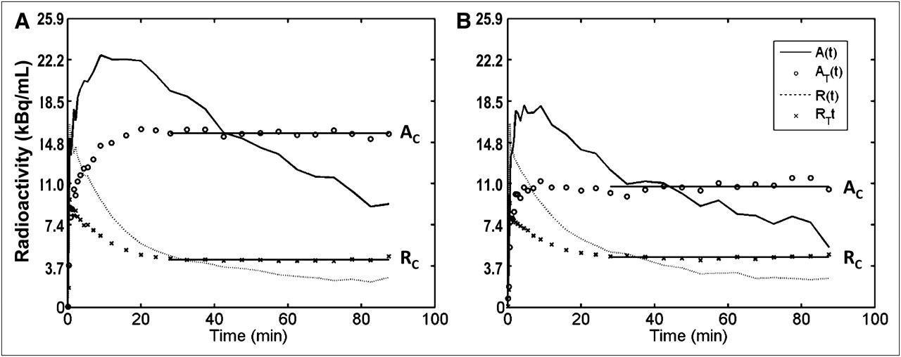
- FIGURE 1.
Observed and transformed regional time–activity curves of anterior putamen, A(t) and AT(t), respectively, and cerebellum, R(t) and RT(t), respectively, of HSA (A) and LSA (B) 11C-raclopride scans. Both AT(t) for all striatal subdivisions and RT(t) became stable by 30 min in all cases. Assumed plateaus (AC and RC) are shown by horizontal solid lines.
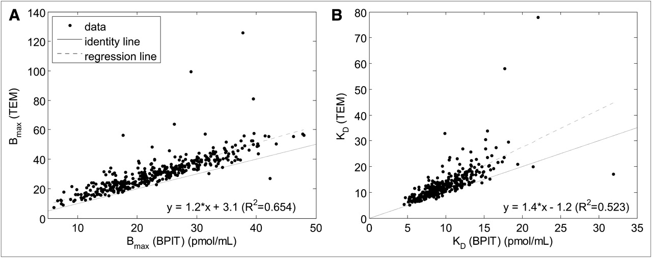
- FIGURE 2.
Scatterplots of regional values of Bmax (A) and KD (B), TEM (=y) vs. proposed BPIT (=x). Data from all subjects (n = 81), 5 subdivisions per subject with left and right VOIs merged, are shown (a total of 405 points). Linear regression equations and coefficients of determination (R2) are shown in each panel.
- FIGURE 3.
Scatterplots of Bmax vs. age for ventral striatum of healthy subjects (n = 47) with left and right VOIs merged, given by BPIT (A) and TEM (B). Linear regression equations and coefficients of determination (R2) are shown in each panel.


Tables
Parameter Anterior putamen Posterior putamen Anterior caudate nucleus Posterior caudate nucleus Ventral striatum Volume (mL) Left 1.93 ± 0.29 1.88 ± 0.39 2.10 ± 0.34 0.67 ± 0.20 0.87 ± 0.21 Right 2.00 ± 0.29 1.76 ± 0.33 2.19 ± 0.33 0.66 ± 0.19 0.85 ± 0.16 Merged 3.93 ± 0.53 3.65 ± 0.69 4.29 ± 0.64 1.22 ± 0.37 1.72 ± 0.34 nRSS (Bq/mL) BPIT 111 ± 77.7 (3.0 ± 2.1) 122.1 ± 88.8 (3.3 ± 2.4) 125.8 ± 99.9 (3.4 ± 2.7) 477.3 ± 336.7 (12.9 ± 9.1) 384.8 ± 333 (10.4 ± 9.0) MRTM2 558.7 ± 436.6 (15.1 ± 11.8) 610.5 ± 547.6 (16.5 ± 14.8) 658.6 ± 577.2 (17.8 ± 15.6) 2,223.7 ± 1,502.2 (60.1 ± 40.6) 2,131.2 ± 2,216.3 (57.6 ± 59.9) Data are mean ± SD, with nCi/mL in parentheses.
- TABLE 2
Effects of TI in Equation 1 and Assumed Scan Length for BPIT (TS) on Bmax and KD Estimates
Correlation equations (R2 = coefficient of determination) TI (min) TS (min) Bmax KD 80 80 y = 1.00x + 0.46 (R2 = 0.989) y = 1.00x + 0.17 (R2 = 0.968) 80 90 y = 1.05x − 0.04 (R2 = 1) y = 1.05x − 0.025 (R2 = 1) 100 90 y = 0.96x − 0.003 (R2 = 1) y = 0.96x − 0.02 (R2 = 1) x= Bmax or KD estimates with both TI and TS set to 90 min; y = estimates obtained with TI and TS set as indicated.
Left Right Merged Striatal subdivisions BPIT TEM BPIT TEM BPIT TEM Anterior putamen Pearson correlation coefficient −0.477 −0.424 −0.417 −0.338 −0.433 −0.375 P 0.000701* 0.00298* 0.00361* 0.0201 0.00239* 0.0095* Rates of decline 0.49% 0.48% 0.45% Not significant 0.47% 0.47% Posterior putamen Pearson correlation coefficient −0.453 −0.463 −0.437 −0.414 −0.461 −0.416 P 0.00137* 0.00105* 0.00214* 0.00386* 0.0011* 0.00363* Rates of decline 0.51% 0.54% 0.50% 0.49% 0.51% 0.49% Anterior caudate nucleus Pearson correlation coefficient −0.508 −0.363 −0.483 −0.428 −0.508 −0.476 P 0.000268* 0.0121 0.000579* 0.00267* 0.000264* 0.000719* Rates of decline 0.62% Not significant 0.60% 0.53% 0.61% 0.58% Posterior caudate nucleus Pearson correlation coefficient −0.34 −0.227 −0.452 −0.285 −0.411 −0.258 P 0.0196 0.126 0.00141* 0.0519 0.00407* 0.0801 Rates of decline Not significant Not significant 0.73% Not significant 0.66% Not significant Ventral striatum Pearson correlation coefficient −0.415 −0.0261 −0.454 −0.224 −0.471 −0.0694 P 0.00377* 0.862 0.00134* 0.130 0.000844* 0.643 Rates of decline 0.62% Not significant 0.68% Not significant 0.67% Not significant ↵* Statistically significant after Bonferroni adjustment.
Rates of decline per year are relative to Bmax value at 20 y.


