


Article Figures & Data
Figures
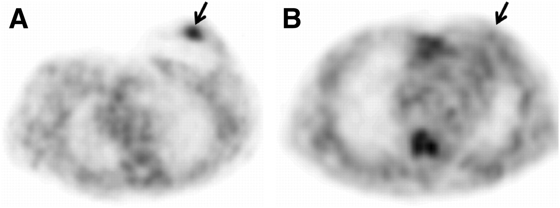
- FIGURE 1.
Representative transverse 18F-FFNP PET images in patient with PR+ breast cancer (A) and another with PR– breast cancer (B). Arrows point to tumor.
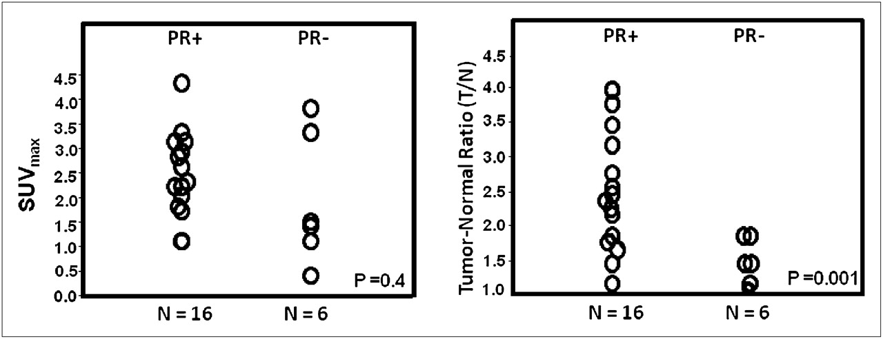
- FIGURE 2.
18F-FFNP uptake assessed by SUVmax (left) and T/N ratio (right) in PR+ and PR– breast cancers.
- FIGURE 3.
Time–activity distribution in liver, tumor, normal breast, and blood (from ventricular chamber). Data were normalized for 370-MBq injection (patient 5, dynamic cohort) with ER+ and PR+ tumor. Uptake of 18F-FFNP in this patient's tumor was twice that in her normal breast.
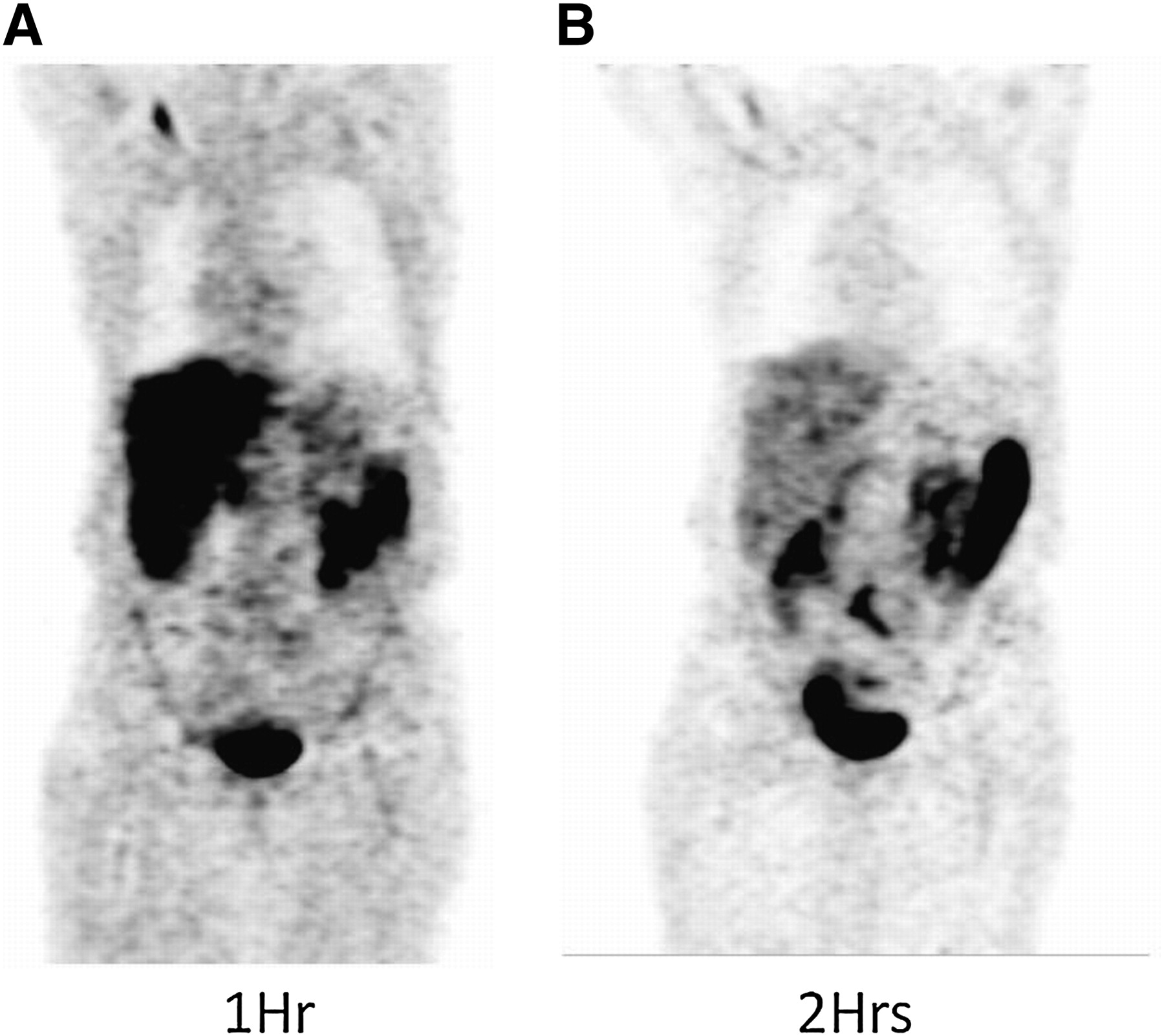
- FIGURE 4.
Typical whole-body coronal images of patient at 1 h (A) and 2 h (B) after injection of 370 MBq (10 mCi) of 18F-FFNP. Accumulation of activity is primarily seen in liver and small intestines
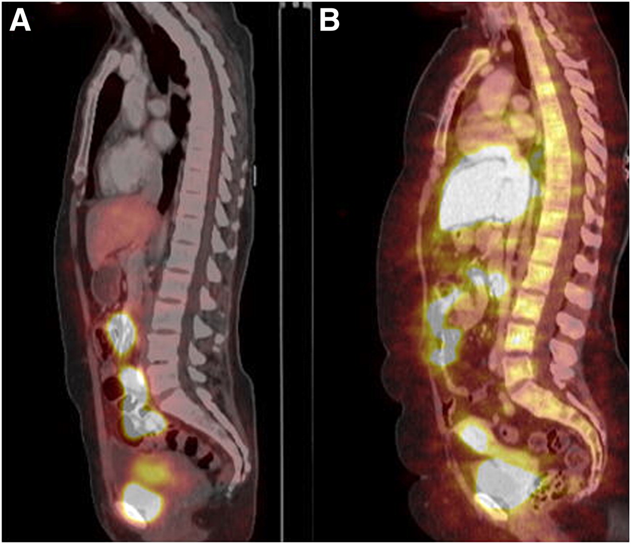
- FIGURE 5.
Sagittal 18F-FFNP images registered to CT images showing accumulation in liver, intestines, uterus, and urinary bladder.


Tables
Patient Age (y) Injected dose (MBq) Mass (μg) ER/PR/HER2 Tumor T/N Tumor SUV DVR Dynamic patients 1 59 359 5.38 –/–/–/ 1.9 3.4 2.4 2 39 281 0.84 +/+/–/, +/+/–/* 2.3, 2.5* 3.0, 3.2* 1.9, 2.7 3 59 222 3.41 –/–/–/ 1.9 1.5 1.4 4 32 178 0.83 +/–/–/ 1.1 3.9 1.4 5 57 366 0.53 +/+/–/, +/+/–/* 3.5, 2.8* 3.4, 2.7* 3.7, 2.8 6 58 148 1.91 +/+/–/ 1.2 2.1 1.1 7 71 366 0.56 +/+/–/ 2.2 2.9 2.0 8 58 370 0.34 +/+/–/ 1.7 1.2 1.6 Dosimetry patients 1 49 89 1.39 +/+/–/ 2.4 1.2 Not applicable 2 52 185 1.87 +/+/–/ 1.9 2.3 Not applicable 3 64 370 2.67 +/+/–/ 1.8 2.4 Not applicable 4 49 104 0.83 –/–/–/ 1.5 1.6 Not applicable 5 53 329 0.75 –/–/–/ 1.5 1.2 Not applicable 6 47 270 1.32 –/–/–/ 1.2 0.5 Not applicable 7 68 359 0.90 +/+/–/ 1.5 1.2 Not applicable 8 64 370 0.31 +/+/–/ 4.0 4.4 Not applicable 9 44 215 0.90 +/+/–/ 2.6 1.8 Not applicable 10 62 352 0.54 +/+/–/ 4.0 3.2 Not applicable 11 55 315 0.76 +/+/–/ 3.2 1.9 Not applicable 12 49 259 0.70 +/+/–/ 3.8 2.3 Not applicable ↵* Second breast cancer in patients with 2 breast cancers.
Organ Residence times (min) Liver 16.3 ± 5.1 Small intestine 20.8 ± 5.2 Gallbladder 4.14 ± 1.04 Bone/marrow 4.60 ± 1.15 Uterus 0.47 ± 0.12 Blood (left ventricle) 11.4 ± 2.8 Data are mean ± SD.
Organ Organ dose human PET (mGy/MBq) Adrenals 0.017 Brain 0.010 Breasts 0.010 Gallbladder 0.113 Lower large intestine 0.020 Small intestine 0.096 Stomach 0.017 Upper large intestine 0.030 Heart wall 0.015 Kidneys 0.017 Liver 0.051 Lungs 0.013 Muscle 0.013 Ovaries 0.023 Pancreas 0.018 Red marrow 0.015 Osteogenic cells 0.024 Skin 0.009 Spleen 0.014 Thymus 0.012 Thyroid 0.011 Urinary bladder wall 0.016 Uterus 0.034 Whole body 0.015 Effective dose (mSv/MBq) 0.020
Supplemental Data
Files in this Data Supplement:


Jump to section
Related Articles
Cited By...
- Longitudinal Molecular Imaging of Progesterone Receptor Reveals Early Differential Response to Endocrine Therapy in Breast Cancer with an Activating ESR1 Mutation
- Recent Advances in Imaging Steroid Hormone Receptors in Breast Cancer
- Measuring Estrogen Receptor Functionality Using Progesterone Receptor PET Imaging: Rising to the (Estradiol) Challenge!
- Sensitivity and Isoform Specificity of 18F-Fluorofuranylnorprogesterone for Measuring Progesterone Receptor Protein Response to Estradiol Challenge in Breast Cancer
- Imaging Diagnostic and Therapeutic Targets: Steroid Receptors in Breast Cancer
- Quo Vadis: PET and Single-Photon Molecular Breast Imaging
- Longitudinal Noninvasive Imaging of Progesterone Receptor as a Predictive Biomarker of Tumor Responsiveness to Estrogen Deprivation Therapy
- Role of Positron Emission Tomography for the Monitoring of Response to Therapy in Breast Cancer
- Small-Animal PET of Steroid Hormone Receptors Predicts Tumor Response to Endocrine Therapy Using a Preclinical Model of Breast Cancer