


Article Figures & Data
Figures
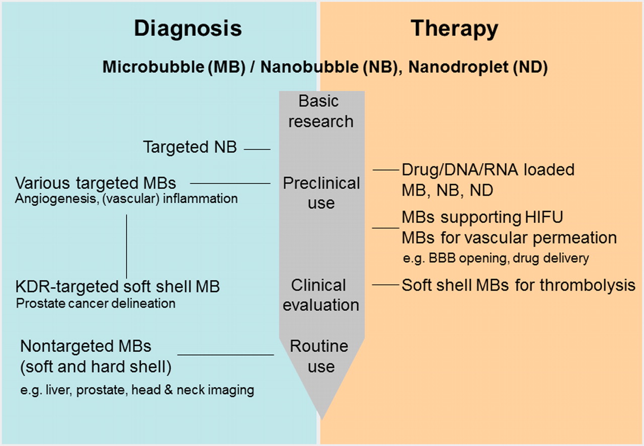
- FIGURE 1.
Diagram illustrating development stage of microbubbles, nanobubbles, and nanodroplets for diagnostic and therapeutic purposes. HIFU = high-intensity focused ultrasound; KDR = kinase domain receptor.
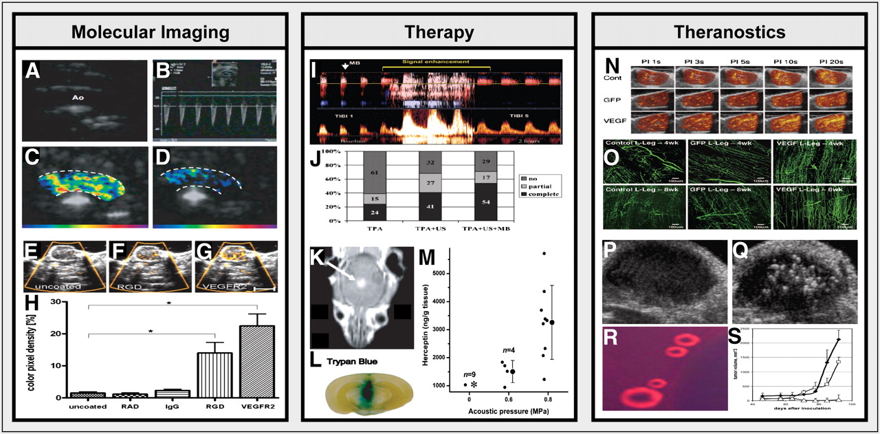
- FIGURE 2.
Microbubbles for molecular imaging, therapy, and theranostics. (A–D) Representative images of the aortic arch of an ApoE-knockout mouse on high-cholesterol diet obtained using 2-dimensional B-mode imaging (A), pulsed-wave Doppler ultrasound (B), and molecular ultrasound using VCAM-1–targeted (C) and control (D) microbubbles (15). (E–H) Binding (E–G) and quantification (H) of control (E), RGD-targeted (F), and VEGFR2-targeted (G) microbubbles to angiogenic tumor endothelium (12). (I) Spectral waveform visualization of arterial recanalization in patient with middle cerebral artery occlusion before (left) and after (right) microbubble-enhanced sonothrombolysis (22). (J) Quantification of complete, partial, and no MCA recanalization on treatment with tPA, tPA plus ultrasound, and tPA plus ultrasound plus microbubbles (22). (K–M) Highly efficient delivery of the MRI contrast agent gadopentetate dimeglumine (K), trypan blue (L) and trastuzumab (M) of the BBB upon microbubble-enhanced focused ultrasound treatment at 2 different acoustic pressures (middle column: 0.6 MPa; right column: 0.8 MPa) (reprinted with permission from Proc Natl Acad Sci USA. 2006;103:11719–11723, Copyright (2012) National Academy of Sciences, USA (23)). (N–O) Combination of microbubbles with ultrasound (at various different pulsing intervals) improves the delivery of plasmid DNA encoding for VEGF to ischemic hind limb muscle in rats, leading to improved perfusion (N) and to dense proliferation of neovessels, with abundant bridging arterioles (O) (26). (P–S) Doxorubicin-containing nanobubbles coalesce into microbubbles at physiologic temperatures (R), they can be visualized upon extravasation into subcutaneous tumor xenografts using B-mode imaging (P: preinjection; Q: 4 h after intravenous injection), and they can be used in combination with focused ultrasound to induce highly efficient tumor growth inhibition (S) (28). Images are adapted from indicated references with permission of publishers.

Tables
- TABLE 1
Common Ultrasound Molecular Imaging Targets, Conjugation Methods, and Applications
Target Ligand (conjugation method) Application Reference VEGF receptor type 2 Anti-VEGFR-2 mAb* Preclinical (10–12) Single-chain VEGF† Preclinical (8) Heterodimeric lipopeptide† Preclinical/clinical (prostate cancer) (4,16–18) avβ3-integrin cRGD peptide* Preclinical (12) cRRL peptide* Preclinical (29) Anti-integrin av-chain mAb* Preclinical (30) Anti-RGD–containing disintegrin echistatin mAb* Preclinical (30) Echistatin* Preclinical (31) Knottin peptide binding avβ3-integrin* Preclinical (32) Cyclic RGD peptide† Preclinical (33) ICAM-1/VCAM-1 Anti-ICAM-1 and -VCAM-1 mAb* Preclinical (15,34) CD105 Anti-CD105 mAb* Preclinical (11,35) P or E selectin Polymeric sulfo-Lewis-x* Preclinical (13,14)


Jump to section
Related Articles
Cited By...
- Efficacy of Affibody-Based Ultrasound Molecular Imaging of Vascular B7-H3 for Breast Cancer Detection
- Targeted enhancement of flotillin-dependent endocytosis augments cellular uptake and impact of cytotoxic drugs
- Advanced Ultrasound Technologies for Diagnosis and Therapy
- Breast Cancer Detection by B7-H3-Targeted Ultrasound Molecular Imaging
- Rhodamine-Loaded Intercellular Adhesion Molecule-1-targeted Microbubbles for Dual-Modality Imaging Under Controlled Shear Stresses