


Article Figures & Data
Figures
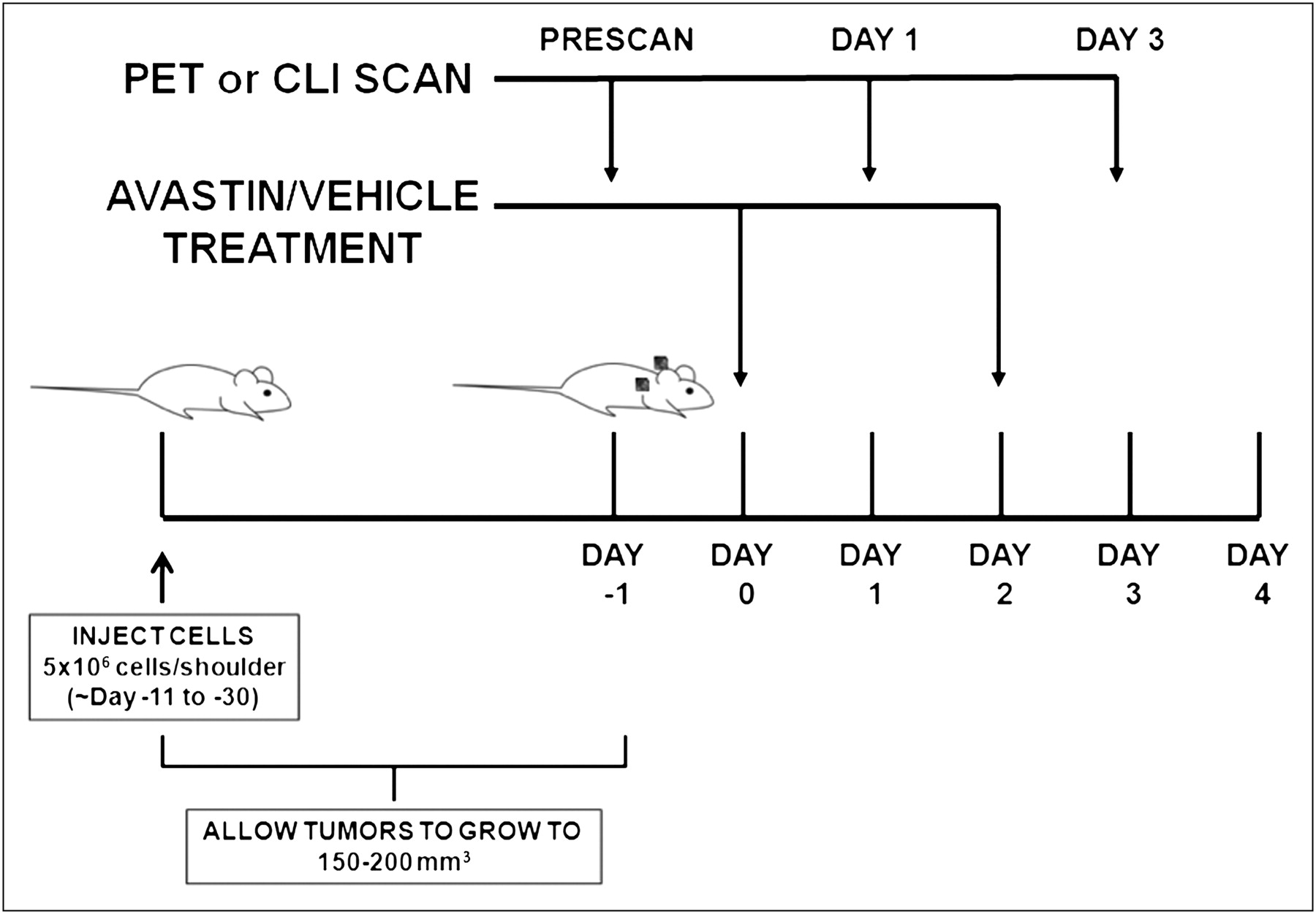
- FIGURE 1.
Schematic of experimental design. Tumors were implanted bilaterally in shoulder region and allowed to grow to 150–200 mm3, and tumor-bearing mice were subjected to in vivo imaging via PET and CLI at day −1, 1, and 3. Bevacizumab treatment was performed by 2 injections of 20 mg/kg at days 0 and 2. For 18F-FDG imaging study, mice were kept fasting overnight before experiment.
- FIGURE 2.
(A) Tumor growth kinetics for H460 xenografts. Measurements were made at days −5, −3, −1, 1, 2, 3, 4, and 5. (B) Tumor growth kinetics for PC3 xenografts. Measurements were made at days −8, −4, −2, 0, 1, 2, 3, 4, and 5. For both figures, day 0 indicates first dose of bevacizumab.
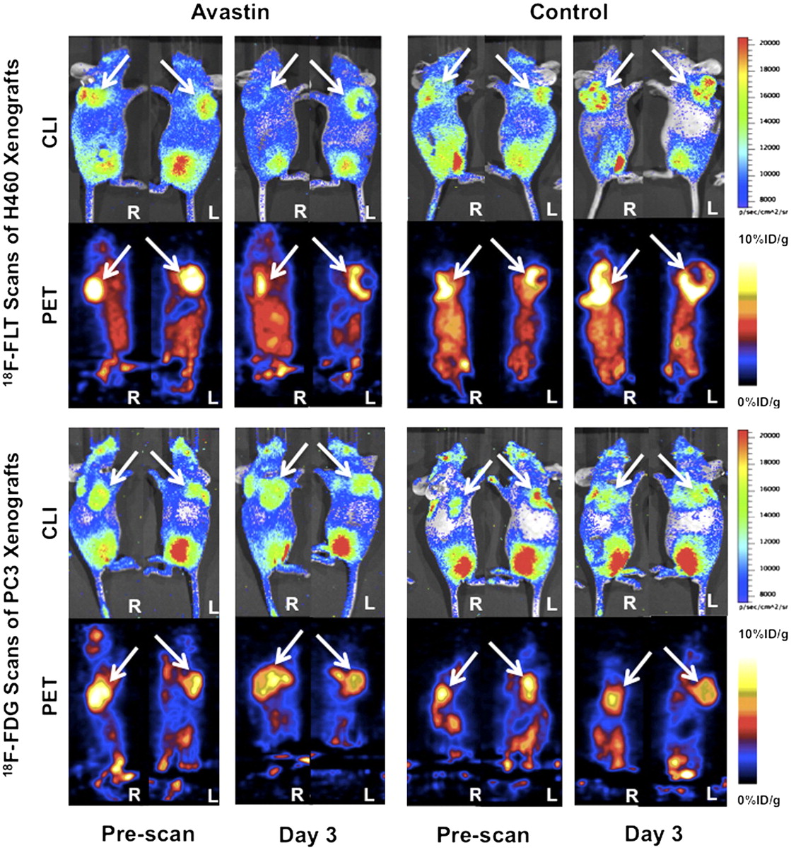
- FIGURE 3.
Comparison between Cerenkov luminescence and PET images. White arrows point to tumors in all panels. (Top left) 18F-FLT scans of representative H460 tumor–bearing mouse during bevacizumab treatment. (Top right) 18F-FLT scans of representative H460 tumor–bearing mouse of control group. (Bottom left) 18F-FDG scans of representative PC3 tumor–bearing mouse during bevacizumab treatment. (Bottom right) 18F-FDG scans of representative PC3 tumor–bearing mouse of control group. In each panel, upper row contains Cerenkov luminescence images, and bottom row contains PET images; for each row, left half contains prescans at day −3, and right half contains scans at day 3.
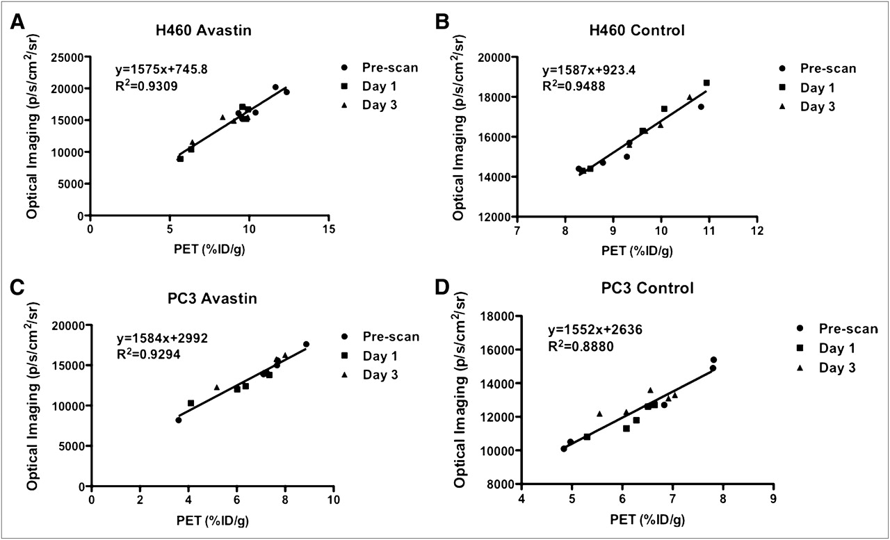
- FIGURE 4.
Quantitative analysis of Cerenkov luminescence and PET images and their correlation calculated through fitting by linear regression. P < 0.0001 for all 4 linear regressions. (A) H460 xenografts with bevacizumab treatment. (B) H460 xenografts of control group. (C) PC3 xenografts with bevacizumab treatment. (D) PC3 xenografts of control group.


Jump to section
Related Articles
Cited By...
- Detection of Shortwave-Infrared Cerenkov Luminescence from Medical Isotopes
- Optimal Scheduling of Bevacizumab and Pemetrexed/cisplatin Dosing in Non-Small Cell Lung Cancer
- {beta}-Radioluminescence Imaging: A Comparative Evaluation with Cerenkov Luminescence Imaging
- Intratherapeutic Biokinetic Measurements, Dosimetry Parameter Estimates, and Monitoring of Treatment Efficacy Using Cerenkov Luminescence Imaging in Preclinical Radionuclide Therapy
- Relaxin receptor antagonist AT-001 synergizes with docetaxel in androgen-independent prostate xenografts
- Clinical Cerenkov Luminescence Imaging of 18F-FDG
- Intraoperative Imaging of Tumors Using Cerenkov Luminescence Endoscopy: A Feasibility Experimental Study