


Article Figures & Data
Figures
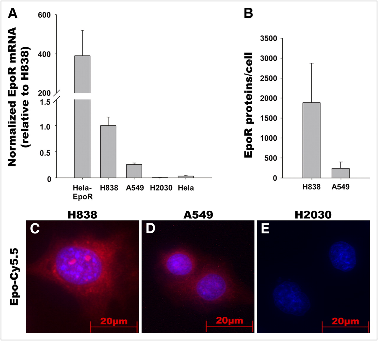
- FIGURE 1.
EpoR expression in NSCLC cell lines and binding of Epo-Cy5.5 to EpoR in vitro. (A) Analysis of EpoR messenger RNA expression in NSCLC cell lines H838, A549, and H2030 by quantitative RT-PCR. Hela cells and EpoR-transfected Hela cells that overexpress EpoR were used as controls. EpoR messenger RNA expression is higher in H838 than in A549, and EpoR is lacking in H2030. (B) Quantitative immunoblotting demonstrates higher EpoR protein expression in H838 than in A549. EpoR molecules were determined by adding recombinant calibrator GST-EpoR in different concentrations. Samples were run in triplicate and are representative of 3 independent experiments. (C–E) Incubation of NSCLC cell lines H838, A549, and H2030 with Epo-Cy5.5 (red) reveals stronger signal for Epo-Cy5.5 (red) in H838 than in A549 and no signal in H2030, thus correlating with EpoR expression levels. Nuclei were counterstained using DAPI (blue); images were acquired with equal exposure time.
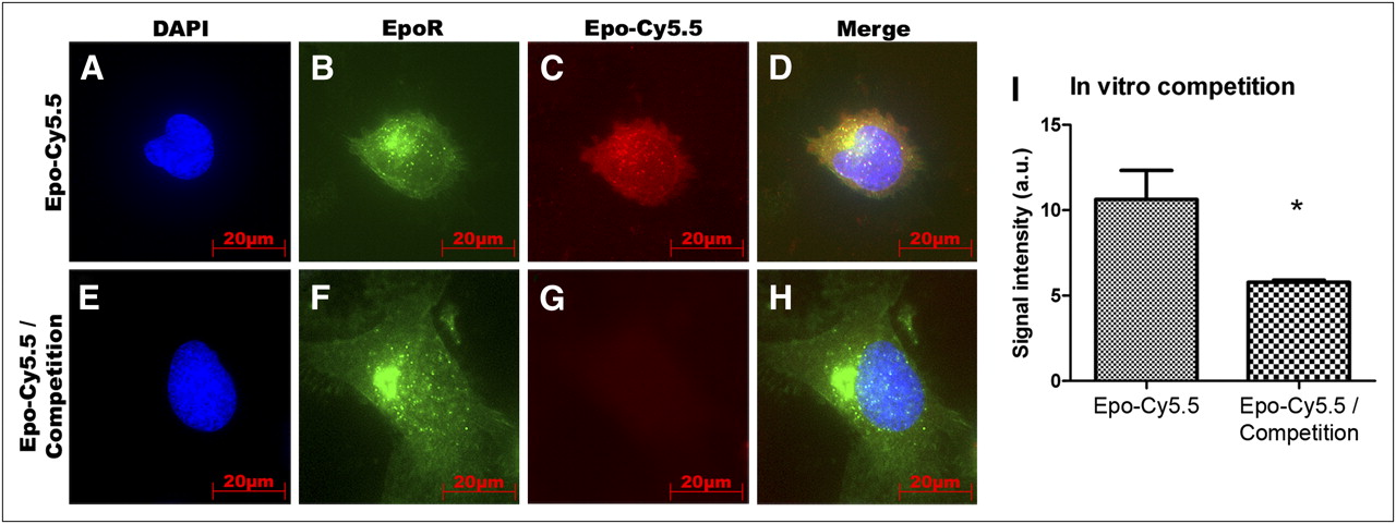
- FIGURE 2.
In vitro competition analysis of Epo-Cy5.5. To test binding specificity, EpoR/EGFP-transfected HeLa cells with EpoR/EGFP overexpression were incubated with Epo-Cy5.5 alone or with 1:20 mixture of Epo-Cy5.5 and unlabeled rhuEpo for competition. (A–D) Colocalization of Epo-Cy5.5 (red) with EGFR/EpoR fusion protein (green) (yellow in D) on EpoR/EGFP-transfected HeLa cells demonstrates specific binding of probe. (E–H) Competition by unlabeled rhuEpo leads to strong signal reduction (G). EGFP/EpoR fusion protein is shown in green, Epo-Cy5.5 in red, and counterstaining of nuclei by DAPI in blue. (I) Comparison of mean fluorescence signal intensities ± SD between control and competition samples of 5 slides shows significantly higher signal intensity in control samples (n = 5, P < 0.05). a.u. = arbitrary units.
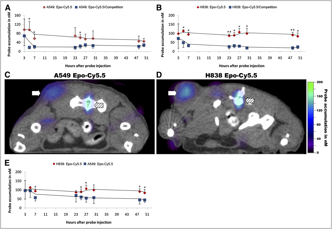
- FIGURE 3.
Epo-Cy5.5 binding specificity in vivo and EpoR expression level in tumors. Epo-Cy5.5 was injected alone or in 1:5 mixture with unlabeled rhuEpo into nude mice bearing subcutaneous A549 and H838 xenografts. Epo-Cy5.5 concentrations in tumors were determined by FMT. Simultaneous micro-CT scans allowed tumor localization and mapping of FMT signals. (A and B) Kinetics of probe accumulation in tumors of A549 control vs. A549 competition mice (A) and H838 control vs. H838 competition mice (B). Reduced binding of Epo-Cy5.5 is seen in competition group (*P < 0.05 and **P < 0.001, n = 4 animals per group). (C and D) Micro-CT/FMT fusion image showing high signal intensity in tumor and bone marrow of A549 and H838 control group. Tumor accumulation of Epo-Cy5.5 is higher in H838 (C) than in A549 (D), whereas signal in bone marrow is similar in both mice. Arrows mark tumor area; hatched arrows mark bone marrow. (E) Comparison of Epo-Cy5.5 tumor accumulation between A549 and H838 control groups, confirming higher probe accumulation in H838 tumors (*P < 0.05, n = 4 animals per group). Trend lines in A, B, and E illustrate almost constant accumulation of probe in tumor over time. Each value represents mean Epo-Cy5.5 concentration in nM ± SD (n = 4 per group).
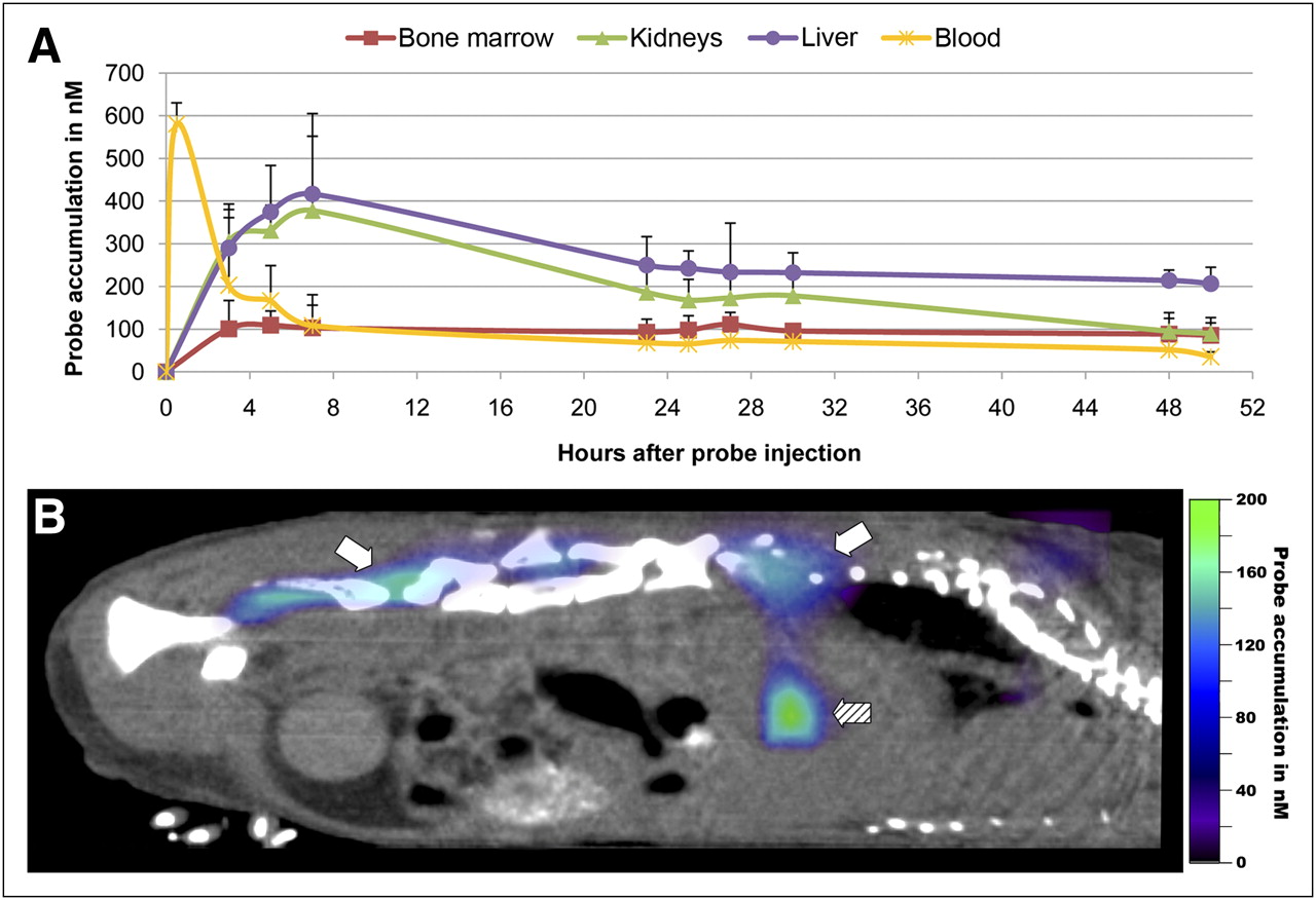
- FIGURE 4.
Biodistribution of Epo-Cy5.5. Organ kinetics were determined in nude mice bearing subcutaneous A549 xenograft tumors after intravenous injection of Epo-Cy5.5 (10 μM). (A) Epo-Cy5.5 concentrations were measured in liver, kidneys, bone marrow, and blood. After bolus intravenous injection of Epo-Cy5.5, high values are measured in blood, followed by rapid decline. Strong accumulation was observed in liver and kidneys starting from 3 h after injection and reaching maximal levels at 7 h; accumulation decreased thereafter. Almost constant accumulation of Epo-Cy5.5 was found in bone marrow, indicating specific receptor binding in organ with highest EpoR level. Shown are mean values of EpoCy5.5 concentrations ± SD for each measuring time point in nanomoles (n = 4). (B) Sagittal micro-CT/FMT fusion image at 48 h after probe injection demonstrating strong accumulation of Epo-Cy5.5 (color signals) in bone marrow (white arrows) and in liver (hatched arrow).
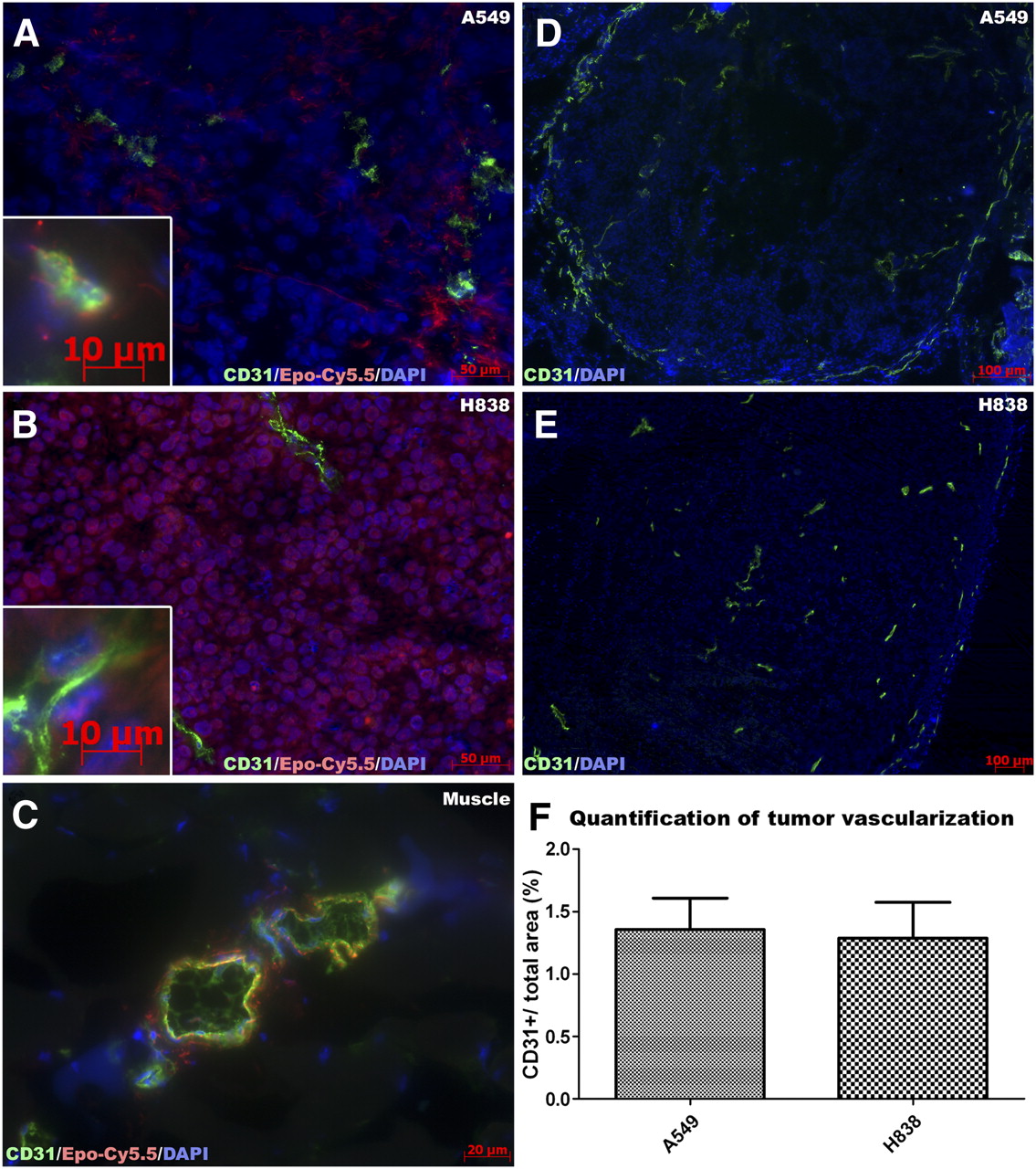
- FIGURE 5.
EpoR expression level–dependent binding to tumor cells and weaker binding to endothelial cells, as shown by Epo-Cy5.5. A549 and H838 tumors have similar vascularization. (A–C) For further validation, cryosections of A549 and H838 tumors were stained with Epo-Cy5.5 (red), followed by vessel (CD31, green) and nucleus (DAPI, blue) staining. Murine muscle tissue was stained as control. Epo-Cy5.5 signal is stronger in H838 tumors (B) than in A549 tumors (A), in line with differences in EpoR expression of cell lines. In muscle tissue, Epo-Cy5.5 binds to muscle endothelial cells (C). (D–E) Immunostaining of CD31 (green) and DAPI (blue) demonstrates similar vascularization of H838 and A549 tumors. (F) Quantification of CD31+ area fraction confirms similar vessel density (P = 0.73, n = 4).


Additional Files
Supplemental Data
Files in this Data Supplement:


Jump to section
Related Articles
Cited By...
- No citing articles found.