


Article Figures & Data
Figures
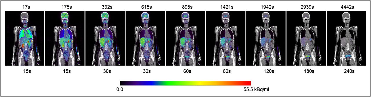
- FIGURE 1.
11C-(+)-PHNO whole-body biodistribution over time for typical subject. PET (color scale) images are coregistered with subjects’ CT image (gray scale). Time, in seconds, above each image is acquisition start time after injection of first bed position of whole-body image, whereas time, also in seconds, below each image is duration of each bed position in whole-body image. Image sequence is not decay-corrected to injection time; decay correction is applied only when each whole-body image is created from its individual-bed-position images.
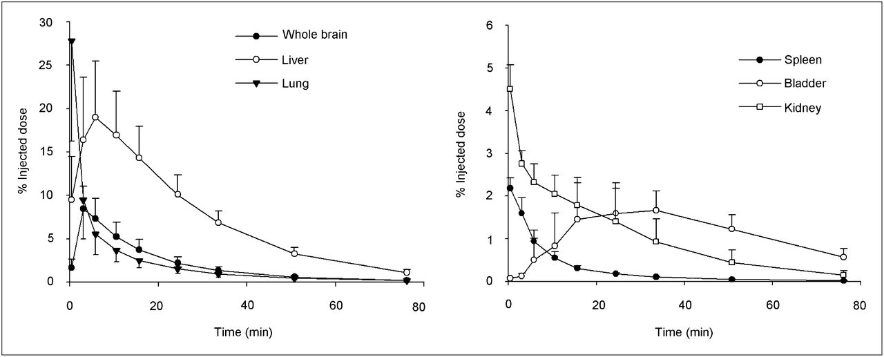
- FIGURE 2.
Mean organ uptake (expressed as percentage of injected dose) for all 6 subjects over time. Vertical bars represent SD, and data are not decay-corrected to time 0.

Tables
- TABLE 1
Normalized Accumulated Activity Measured Assuming Organ Volume for Standard Male and Female
Organ All subjects (n = 6) as males Females (n = 3) as females Whole brain 0.027 ± 0.007 0.029 ± 0.005 Lung 0.027 ± 0.009 0.027 ± 0.007 Liver 0.102 ± 0.024 0.073 ± 0.011 Spleen 0.003 ± 0.001 0.003 ± 0.000 Kidneys 0.014 ± 0.004 0.014 ± 0.005 Bladder 0.017 ± 0.005 0.011 ± 0.003 Remainder 0.300 ± 0.025 0.333 ± 0.009 Data are organ MBq⋅h divided by injected MBq (mean ± SD).
- TABLE 2
Organ Dose as Calculated by OLINDA/EXM 1.1 for Standard Adult Hermaphrodite Male Model as Applied to All 6 Subjects and Standard Female Model as Applied Only to the 3 Female Subjects
Parameter SAHMM SAFM Organ dose (μGy/MBq) Adrenals 3.5 ± 0.1 4.3 ± 0.1 Brain 6.4 ± 1.7 8.2 ± 1.2 Breasts 2.0 ± 0.1 2.6 ± 0.0 Gallbladder wall 4.4 ± 0.4 4.8 ± 0.2 Lower large intestine wall 2.4 ± 0.2 3.2 ± 0.1 Small intestine 2.6 ± 0.1 3.2 ± 0.1 Stomach wall 2.6 ± 0.1 3.4 ± 0.0 Upper large intestine wall 2.7 ± 0.1 3.5 ± 0.1 Heart wall 2.9 ± 0.0 3.7 ± 0.1 Kidneys 14.3 ± 3.6 15.8 ± 4.5 Liver 17.9 ± 3.9 17.4 ± 2.4 Lungs 8.2 ± 2.2 10.1 ± 2.3 Muscle 2.2 ± 0.1 2.9 ± 0.1 Ovaries 2.5 ± 0.2 3.3 ± 0.1 Pancreas 3.4 ± 0.1 4.2 ± 0.1 Red marrow 2.1 ± 0.1 2.7 ± 0.0 Osteogenic cells 3.1 ± 0.2 4.4 ± 0.1 Skin 1.7 ± 0.1 2.3 ± 0.0 Spleen 6.4 ± 0.8 7.6 ± 0.5 Testes 2.0 ± 0.1 — Thymus 2.2 ± 0.1 3.1 ± 0.1 Thyroid 2.1 ± 0.1 2.6 ± 0.1 Urinary bladder wall 13.5 ± 3.7 12.8 ± 3.2 Uterus 2.8 ± 0.2 3.5 ± 0.2 Total body 2.9 ± 0.0 3.6 ± 0.0 Effective dose (μSv/MBq) Effective dose equivalent 5.8 ± 0.4 6.7 ± 0.2 Effective dose 4.5 ± 0.3 5.2 ± 0.2 SAHMM = standard adult hermaphrodite male model; SAFM = standard female model.


Jump to section
Related Articles
Cited By...
- No citing articles found.