


Article Figures & Data
Figures
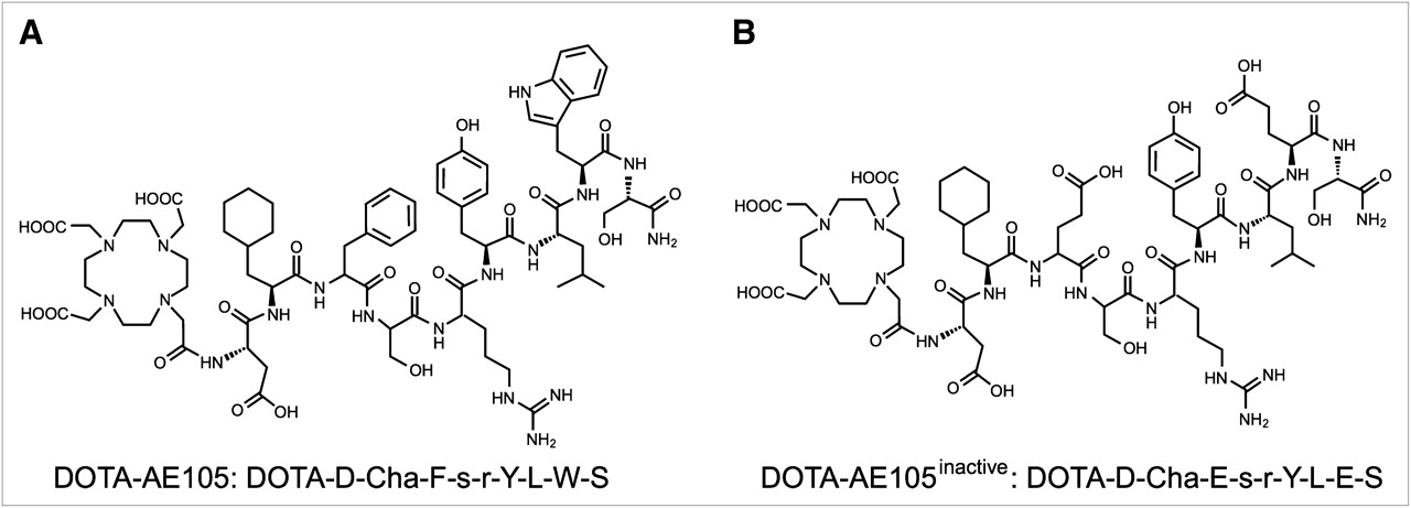
- FIGURE 1.
Chemical structures of DOTA-AE105 (A) and control peptide inactive DOTA-AE105 (B), in which 2 essential amino acids for uPAR binding (Phe → Glu and Trp → Glu) are substituted.
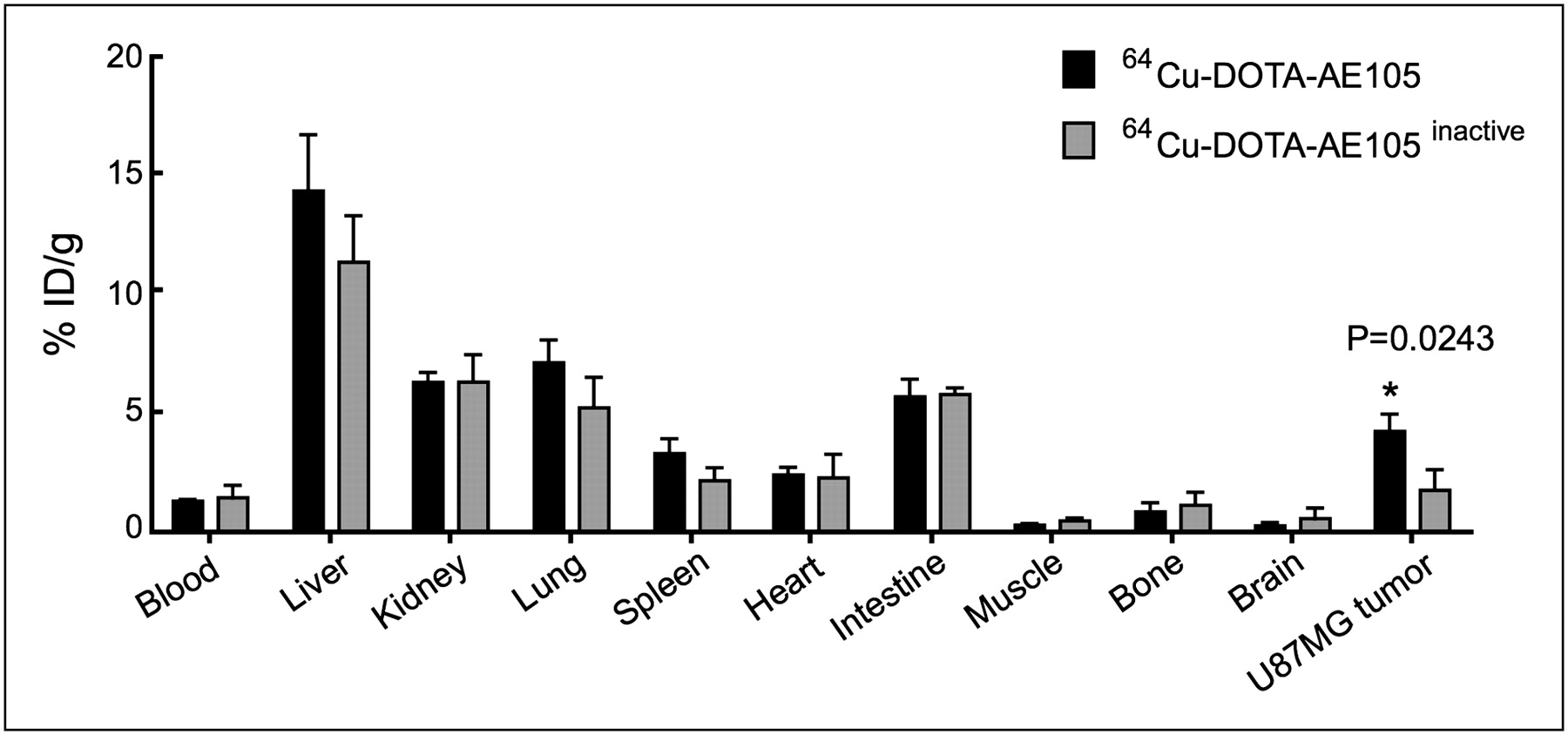
- FIGURE 2.
Biodistribution results for 64Cu-DOTA-AE105 and inactive 64Cu-DOTA-AE105 in nude mice bearing subcutaneously xenotransplanted U87MG human glioblastoma at 4.5 h after injection. Results are shown as %ID/g ± SEM (n = 3 mice/group).
- FIGURE 3.
Representative decay-corrected transverse images at 4.5 h after injection of 64Cu-DOTA-AE105, inactive 64Cu-DOTA-AE105, and 64Cu-DOTA-AE105 preinjected with excess of DOTA-AE105 (blocking) (A). Representative images shown are static scans of single mouse (arrows indicate tumors), which is representative of 3 mice tested in each group, with corresponding quantitative tumor ROI analysis (B). Results are shown as %ID/g ± SEM (n = 3 mice/group).
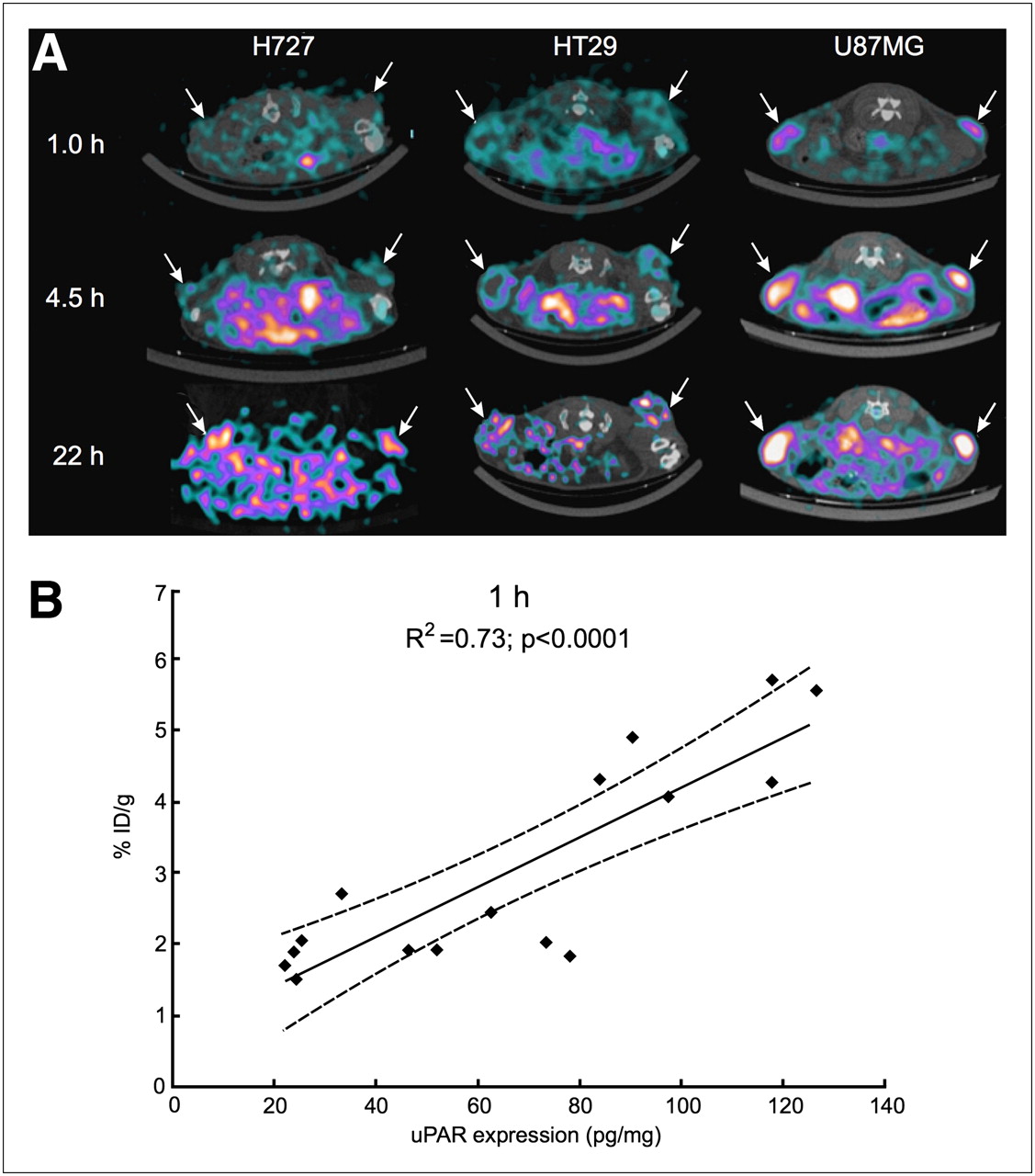
- FIGURE 4.
Decay-corrected transverse images of subcutaneously xenotransplanted U87MG, HT-29, and H727 mice at 1, 4.5, and 22 h after injection of 64Cu-DOTA-AE105 (A). Images shown are static scans of single mouse, which is representative of 4 mice tested in each group. Arrows indicate tumors. Correlation between tumor uptake at 1 h after injection of 64Cu-DOTA-AE105 (%ID/g) and corresponding uPAR expression (pg/mg) is depicted (n = 16 tumors) (B).
- FIGURE 5.
Representative decay-corrected coronal (top) and transversal (bottom) images of same mouse at 1 h after injection of either 18F-FDG or 64Cu-DOTA-AE105 (A). Mouse was inoculated with 2 different xenotransplanted tumors on each flank (H727 and U87MG). Arrows indicate tumor. Comparison of quantified tumor uptake of 18F-FDG and 64Cu-DOTA-AE105 in 3 combination mice bearing two different human cancer xenografts (combi-mice) is shown. Results are shown as %ID/g ± SEM (n = 3) (B).
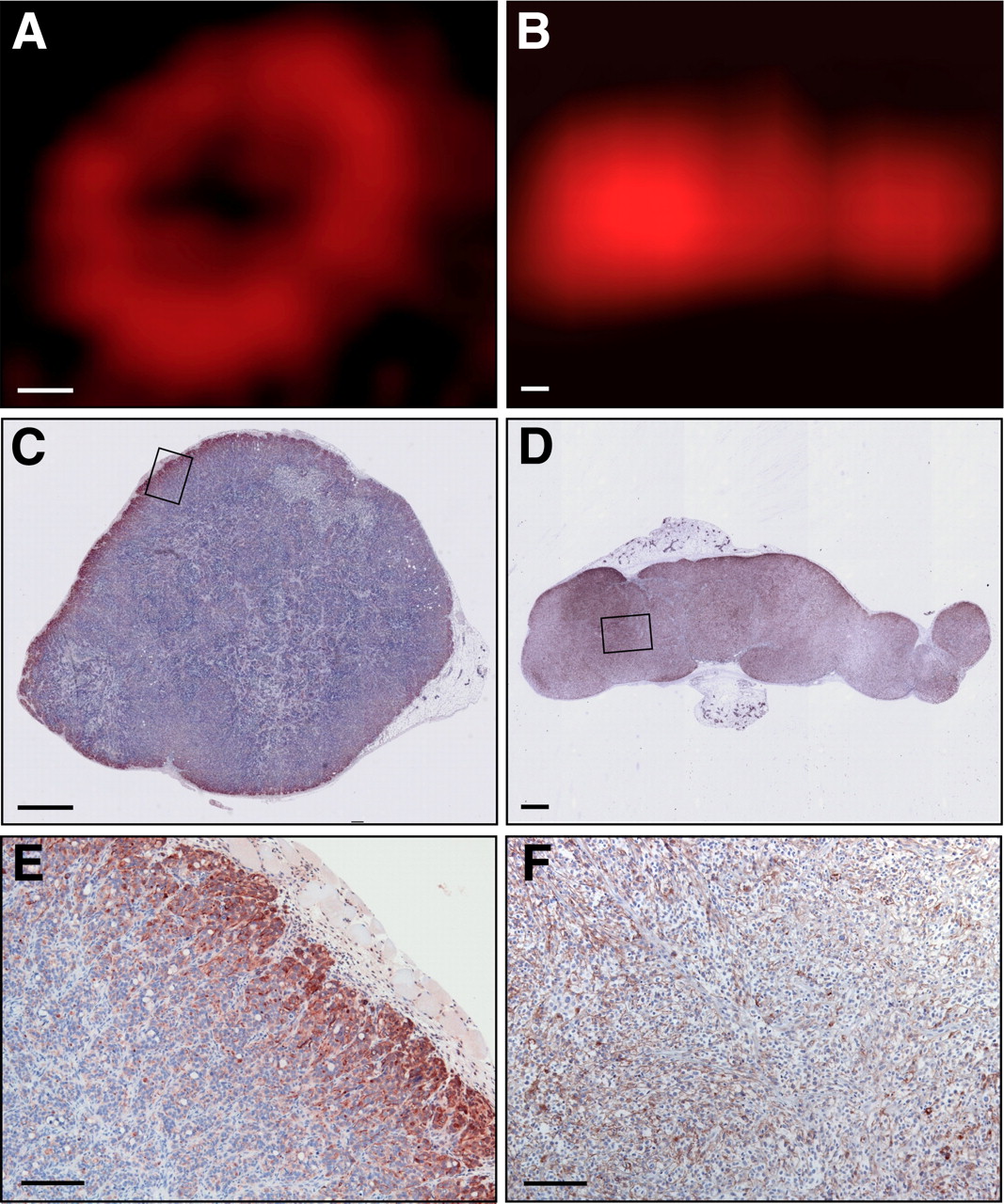
- FIGURE 6.
High-resolution uPAR PET images of tumor and corresponding immunohistologic slides for HT-29 (A, C, and E) and U87MG (B, D, and F). One tumor for each type was PET-scanned and afterward resected and stained for human uPAR. High degree of correlation between uPAR expression pattern and uptake of 64Cu-DOTA-AE105 in each tumor was observed. Bar = 1 mm in A–D and 40 μm in E–F.
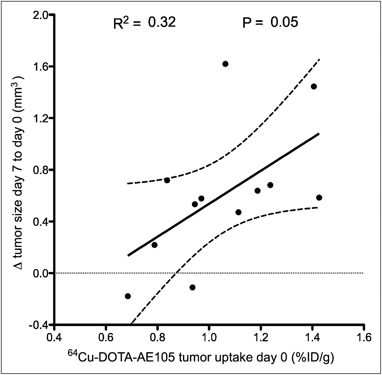
- FIGURE 7.
Correlation between baseline uPAR expression levels detected by 64Cu-DOTA-AE105 PET at day 0 and sensitivity toward 5-fluorouracil chemotherapy treatment (P = 0.05, r2 = 0.32) in nude mice bearing human colorectal HT-29 cancer xenografts (n = 12 tumors).


Additional Files
Supplemental Data
Files in this Data Supplement:


Jump to section
Related Articles
Cited By...
- uPAR Immuno-PET in Pancreatic Cancer, Aging, and Chemotherapy-Induced Senescence
- Prognostic Value of Urokinase-Type Plasminogen Activator Receptor PET/CT in Head and Neck Squamous Cell Carcinomas and Comparison with 18F-FDG PET/CT: A Single-Center Prospective Study
- Urokinase-Type Plasminogen Activator Receptor (uPAR) PET/MRI of Prostate Cancer for Noninvasive Evaluation of Aggressiveness: Comparison with Gleason Score in a Prospective Phase 2 Clinical Trial
- Safety, Dosimetry, and Tumor Detection Ability of 68Ga-NOTA-AE105: First-in-Human Study of a Novel Radioligand for uPAR PET Imaging
- Urokinase-Type Plasminogen Activator Receptor as a Potential PET Biomarker in Glioblastoma
- uPAR as a Glioma Imaging Target
- Interrogating Tumor Metabolism and Tumor Microenvironments Using Molecular Positron Emission Tomography Imaging. Theranostic Approaches to Improve Therapeutics
- A Flexible Multidomain Structure Drives the Function of the Urokinase-type Plasminogen Activator Receptor (uPAR)