
Main menu

  • Home
  • Content
    • Current
    • Ahead of print
    • Past Issues
    • JNM Supplement
    • SNMMI Annual Meeting Abstracts
    • Continuing Education
    • JNM Podcasts
  • Subscriptions
    • Subscribers
    • Institutional and Non-member
    • Rates
    • Journal Claims
    • Corporate & Special Sales
  • Authors
    • Submit to JNM
    • Information for Authors
    • Assignment of Copyright
    • AQARA requirements
  • Info
    • Reviewers
    • Permissions
    • Advertisers
  • About
    • About Us
    • Editorial Board
    • Contact Information
  • More
    • Alerts
    • Feedback
    • Help
    • SNMMI Journals
  • SNMMI
    • JNM
    • JNMT
    • SNMMI Journals
    • SNMMI

User menu

  • Subscribe
  • My alerts
  • Log in
  • Log out
  • My Cart

Search

  • Advanced search
Journal of Nuclear Medicine
  • SNMMI
    • JNM
    • JNMT
    • SNMMI Journals
    • SNMMI
  • Subscribe
  • My alerts
  • Log in
  • Log out
  • My Cart
Journal of Nuclear Medicine

Advanced Search

  • Home
  • Content
    • Current
    • Ahead of print
    • Past Issues
    • JNM Supplement
    • SNMMI Annual Meeting Abstracts
    • Continuing Education
    • JNM Podcasts
  • Subscriptions
    • Subscribers
    • Institutional and Non-member
    • Rates
    • Journal Claims
    • Corporate & Special Sales
  • Authors
    • Submit to JNM
    • Information for Authors
    • Assignment of Copyright
    • AQARA requirements
  • Info
    • Reviewers
    • Permissions
    • Advertisers
  • About
    • About Us
    • Editorial Board
    • Contact Information
  • More
    • Alerts
    • Feedback
    • Help
    • SNMMI Journals
  • View or Listen to JNM Podcast
  • Visit JNM on Facebook
  • Join JNM on LinkedIn
  • Follow JNM on Twitter
  • Subscribe to our RSS feeds
Meeting ReportOncology: Clinical Diagnosis: Breast Cancer

Interpretation of FDG PET and CT patterns in follow up of bone metastases (bmets) in breast cancer (BrCa)

Marina Orevi, Beatrice Uziely, Nanette Freedman, Roland Chisin and Martine Klein
Journal of Nuclear Medicine May 2011, 52 (supplement 1) 1806;
Marina Orevi
1Nuclear Medicine, Hadassah Hebrew Univ Med Ctr, Jerusalem, Israel
  • Find this author on Google Scholar
  • Find this author on PubMed
  • Search for this author on this site
Beatrice Uziely
2Oncology, Hadassah Hebrew Univ Med Ctr, Jerusalem, Israel
  • Find this author on Google Scholar
  • Find this author on PubMed
  • Search for this author on this site
Nanette Freedman
1Nuclear Medicine, Hadassah Hebrew Univ Med Ctr, Jerusalem, Israel
  • Find this author on Google Scholar
  • Find this author on PubMed
  • Search for this author on this site
Roland Chisin
1Nuclear Medicine, Hadassah Hebrew Univ Med Ctr, Jerusalem, Israel
  • Find this author on Google Scholar
  • Find this author on PubMed
  • Search for this author on this site
Martine Klein
1Nuclear Medicine, Hadassah Hebrew Univ Med Ctr, Jerusalem, Israel
  • Find this author on Google Scholar
  • Find this author on PubMed
  • Search for this author on this site
  • Article
  • Figures & Data
  • Info & Metrics
Loading

Abstract

1806

Objectives FDG PET/CT is useful to assess changes in bmets in BrCa patients (pts) under treatment (Tx). However validation is problematic due to absence of tissue diagnosis from bmets. We examined combined changes in FDG uptake and CT results over follow up (f/u) scans, aiming to identify prognostic patterns.

Methods We retrospectively analyzed PET/CT scans of 42 BrCa pts with increased FDG uptake (PET+) in bmets, and with 2-9 scans over 6-57 months f/u. All pts received chemo/hormone and bisphosphonate Tx. CT bone lesions corresponding to PET foci were categorized as blastic (bl), lytic (lyt) or negative (CT-). Multiple bone foci with the same PET and CT characteristics in a single scan were grouped together as one finding.

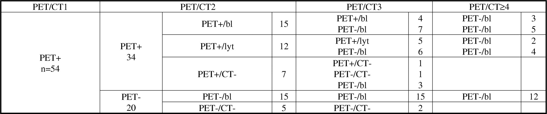
Results Mean time intervals between scans were 6 months. On scan 1, 54 PET+ bone findings were identified. On scan 2, 34/54 were PET+ (15 bl, 12 lyt, 7 CT-) and 20 PET- (15 bl, 0 lyt, 5 CT-) which remained PET- on all available f/u. Of the 34 PET+ findings on scan 2, 10 (4 bl, 5 lyt, 1 CT-) remained PET+ on scan 3, and 5 became PET- on scans 4-9 with CT changing from lytic to blastic in 2; 17 (7 bl, 6 lyt, 4 CT-) became PET- on scan 3, with CT changing from lytic (6) and CT- (3) to blastic. In all, 36/54 findings became PET-/bl and remained PET- on f/u. There were no PET-/lyt findings.

Conclusions Three main patterns of behavior of BrCa bone mets under Tx emerged: lesions becoming PET-/bl are compatible with response to Tx; a flare phenomenon can explain transient ongoing FDG uptake in PET+/bl lesions; lytic lesions remaining PET+ do not respond to Tx. Larger studies with clinical and biologic correlates are required to validate the significance of these combined PET/CT results

Figure
  • Download figure
  • Open in new tab
  • Download powerpoint
Back to top

In this issue

Journal of Nuclear Medicine
Vol. 52, Issue supplement 1
May 2011
  • Table of Contents
  • Index by author
Article Alerts
Sign In to Email Alerts with your Email Address
Email Article

Thank you for your interest in spreading the word on Journal of Nuclear Medicine.

NOTE: We only request your email address so that the person you are recommending the page to knows that you wanted them to see it, and that it is not junk mail. We do not capture any email address.

Enter multiple addresses on separate lines or separate them with commas.
Interpretation of FDG PET and CT patterns in follow up of bone metastases (bmets) in breast cancer (BrCa)
(Your Name) has sent you a message from Journal of Nuclear Medicine
(Your Name) thought you would like to see the Journal of Nuclear Medicine web site.
Citation Tools
Interpretation of FDG PET and CT patterns in follow up of bone metastases (bmets) in breast cancer (BrCa)
Marina Orevi, Beatrice Uziely, Nanette Freedman, Roland Chisin, Martine Klein
Journal of Nuclear Medicine May 2011, 52 (supplement 1) 1806;

Citation Manager Formats

  • BibTeX
  • Bookends
  • EasyBib
  • EndNote (tagged)
  • EndNote 8 (xml)
  • Medlars
  • Mendeley
  • Papers
  • RefWorks Tagged
  • Ref Manager
  • RIS
  • Zotero
Share
Interpretation of FDG PET and CT patterns in follow up of bone metastases (bmets) in breast cancer (BrCa)
Marina Orevi, Beatrice Uziely, Nanette Freedman, Roland Chisin, Martine Klein
Journal of Nuclear Medicine May 2011, 52 (supplement 1) 1806;
Twitter logo Facebook logo LinkedIn logo Mendeley logo
  • Tweet Widget
  • Facebook Like
  • Google Plus One
Bookmark this article

Jump to section

  • Article
  • Figures & Data
  • Info & Metrics

Related Articles

  • No related articles found.
  • Google Scholar

Cited By...

  • No citing articles found.
  • Google Scholar

More in this TOC Section

Oncology: Clinical Diagnosis: Breast Cancer

  • Investigation of 18F-FDG PET in the selection of patients with breast cancer candidate to sentinel node biopsy after neoadjuvant therapy - Update of an ongoing study
  • Volume-based parameters of primary tumor in F-18 FDG PET/CT in invasive ductal breast cancer for the prediction of lymph node metastasis
  • Is FDG PET/CT an accurate tool for identifying metastases of lobular breast cancer?
Show more Oncology: Clinical Diagnosis: Breast Cancer

Breast Cancer Posters

  • The importance of 18F-FDG PET/CT in staging male breast carcinoma
  • Correlation between 18F-FDG PET/CT standardized uptake value with prognostic factors and gene expression profiles in breast cancer: Single center study from Azerbaijan
  • Relationship between tumor markers, Ca 15-3 and Ca 125, levels and Sodium-18 Fluoride Positron Emission Tomography Scan in breast cancer bone metastasis
Show more Breast Cancer Posters

Similar Articles

SNMMI

© 2025 SNMMI

Powered by HighWire