


Article Figures & Data
Figures
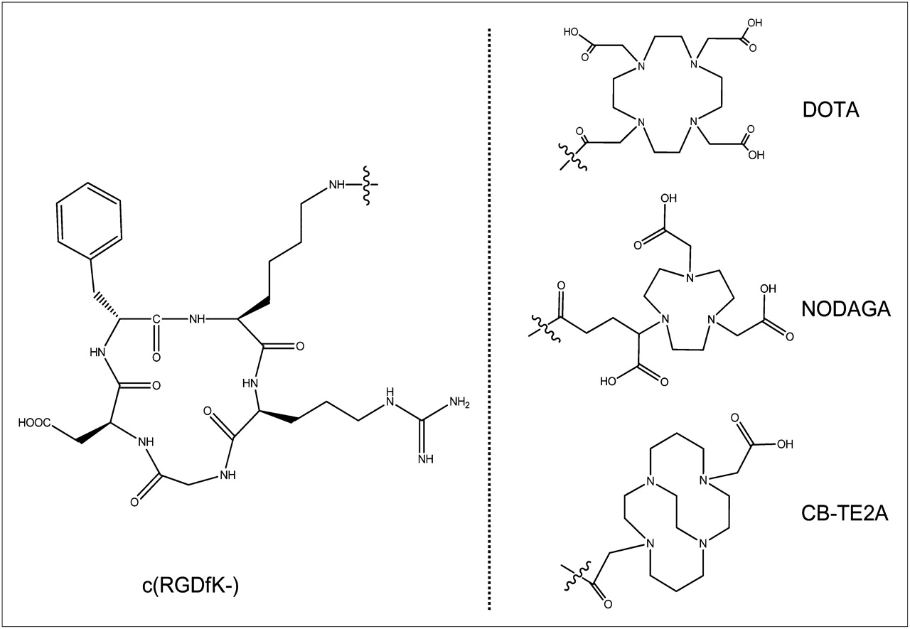
- FIGURE 1.
Structures of DOTA-c(RGDfK), NODAGA-c(RGDfK), and CB-TE2A-c(RGDfK).
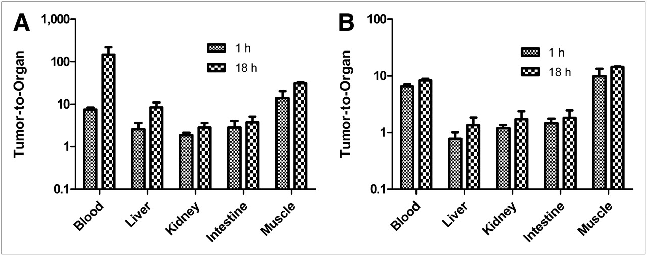
- FIGURE 2.
Tumor-to-organ ratios for 64Cu-CB-TE2A-c(RGDfK) (A) and 64Cu-DOTA-c(RGDfK) (B) at 1 and 18 h after injection. Graphs are in logarithmic scale.
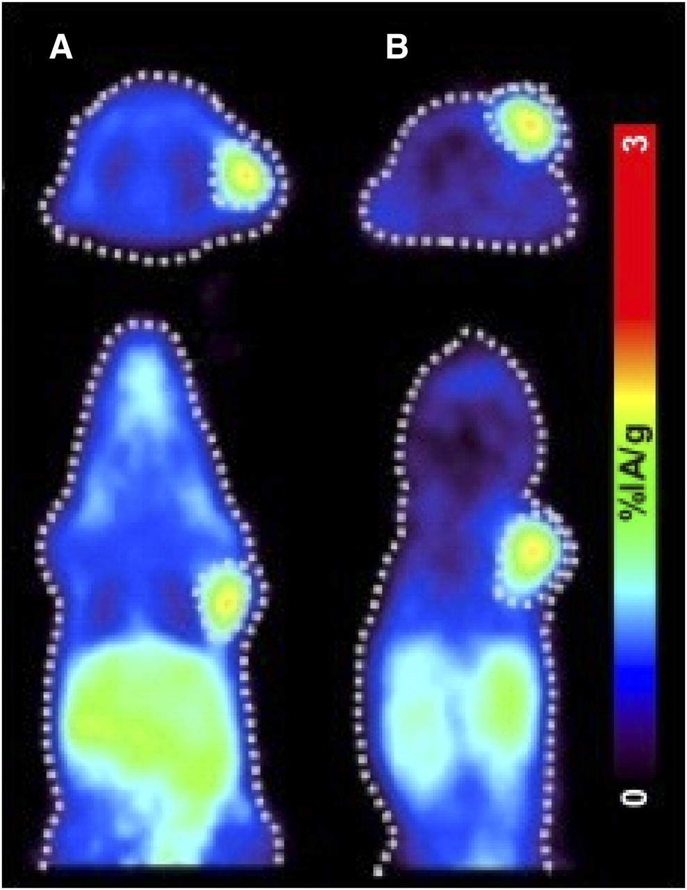
- FIGURE 3.
Transverse (top) and coronal (bottom) small-animal PET images of 68Ga-DOTA-c(RGDfK) (A) and 68Ga-NODAGA-c(RGDfK) (B) in nude mice bearing U87MG tumors at 1 h after injection.
- FIGURE 4.
Small-animal PET images of 64Cu-DOTA-c(RGDfK) at 1 h after injection (A) and 18 h after injection (D), 64Cu-NODAGA-c(RGDfK) at 1 h after injection (B) and 18 h after injection (E), and 64Cu-CB-TE2A-c(RGDfK) at 1 h after injection (C) and 18 h after injection (F) in nude mice bearing U87MG tumors.


Tables
- TABLE 1
Biodistribution Results and Tumor–to–Normal Tissue Ratios of 64Cu-CB-TE2A-c(RGDfK) in Nude Mice Bearing U87MG Tumors
Organ 1 h 1-h blocking* 18 h Blood 0.49 ± 0.05 0.03 ± 0.01 0.02 ± 0.01 Heart 0.33 ± 0.11 0.04 ± 0.01 0.12 ± 0.05 Liver 1.57 ± 0.54 0.48 ± 0.10 0.37 ± 0.16 Spleen 1.33 ± 0.32 0.12 ± 0.03 0.63 ± 0.22 Lung 0.83 ± 0.21 0.14 ± 0.04 0.26 ± 0.07 Kidney 2.14 ± 0.53 1.18 ± 0.23 1.10 ± 0.38 Stomach 1.52 ± 0.63 0.15 ± 0.04 0.45 ± 0.14 Intestine 1.45 ± 0.55 0.30 ± 0.09 0.82 ± 0.19 Adrenal 2.63 ± 0.72 0.10 ± 0.04 1.45 ± 0.52 Pancreas 0.40 ± 0.25 0.09 ± 0.04 0.09 ± 0.04 Muscle 0.31 ± 0.13 0.02 ± 0.01 0.10 ± 0.03 Bone 2.57 ± 0.23 0.10 ± 0.01 0.22 ± 0.04 Tumor 3.66 ± 0.58 0.35 ± 0.29 2.99 ± 0.79 Tumor-to-nontumor ratios Tumor-to-blood 7.48 ± 0.90 146.07 ± 71.08 Tumor-to-liver 2.56 ± 1.06 8.47 ± 2.43 Tumor-to-kidney 1.87 ± 0.26 2.83 ± 0.80 Tumor-to-intestine 2.84 ± 1.22 3.75 ± 1.35 Tumor-to-muscles 13.61 ± 6.65 30.75 ± 2.03 ↵* Coinjection of c(RGDfV) (5 mg/kg).
Data are %IA/g ± SD (n = 4–7).
- TABLE 2
Biodistribution Results and Tumor–to–Normal Tissue Ratios of 64Cu- and 68Ga-NODAGA-c(RGDfK) in Nude Mice Bearing U87MG Tumors
64Cu 68Ga Organ 1 h 1-h blocking* 4 h 18 h 1 h 1-h blocking* Blood 0.31 ± 0.09 0.15 ± 0.03 0.10 ± 0.03 0.07 ± 0.01 0.16 ± 0.03 0.03 ± 0.01 Heart 0.43 ± 0.04 0.20 ± 0.08 0.29 ± 0.08 0.18 ± 0.04 0.33 ± 0.07 0.03 ± 0.01 Liver 1.67 ± 0.33 0.42 ± 0.14 1.03 ± 0.34 0.58 ± 0.20 1.86 ± 0.23 0.28 ± 0.04 Spleen 1.63 ± 0.42 0.38 ± 0.12 1.02 ± 0.12 0.50 ± 0.11 1.73 ± 0.44 0.15 ± 0.04 Lung 1.08 ± 0.09 0.23 ± 0.01 0.66 ± 0.22 0.38 ± 0.06 0.80 ± 0.07 0.09 ± 0.00 Kidney 2.21 ± 0.27 0.98 ± 0.03 1.57 ± 0.32 0.89 ± 0.23 1.98 ± 0.51 1.07 ± 0.19 Stomach 1.63 ± 0.43 0.49 ± 0.20 0.92 ± 0.20 0.54 ± 0.12 1.40 ± 0.34 0.19 ± 0.09 Intestine 2.12 ± 0.40 0.73 ± 0.19 1.26 ± 0.02 0.95 ± 0.22 1.83 ± 0.51 0.18 ± 0.05 Adrenal 4.67 ± 1.07 0.54 ± 0.15 3.47 ± 0.58 2.78 ± 1.26 5.88 ± 1.06 0.25 ± 0.10 Pancreas 0.53 ± 0.36 0.18 ± 0.09 0.21 ± 0.06 0.16 ± 0.02 0.23 ± 0.08 0.04 ± 0.01 Muscle 0.31 ± 0.03 0.44 ± 0.10 0.16 ± 0.03 0.10 ± 0.02 0.49 ± 0.29 0.07 ± 0.03 Bone 0.58 ± 0.08 0.33 ± 0.11 0.28 ± 0.06 0.34 ± 0.10 0.45 ± 0.21 0.08 ± 0.02 Tumor 3.77 ± 0.52 0.89 ± 0.10 3.62 ± 0.95 3.05 ± 0.62 5.19 ± 1.45 0.41 ± 0.11 Tumor-to-nontumor ratios Tumor-to-blood 10.15 ± 5.19 36.65 ± 6.46 50.69 ± 8.13 27.67 ± 7.01 Tumor-to-liver 2.67 ± 0.53 3.75 ± 1.07 5.64 ± 1.47 2.75 ± 0.31 Tumor-to-kidney 1.72 ± 0.24 2.37 ± 0.46 3.54 ± 0.67 2.64 ± 0.31 Tumor-to-intestine 1.83 ± 0.46 2.88 ± 0.05 3.42 ± 1.23 2.87 ± 0.49 Tumor-to-muscle 12.16 ± 1.48 23.34 ± 4.41 31.83 ± 3.17 12.80 ± 5.25 ↵* Coinjection of c(RGDfV) (5 mg/kg).
Data are %IA/g ± SD (n = 4–7).
- TABLE 3
Biodistribution Results and Tumor–to–Normal Tissue Ratios of 64Cu- and 68Ga-DOTA-c(RGDfK) in Nude Mice Bearing U87MG Tumors
64Cu 68Ga Organ 1 h 1-h blocking* 18 h 1 h 1-h blocking* Blood 0.63 ± 0.14 0.56 ± 0.23 0.42 ± 0.15 0.38 ± 0.07 0.60 ± 0.14 Heart 1.01 ± 0.21 0.70 ± 0.01 0.93 ± 0.16 0.35 ± 0.08 0.20 ± 0.05 Liver 5.46 ± 1.31 3.14 ± 0.44 2.59 ± 0.81 1.60 ± 0.27 0.48 ± 0.10 Spleen 1.71 ± 0.27 0.73 ± 0.01 1.30 ± 0.27 1.34 ± 0.21 0.36 ± 0.09 Lung 1.58 ± 0.30 2.38 ± 1.07 1.51 ± 0.44 0.87 ± 0.12 0.58 ± 0.11 Kidney 3.31 ± 0.12 2.96 ± 0.31 2.03 ± 0.65 2.24 ± 0.34 1.95 ± 0.68 Stomach 2.65 ± 0.50 1.31 ± 0.32 1.35 ± 0.62 1.50 ± 0.36 0.57 ± 0.11 Intestine 2.79 ± 0.52 1.40 ± 0.32 1.92 ± 0.58 1.79 ± 0.30 0.53 ± 0.08 Adrenal 4.02 ± 1.40 1.03 ± 0.24 2.31 ± 0.72 4.87 ± 1.21 0.71 ± 0.04 Pancreas 0.68 ± 0.08 0.34 ± 0.13 0.53 ± 0.15 0.29 ± 0.05 0.18 ± 0.01 Muscle 0.42 ± 0.08 0.23 ± 0.08 0.23 ± 0.01 0.29 ± 0.04 0.18 ± 0.02 Bone 0.53 ± 0.09 0.51 ± 0.08 0.39 ± 0.14 0.35 ± 0.05 0.11 ± 0.05 Tumor 3.97 ± 0.48 1.57 ± 0.13 3.23 ± 0.75 3.47 ± 0.78 0.89 ± 0.05 Tumor-to-nontumor ratios Tumor-to-blood 6.46 ± 1.57 8.30 ± 2.58 9.24 ± 1.12 Tumor-to-liver 0.77 ± 0.24 1.35 ± 0.50 2.25 ± 0.37 Tumor-to-kidney 1.20 ± 0.15 1.73 ± 0.65 1.57 ± 0.14 Tumor-to-intestine 1.46 ± 0.29 1.82 ± 0.66 1.95 ± 0.16 Tumor-to-muscles 9.94 ± 3.44 14.30 ± 0.40 12.37 ± 1.81 ↵* Coinjection of c(RGDfV) (5 mg/kg).
Data are %IA/g ± SD (n = 4–7).
Supplemental Data
Files in this Data Supplement:


Jump to section
Related Articles
Cited By...
- 61Cu-PSMA-Targeted PET for Prostate Cancer: From Radiotracer Development to First-in-Human Imaging
- Variation of Specific Activities of 68Ga-Aquibeprin and 68Ga-Avebetrin Enables Selective PET Imaging of Different Expression Levels of Integrins {alpha}5{beta}1 and {alpha}v{beta}3
- Does Imaging {alpha}v{beta}3 Integrin Expression with PET Detect Changes in Angiogenesis During Bevacizumab Therapy?
- Can 111In-RGD2 Monitor Response to Therapy in Head and Neck Tumor Xenografts?
- Noninvasive positron emission tomography and fluorescence imaging of CD133+ tumor stem cells
- Inflammatory neovascularization during graft-versus-host disease is regulated by {alpha}v integrin and miR-100