


Article Figures & Data
Figures
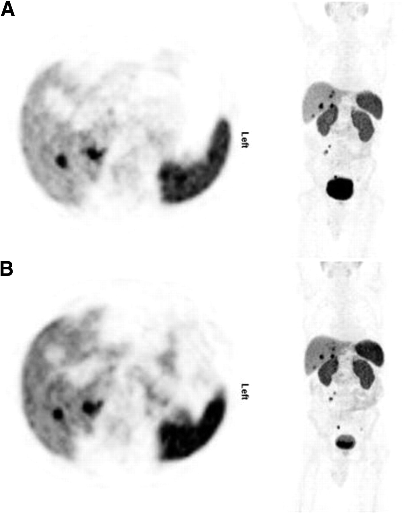
- FIGURE 1.
Axial views and multiple-intensity projections of 2 PET/CT scans from same patient: scan under treatment with long-acting octreotide (A) and scan without treatment (B). Although SUVmax of metastases was not different (32.9 and 31.6 with and without treatment, respectively), uptake of both spleen (20.2 and 25.3, respectively) and liver (9.5 and 11.5, respectively) was notably lower under long-acting octreotide treatment. Between the 2 examinations, no disease progression was evident and no treatment was performed.
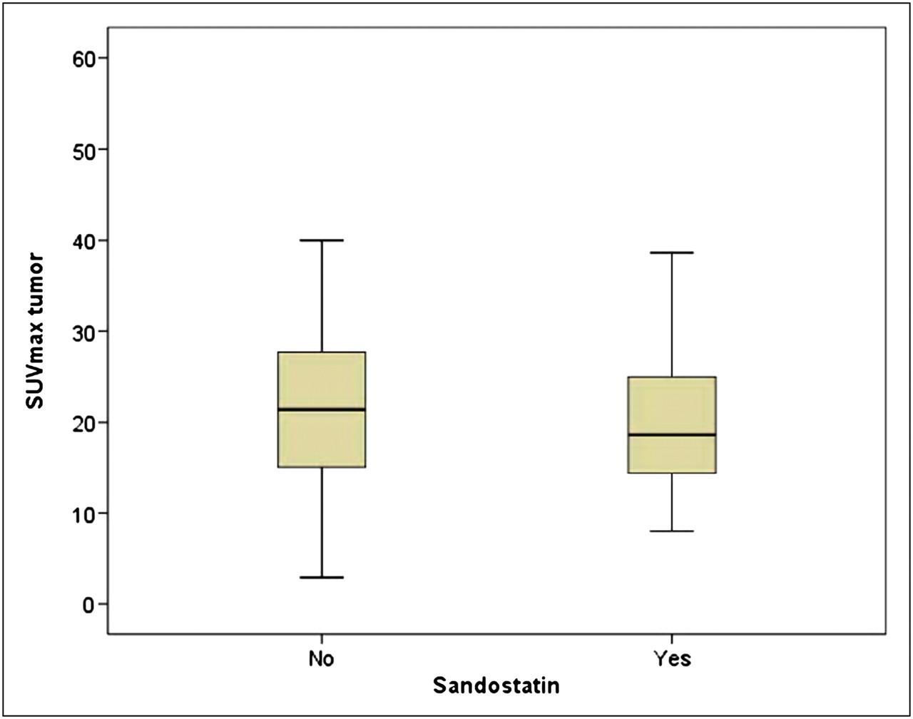
- FIGURE 2.
SUVmax measurements of tumor with highest value per patient. Mann–Whitney U test revealed no difference between groups with and without octreotide medication (P = 0.70).
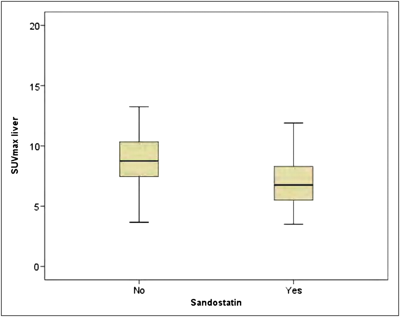
- FIGURE 3.
SUVmax measurements of liver. Mann–Whitney U test revealed significantly lower SUVmax in group with octreotide treatment than in group without (P < 0.001).
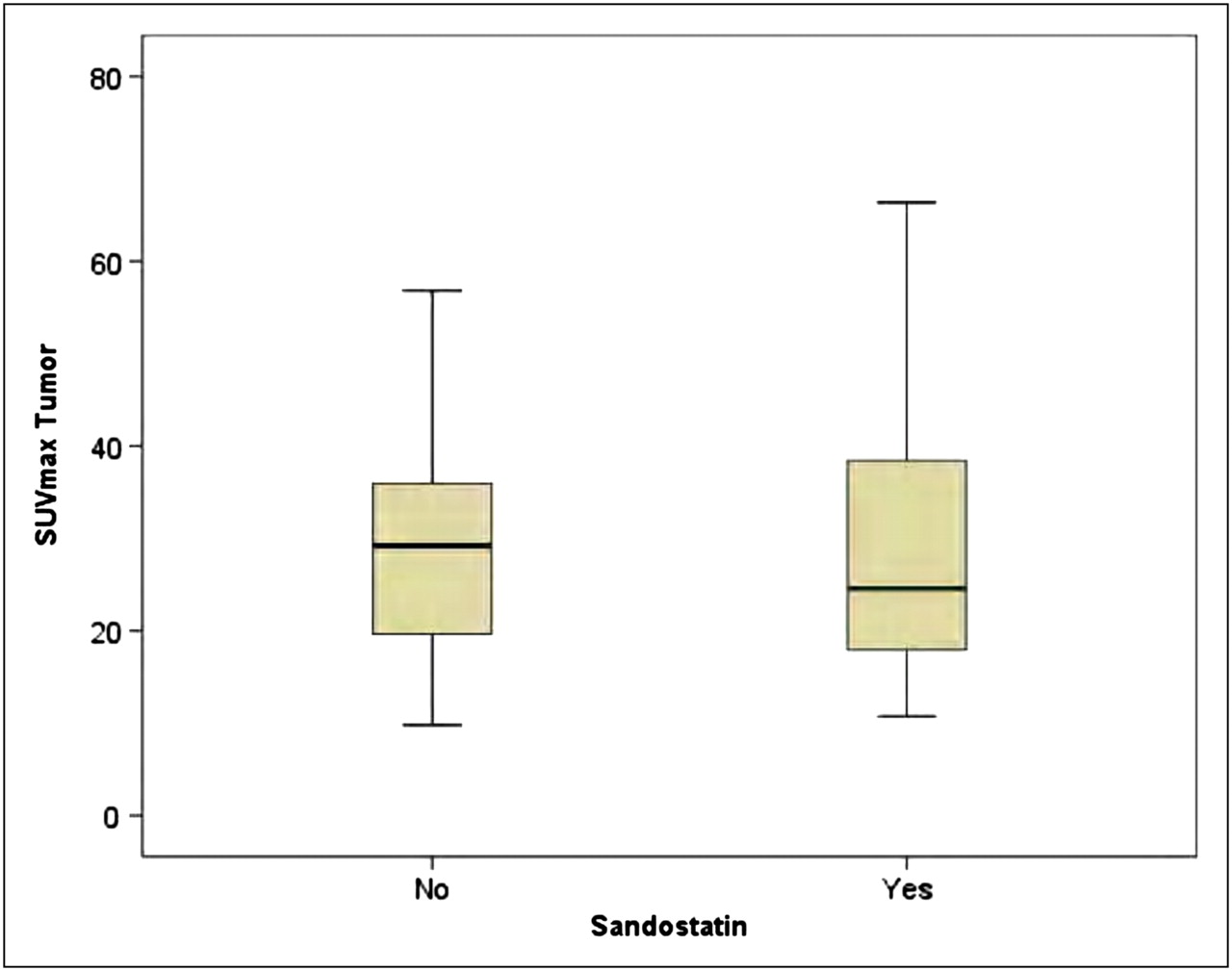
- FIGURE 4.
SUVmax measurements of up to 5 tumors per organ and patient in subgroup of 9 patients with successive PET scans with and without octreotide medication. Wilcoxon test revealed no statistical difference (P = 0.93).


Tables
Parameter Octreotide (n = 35) No octreotide (n = 70) P Sex Male 18 (51.4%) 38 (54.3%) NS Female 17 (48.6%) 32 (45.7%) Age 55.7 ± 13.7 58.8 ± 12.0 NS Primary tumor NS Ileum 26 (74.3%) 23 (32.9%) Lung 2 (5.7%) 8 (11.4%) Pancreas 3 (8.6%) 11 (15.7%) Stomach 1 (2.8%) 3 (4.3%) Rectum 2 (5.7%) 2 (2.9%) Other 1 (2.8%) 23 (32.9%) Primary tumor present 2 (6%) 11 (16%) NS Metastases Total 35 (100%) 34 (49%) <0.001 Liver 28 (80%) 28 (40%) <0.001 Lymph node 24 (69%) 19 (27%) <0.001 Bone 17 (49%) 9 (13%) <0.001 Pulmonary 0 (0%) 2 (3%) NS NS = not statistically significant.
- TABLE 2
SUVmax and SUVmean of Organs, Primary Tumors, and Metastases in Treated and Untreated Patients
Parameter Octreotide No octreotide P Most intense metastases per patient SUVmax 32.5 ± 16.6 (10.8–70.1) 33.0 ± 23.0 (6.3–117.1) 0.73 SUVmean 11.2 ± 6.8 (3.7–25.6) 10.6 ± 5.1 (3.6–21.8) 0.83 Primary tumor SUVmax 28.6 ± 6.8 (23.8–33.4) 32.9 ± 31.5 (6.3–117.1) 0.69 SUVmean 19.8 ± 6.7 (15.1–24.5) 21.3 ± 19.5 (4.1–72.7) 0.55 Liver metastases SUVmax 27.2 ± 14.8 (8.1–66.4) 25.7 ± 10.7 (9.8–56.8) 0.92 SUVmean 18.5 ± 10.7 (5.2–46.3) 16.9 ± 7.5 (6.9–39.9) 0.83 Lymph node metastases SUVmax 41.4 ± 19.5 (5.6–68.8) 25.0 ± 6.3 (18.1–33.0) 0.20 SUVmean 27.1 ± 13.1 (3.6–45.7) 16.4 ± 4.8 (11.2–22.0) 0.17 Osseous metastases SUVmax 39.5 ± 22.0 (11.5–70.1) 15.4 ± 7.8 (10.2–27.0) 0.49 SUVmean 26.9 ± 15.7 (6.6–48.6) 10.2 ± 5.1 (6.7–17.8) 0.38 Lung metastases SUVmax None 21.4 ± 11.0 (13.7–29.2) NA SUVmean None 14.0 ± 7.1 (9.0–19.0) NA Liver SUVmax 7.1 ± 2.1 (3.5–11.9) 9.3 ± 2.9 (3.7–19.2) <0.001 SUVmean 4.9 ± 1.4 (2.8–7.4) 6.6 ± 1.9 (2.8–12.8) <0.001 Spleen SUVmax 18.4 ± 6.4 (9.5–31.8) 24.9 ± 6.7 (10.4–43.8) <0.001 SUVmean 12.8 ± 4.4 (6.4–23.5) 18.2 ± 5.0 (7.2–32.8) <0.001 Adrenal gland SUVmax 18.3 ± 5.7 (9.4–28.0) 20.0 ± 5.6 (10.1–41.2) 0.70 SUVmean 11.7 ± 3.7 (6.0–17.7) 14.9 ± 16.4 (7.2–18.5) 0.76 Pituitary gland SUVmax 3.9 ± 4.8 (0.4–19.3) 2.0 ± 0.8 (0.5–3.5) 0.12 SUVmean 2.6 ± 3.2 (0.3–13.0) 1.3 ± 0.5 (0.3–2.4) 0.08 Kidney SUVmax 14.5 ± 6.0 (5.9–31.0) 15.8 ± 4.6 (8.3–35.0) 0.24 SUVmean 9.5 ± 3.3 (4.1–17.3) 10.4 ± 2.8 (5.4–22.5) 0.32 NA = not applicable.
Data are mean ± SD, with range in parentheses.


Jump to section
Related Articles
Cited By...
- A Prospective Observational Study to Evaluate the Effects of Long-Acting Somatostatin Analogs on 68Ga-DOTATATE Uptake in Patients with Neuroendocrine Tumors
- Long-Acting Somatostatin Analog Therapy Differentially Alters 68Ga-DOTATATE Uptake in Normal Tissues Compared with Primary Tumors and Metastatic Lesions
- Current Concepts in 68Ga-DOTATATE Imaging of Neuroendocrine Neoplasms: Interpretation, Biodistribution, Dosimetry, and Molecular Strategies
- Prospective Study of 68Ga-DOTATATE Positron Emission Tomography/Computed Tomography for Detecting Gastro-Entero-Pancreatic Neuroendocrine Tumors and Unknown Primary Sites
- Impact of 68Ga-DOTATATE PET/CT on the Management of Neuroendocrine Tumors: The Referring Physician's Perspective
- Quantitative and Qualitative Intrapatient Comparison of 68Ga-DOTATOC and 68Ga-DOTATATE: Net Uptake Rate for Accurate Quantification
- Free Somatostatin Receptor Fraction Predicts the Antiproliferative Effect of Octreotide in a Neuroendocrine Tumor Model: Implications for Dose Optimization
- Neuroendocrine tumor disease: an evolving landscape