


Article Figures & Data
Figures
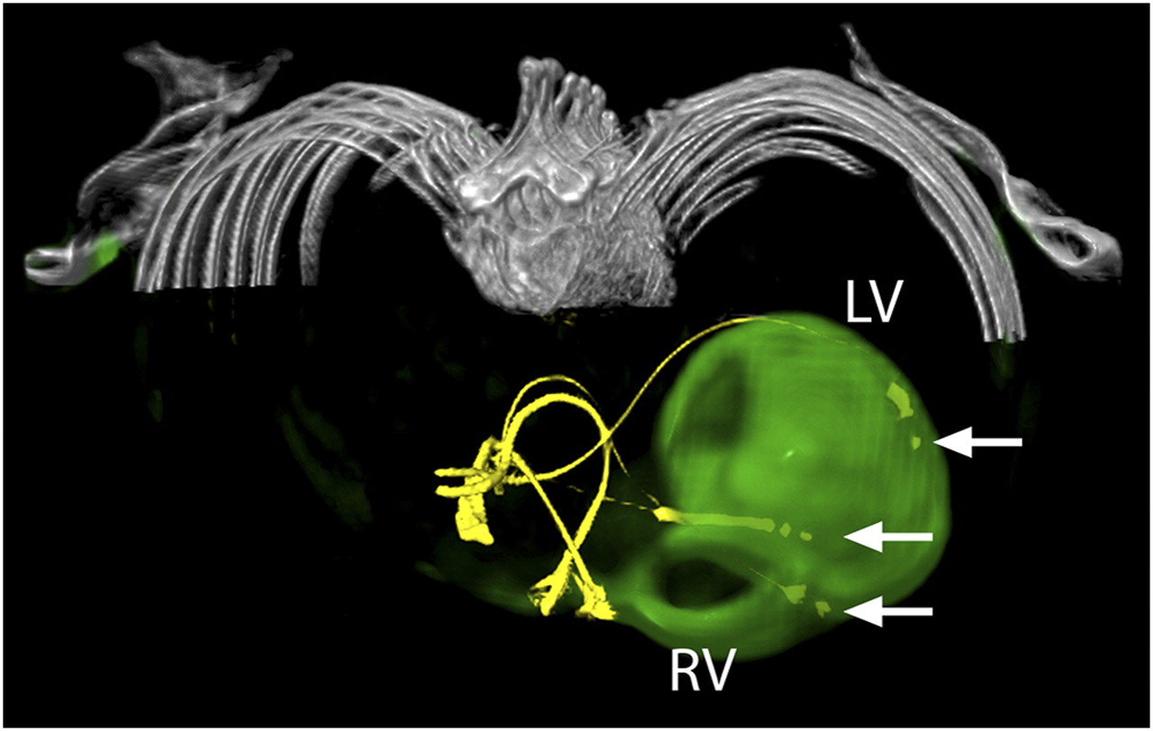
- FIGURE 1.
LV lead position in viable myocardium. CT and 3D fused 18F-FDG PET/CT image of patient 5, who was rated as responder to CRT. Image shows attenuation-corrected CT images, with leads of both implantable cardioverter defibrillator and biventricular pacemaker in yellow and PET information in green. There is no relevant scar burden close to catheter lead in lateral wall of left ventricle. Right ventricle is enlarged and shows slight 18F-FDG uptake. Tips of pacemaker leads are marked with arrows. LV = left ventricle; RV = right ventricle.
- FIGURE 2.
LV lead position in nonviable myocardium. CT and 3D fused PET/CT image of patient 13, who was rated as nonresponder to CRT. Image shows attenuation-corrected CT images, with leads of biventricular pacemaker in yellow and PET information in green. Huge amount of left ventricle, especially lateral wall where tip of LV lead was located, shows absent glucose metabolism. right ventricle shows slight 18F-FDG uptake. Catheter leads are marked with arrows. LV = left ventricle; RV = right ventricle.
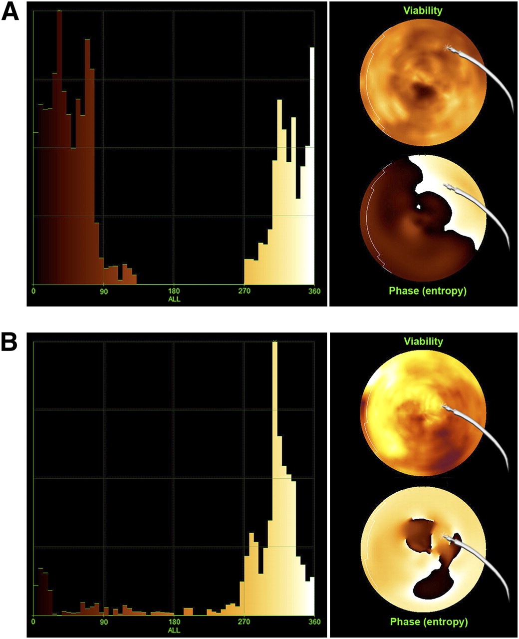
- FIGURE 3.
Shown are phase histogram (left) and bull's eye (right) of LV glucose metabolism (upper polar map) and global phase entropy (lower polar map) in relation to LV pacemaker lead position in nonresponder (patient 2; A) and responder (patient 5; B) to CRT. Compared with responder patient, nonresponder shows broader bandwidth in phase histogram and LV lead located in area of nonviable myocardium and high phase entropy.

Tables
- TABLE 1
Baseline Characteristics for Study Cohort and Subcohorts of Responders and Nonresponders
Parameter All patients (n = 14) Responders (n = 7) Nonresponders (n = 7) P Age (y) 67.9 ± 8.4 68.0 ± 8.8 67.7 ± 8.7 NS Men 12 (86) 6 (86) 6 (86) NS Angiotensin-converting enzyme inhibitor 13 (93) 7 (100) 6 (86) NS β-blocker 13 (93) 6 (86) 7 (100) NS Diuretics 10 (71) 4 (57) 6 (86) NS Aldosterone receptor antagonist 8 (57) 4 (50) 4 (50) NS Clinical evaluation NYHA class before CRT 3.0 ± 0.0 3.0 ± 0.0 3.0 ± 0.0 NS NYHA class after CRT 2.6 ± 0.6 2.0 ± 0.0 3.1 ± 0.4 <0.05 Δ NYHA class* −0.4 ± 0.6 −1.0 ± 0.3 +0.1 ± 0.4 <0.05 Brain natriuretic peptide before CRT (pg/mL) 625 ± 514 691 ± 582 560 ± 473 NS Brain natriuretic peptide after CRT (pg/mL) 432 ± 533 236 ± 257 628 ± 679 NS Δ Brain natriuretic peptide (pg/mL) −193 ± 467 −454 ± 416 +68 ± 373 <0.05 Echocardiographic parameters LVEF before CRT (%) 25 ± 8 24 ± 9 27 ± 8 NS LVEF after CRT (%) 32 ± 13 39 ± 14 26 ± 8 NS Δ LVEF (%) +7 ± 12 +15 ± 11 −1 ± 7 <0.05 LVESV before CRT (mL) 219 ± 57 231 ± 64 206 ± 52 NS LVESV after therapy (mL) 177 ± 65 150 ± 51 205 ± 70 0.073 Δ LVESV (mL)* −41 ± 51 −82 ± 37 +1 ± 24 <0.05 Residual dyssynchrony 7 (50) 2 (29) 5 (71) PET/CT including phase analysis Scar burden (%) 20 ± 19 10 ± 8 30 ± 21 <0.05 Lead over scar 4 (29) 0 (0) 4 (57) Bandwidth (°) 90 ± 38 91 ± 28 89 ± 48 NS Phase SD (°) 38 ± 11 42 ± 8 34 ± 14 NS Phase entropy (°) 80 ± 5 77 ± 4 83 ± 3 <0.05 ↵* Prespecified response criteria.
NS = not significant.
Values are expressed as n, with percentages in parentheses, or mean ± SD.
Supplemental Data
Files in this Data Supplement:


