
Main menu

  • Home
  • Content
    • Current
    • Ahead of print
    • Past Issues
    • JNM Supplement
    • SNMMI Annual Meeting Abstracts
  • Subscriptions
    • Subscribers
    • Institutional and Non-member
    • Rates
    • Corporate & Special Sales
    • Journal Claims
  • Authors
    • Submit to JNM
    • Information for Authors
    • Assignment of Copyright
    • AQARA requirements
  • Info
    • Permissions
    • Advertisers
    • Continuing Education
  • About
    • About Us
    • Editorial Board
    • Contact Information
  • More
    • Alerts
    • Feedback
    • Help
    • SNMMI Journals
  • SNMMI
    • JNM
    • JNMT
    • SNMMI Journals
    • SNMMI

User menu

  • Subscribe
  • My alerts
  • Log in
  • My Cart

Search

  • Advanced search
Journal of Nuclear Medicine
  • SNMMI
    • JNM
    • JNMT
    • SNMMI Journals
    • SNMMI
  • Subscribe
  • My alerts
  • Log in
  • My Cart
Journal of Nuclear Medicine

Advanced Search

  • Home
  • Content
    • Current
    • Ahead of print
    • Past Issues
    • JNM Supplement
    • SNMMI Annual Meeting Abstracts
  • Subscriptions
    • Subscribers
    • Institutional and Non-member
    • Rates
    • Corporate & Special Sales
    • Journal Claims
  • Authors
    • Submit to JNM
    • Information for Authors
    • Assignment of Copyright
    • AQARA requirements
  • Info
    • Permissions
    • Advertisers
    • Continuing Education
  • About
    • About Us
    • Editorial Board
    • Contact Information
  • More
    • Alerts
    • Feedback
    • Help
    • SNMMI Journals
  • Follow JNM on Twitter
  • Visit JNM on Facebook
  • Join JNM on LinkedIn
  • Subscribe to our RSS feeds

Table of Contents

January 1, 2011; Volume 52,Issue 1

Cover image

Cover image expansion

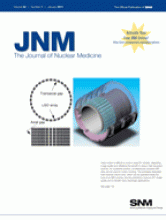
Head motion is difficult to avoid in long PET studies, degrading image quality and offsetting the benefit of using a high-resolution scanner. As a potential solution, simultaneously acquired MRI data can be used for motion tracking. The prototype dedicated brain scanner shown here, which can be operated inside the bore of an MRI scanner, has the potential to improve PET image quality and to benefit many neurologic applications.
See page 154.

Back to top
PreviousNext

In this issue

Journal of Nuclear Medicine: 52 (1)
Journal of Nuclear Medicine
Vol. 52, Issue 1
January 1, 2011
  • Table of Contents
  • Table of Contents (PDF)
  • About the Cover
  • Index by author
Sign up for alerts

Jump to

  • This Month in JNM
  • SNM Newsline
  • Focus on Molecular Imaging
  • Invited Perspectives
  • Clinical Investigations
  • Brief Communications
  • Continuing Education
  • Erratum
  • Basic Science Investigations
  • Book Review
  • Most Cited
  • Most Read
Loading
  • Performance Evaluation of the uEXPLORER Total-Body PET/CT Scanner Based on NEMA NU 2-2018 with Additional Tests to Characterize PET Scanners with a Long Axial Field of View
  • Slow but Evident Recovery from Neocortical Dysfunction and Cognitive Impairment in a Series of Chronic COVID-19 Patients
  • Impact of 68Ga-FAPI PET/CT Imaging on the Therapeutic Management of Primary and Recurrent Pancreatic Ductal Adenocarcinomas
  • Tumor Response to Radiopharmaceutical Therapies: The Knowns and the Unknowns
  • Molecular Imaging Findings on Acute and Long-Term Effects of COVID-19 on the Brain: A Systematic Review
More...
SNMMI

© 2023 Journal of Nuclear Medicine

Powered by HighWire