
Main menu

  • Home
  • Content
    • Current
    • Ahead of print
    • Past Issues
    • JNM Supplement
    • SNMMI Annual Meeting Abstracts
    • Continuing Education
    • JNM Podcasts
  • Subscriptions
    • Subscribers
    • Institutional and Non-member
    • Rates
    • Journal Claims
    • Corporate & Special Sales
  • Authors
    • Submit to JNM
    • Information for Authors
    • Assignment of Copyright
    • AQARA requirements
  • Info
    • Reviewers
    • Permissions
    • Advertisers
  • About
    • About Us
    • Editorial Board
    • Contact Information
  • More
    • Alerts
    • Feedback
    • Help
    • SNMMI Journals
  • SNMMI
    • JNM
    • JNMT
    • SNMMI Journals
    • SNMMI

User menu

  • Subscribe
  • My alerts
  • Log in
  • My Cart

Search

  • Advanced search
Journal of Nuclear Medicine
  • SNMMI
    • JNM
    • JNMT
    • SNMMI Journals
    • SNMMI
  • Subscribe
  • My alerts
  • Log in
  • My Cart
Journal of Nuclear Medicine

Advanced Search

  • Home
  • Content
    • Current
    • Ahead of print
    • Past Issues
    • JNM Supplement
    • SNMMI Annual Meeting Abstracts
    • Continuing Education
    • JNM Podcasts
  • Subscriptions
    • Subscribers
    • Institutional and Non-member
    • Rates
    • Journal Claims
    • Corporate & Special Sales
  • Authors
    • Submit to JNM
    • Information for Authors
    • Assignment of Copyright
    • AQARA requirements
  • Info
    • Reviewers
    • Permissions
    • Advertisers
  • About
    • About Us
    • Editorial Board
    • Contact Information
  • More
    • Alerts
    • Feedback
    • Help
    • SNMMI Journals
  • View or Listen to JNM Podcast
  • Visit JNM on Facebook
  • Join JNM on LinkedIn
  • Follow JNM on Twitter
  • Subscribe to our RSS feeds
Research ArticleBasic Science Investigation

Multimodality Imaging of Gene Transfer with a Receptor-Based Reporter Gene

Ron Chen, Jesse J. Parry, Walter J. Akers, Mikhail Y. Berezin, Issam M. El Naqa, Samuel Achilefu, W. Barry Edwards and Buck E. Rogers
Journal of Nuclear Medicine September 2010, 51 (9) 1456-1463; DOI: https://doi.org/10.2967/jnumed.109.063586
Ron Chen
  • Find this author on Google Scholar
  • Find this author on PubMed
  • Search for this author on this site
Jesse J. Parry
  • Find this author on Google Scholar
  • Find this author on PubMed
  • Search for this author on this site
Walter J. Akers
  • Find this author on Google Scholar
  • Find this author on PubMed
  • Search for this author on this site
Mikhail Y. Berezin
  • Find this author on Google Scholar
  • Find this author on PubMed
  • Search for this author on this site
Issam M. El Naqa
  • Find this author on Google Scholar
  • Find this author on PubMed
  • Search for this author on this site
Samuel Achilefu
  • Find this author on Google Scholar
  • Find this author on PubMed
  • Search for this author on this site
W. Barry Edwards
  • Find this author on Google Scholar
  • Find this author on PubMed
  • Search for this author on this site
Buck E. Rogers
  • Find this author on Google Scholar
  • Find this author on PubMed
  • Search for this author on this site
  • Article
  • Figures & Data
  • Info & Metrics
  • PDF
Loading

Article Figures & Data

Figures

  • Tables
  • FIGURE 1.
    • Download figure
    • Open in new tab
    • Download powerpoint
    FIGURE 1.

    In vitro evaluation of AdSSTR2, AdSSTR2-EGFP, and AdEGFP. (A) Representative saturation binding curves for 111In-DTPA-Y3-octreotate on membrane preparations from SCC-9 cells infected with AdSSTR2 or AdSSTR2-EGFP at 100 PFU per cell. Each data point represents mean ± SEM of triplicate measurements. (B) Fluorescence spectra of SCC-9 cells infected with AdEGFP or AdSSTR2-EGFP at 100 PFU per cell. norm = normalized.

  • FIGURE 2.
    • Download figure
    • Open in new tab
    • Download powerpoint
    FIGURE 2.

    Specific internalization of 111In-DTPA-Y3-octreotate at 37°C into SCC-9 cells infected with AdSSTR2 or AdSSTR2-EGFP at 100 PFU per cell. 111In-DTPA-Y3-octreotate (∼1.5 nM) was incubated with cells for various times in presence or absence of inhibitor. Cells were acid washed to remove surface-bound radioactivity and then harvested to determine internalized radioactivity. Data for each time point are presented as mean ± SEM of 3 experiments each performed in triplicate.
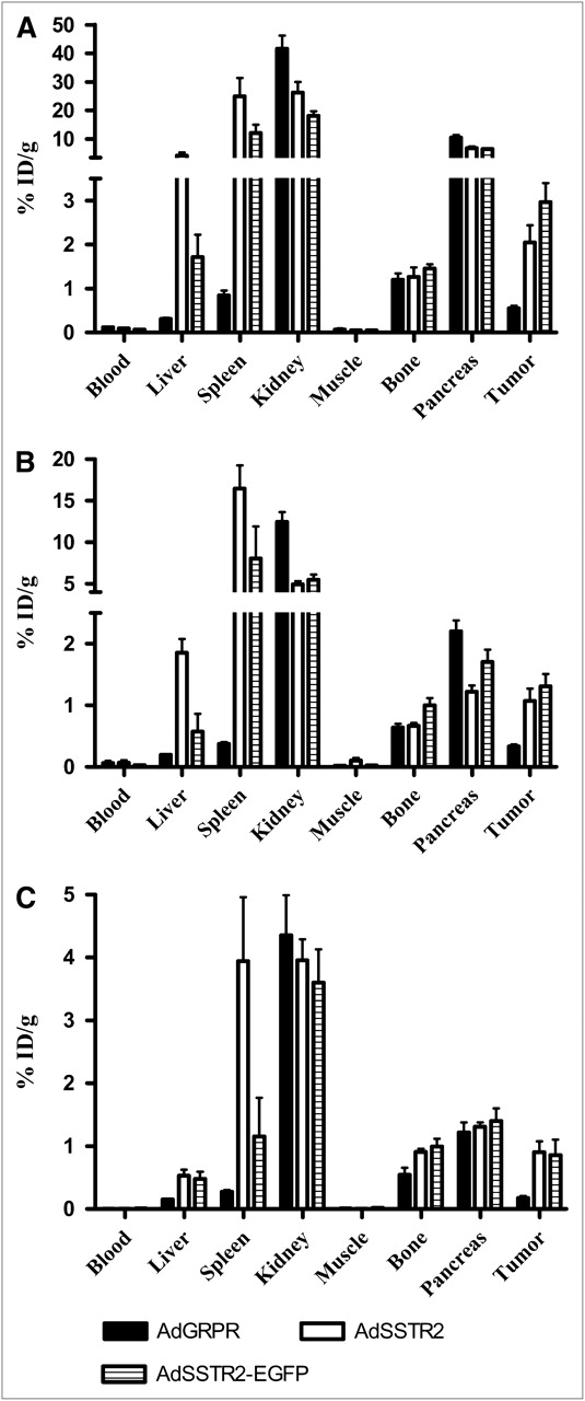
  • FIGURE 3.
    • Download figure
    • Open in new tab
    • Download powerpoint
    FIGURE 3.

    Biodistribution of 111In-DTPA-Y3-octreotate in mice bearing SCC-9 tumor xenografts. Tumors were injected directly with adenovirus vectors, and 111In-DTPA-Y3-octreotate was injected via tail vein 2 d later. Mice were sacrificed 4 h (A), 24 h (B), and 48 h (C) later (n = 5 for each group). Data are presented as % ID/g ± SEM.

  • FIGURE 4.
    • Download figure
    • Open in new tab
    • Download powerpoint
    FIGURE 4.

    Representative coronal nano-SPECT/CT images of SCC-9 tumor–bearing mice at 4, 24, and 48 h after injection of 111In-DTPA-Y3-octreotate. Axillary tumors were injected directly with Opti-MEM (control), AdSSTR2, or AdSSTR2-EGFP 2 d before administration of 111In-DTPA-Y3-octreotate. Coronal images show uptake of 111In-DTPA-Y3-octreotate in AdSSTR2- and AdSSTR2-EGFP-injected tumors but not control tumors (white arrows) and clearance through kidneys (yellow arrows).

  • FIGURE 5.
    • Download figure
    • Open in new tab
    • Download powerpoint
    FIGURE 5.

    Representative in vivo (A–C) and ex vivo (D–F) fluorescence imaging of mice bearing SCC-9 tumors injected directly with AdSSTR2 (A and D), AdSSTR2-EGFP (B and E), or AdEGFP (C and F) 2 d earlier. Tumor (arrow) was not visualized in vivo after injection of AdSSTR2 (A, negative control) but could be seen after injection of AdSSTR2-EGFP (B) or AdEGFP (C). In ex vivo studies, tumor had higher autofluorescence than liver and spleen (E), and fluorescence was observed in tumor injected with AdSSTR2-EGFP (D) or AdEGFP (F). au = arbitrary units.

Tables

  • Figures
    • View popup
    TABLE 1.

    Ex Vivo Fluorescent Imaging of Tissues 48 Hours After Direct Injection of Adenovirus Vectors into SCC-9 Tumors

    TissueAdSSTR2 (n = 6)AdSSTR2-EGFP (n = 8)AdEGFP (n = 5)
    Tumor261* ± 69687 ± 161997* ± 306
    Liver83† ± 12560†,‡ ± 15286‡ ± 40
    Spleen9 ± 311 ± 34 ± 1
    • ↵* Values were significantly different at P ≤ 0.05.

    • ↵† Values were significantly different at P ≤ 0.05.

    • ↵‡ Values were significantly different at P ≤ 0.05.

    • Data are expressed as mean ± SEM for each tissue in arbitrary units.

PreviousNext
Back to top

In this issue

Journal of Nuclear Medicine: 51 (9)
Journal of Nuclear Medicine
Vol. 51, Issue 9
September 1, 2010
  • Table of Contents
  • Table of Contents (PDF)
  • About the Cover
  • Index by author
Print
Download PDF
Article Alerts
Sign In to Email Alerts with your Email Address
Email Article

Thank you for your interest in spreading the word on Journal of Nuclear Medicine.

NOTE: We only request your email address so that the person you are recommending the page to knows that you wanted them to see it, and that it is not junk mail. We do not capture any email address.

Enter multiple addresses on separate lines or separate them with commas.
Multimodality Imaging of Gene Transfer with a Receptor-Based Reporter Gene
(Your Name) has sent you a message from Journal of Nuclear Medicine
(Your Name) thought you would like to see the Journal of Nuclear Medicine web site.
Citation Tools
Multimodality Imaging of Gene Transfer with a Receptor-Based Reporter Gene
Ron Chen, Jesse J. Parry, Walter J. Akers, Mikhail Y. Berezin, Issam M. El Naqa, Samuel Achilefu, W. Barry Edwards, Buck E. Rogers
Journal of Nuclear Medicine Sep 2010, 51 (9) 1456-1463; DOI: 10.2967/jnumed.109.063586

Citation Manager Formats

  • BibTeX
  • Bookends
  • EasyBib
  • EndNote (tagged)
  • EndNote 8 (xml)
  • Medlars
  • Mendeley
  • Papers
  • RefWorks Tagged
  • Ref Manager
  • RIS
  • Zotero
Share
Multimodality Imaging of Gene Transfer with a Receptor-Based Reporter Gene
Ron Chen, Jesse J. Parry, Walter J. Akers, Mikhail Y. Berezin, Issam M. El Naqa, Samuel Achilefu, W. Barry Edwards, Buck E. Rogers
Journal of Nuclear Medicine Sep 2010, 51 (9) 1456-1463; DOI: 10.2967/jnumed.109.063586
Twitter logo Facebook logo LinkedIn logo Mendeley logo
  • Tweet Widget
  • Facebook Like
  • Google Plus One
Bookmark this article

Jump to section

  • Article
    • Abstract
    • MATERIALS AND METHODS
    • RESULTS
    • DISCUSSION
    • CONCLUSION
    • Acknowledgments
    • REFERENCES
  • Figures & Data
  • Info & Metrics
  • PDF

Related Articles

  • This Month in JNM
  • PubMed
  • Google Scholar

Cited By...

  • Molecular Imaging with Reporter Genes: Has Its Promise Been Delivered?
  • Google Scholar

More in this TOC Section

  • 225Ac α-Pretargeted Radioimmunotherapy of Human Epidermal Growth Factor Receptor 2–Expressing Breast Cancer
  • 212Bi-Macroaggregated Albumin Inhibited Mouse Melanoma Growth by Regulating Cell Cycle Checkpoint Markers Without Promoting Living Cell Repopulation
  • In Vivo Visualization and Quantification of Brain Heat Shock Protein 90 with [11C]HSP990 in Healthy Aging and Neurodegeneration
Show more Basic Science Investigation

Similar Articles

SNMMI

© 2025 SNMMI

Powered by HighWire