


Article Figures & Data
Figures
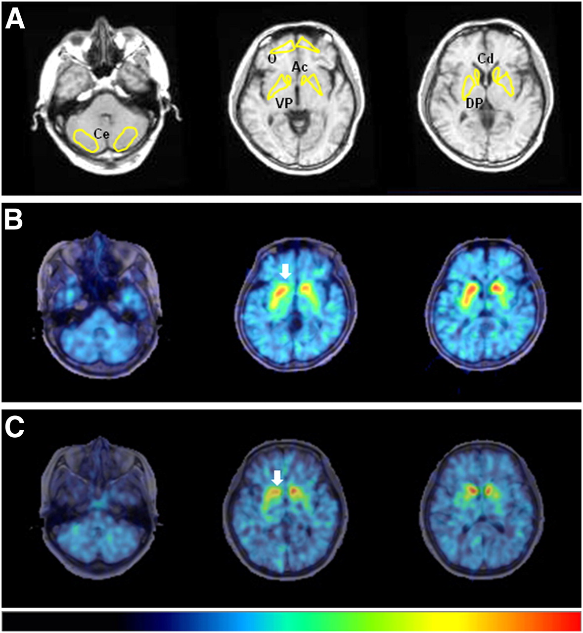
- FIGURE 1.
11C-CFT PET images and ROI setting. (A) Irregular ROIs, drawn bilaterally on concerned regions on MR images, were placed on corresponding PET images. (B) PD patient with longer conversion period from HY stage 1 to HY stage 2. (C) PD patient with shorter conversion period from HY stage 1 to HY stage 2. Arrow indicates reduction in 11C-CFT binding in nucleus accumbens. Color bar indicates quantified level of RI (from 0 to 3). Ac = nucleus accumbens; Cd = caudate; Ce = cerebellum; DP = dorsal putamen; O = orbitofrontal cortex; VP = ventral putamen.
- FIGURE 2.
Correlations between UPDRS score at HY stage 1 and 11C-CFT RI values. Dashed lines for ○ and chain lines for ▲ show all tendencies of negative correlations. ○ = affected side; ▲ = unaffected side.
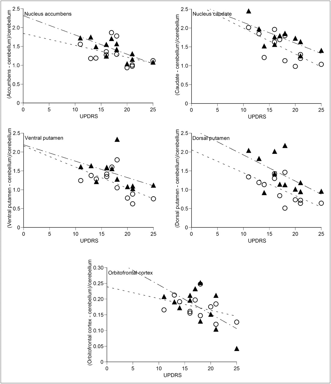
- FIGURE 3.
Correlations between conversion interval (y) and 11C-CFT RI values. Straight lines for ▲ show significant correlations (P < 0.05). ○ = affected side; ▲ = unaffected side.

Tables
Patient no. Sex Age (y) Duration of illness (mo)* UPDRS total Affected side Initial symptom 1 F 44 18 11 (2/3/6) L Rigidity 2 F 55 12 13 (2/3/8) L Rigidity/tremor (UL/LL) 3 M 67 14 14 (1/2/11) R Tremor (UL) 4 F 60 24 16 (2/3/11) R Tremor (UL/LL) 5 M 58 25 16 (4/6/6) R Rigidity/tremor (UL) 6 F 58 10 17 (2/5/10) R Rigidity/tremor (UL) 7 F 71 9 18 (2/4/12) R Rigidity/tremor (UL) 8 M 56 33 18 (4/4/10) R Rigidity/tremor (UL/LL) 9 F 73 4 20 (2/2/16) L Rigidity/tremor (UL/LL) 10 M 64 6 21 (2/4/15) R Rigidity 11 M 43 12 21 (2/6/13) L Rigidity/tremor (UL) 12 F 57 28 25 (4/5/16) R Rigidity/tremor (UL) ↵* Duration between disease onset and PET examination.
UPDRS scores in parentheses are mentation/activities of daily living/motor examination.
UL = upper limb; LL = lower limb.
Putamen Nucleus accumbens Caudate Ventral Dorsal Orbitofrontal Subject Affected side Unaffected side Affected side Unaffected side Affected side Unaffected side Affected side Unaffected side Affected side Unaffected side Healthy control (n = 8) 2.08 ± 0.20 2.23 ± 0.24 2.36 ± 0.29 2.51 ± 0.33 0.25 ± 0.08 PD patient (n = 12) 1.29 ± 0.30 1.42 ± 0.25 1.48 ± 0.38 1.75 ± 0.30 1.18 ± 0.36 1.43 ± 0.37 0.99 ± 0.35 1.40 ± 0.47 0.17 ± 0.04 0.18 ± 0.06 % reduction 38% 32% 34% 22% 50% 39% 61% 45% 34% 28% Each value is expressed as ratio index. % reduction denotes level of change in ratio index of PD patient compared with healthy control.


