


Article Figures & Data
Figures
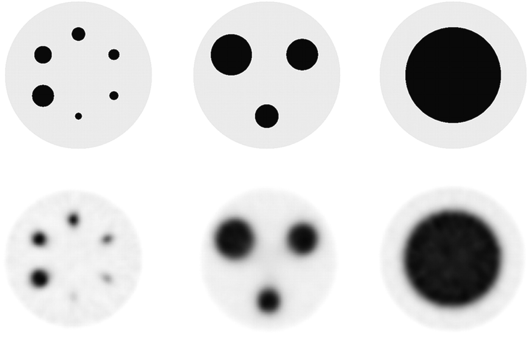
- FIGURE 1.
True (top) and example reconstructed images (bottom; LEHR collimation, 2.4-mm voxel, 32 OSEM updates) of simulated spheres of different diameters in 10% background.
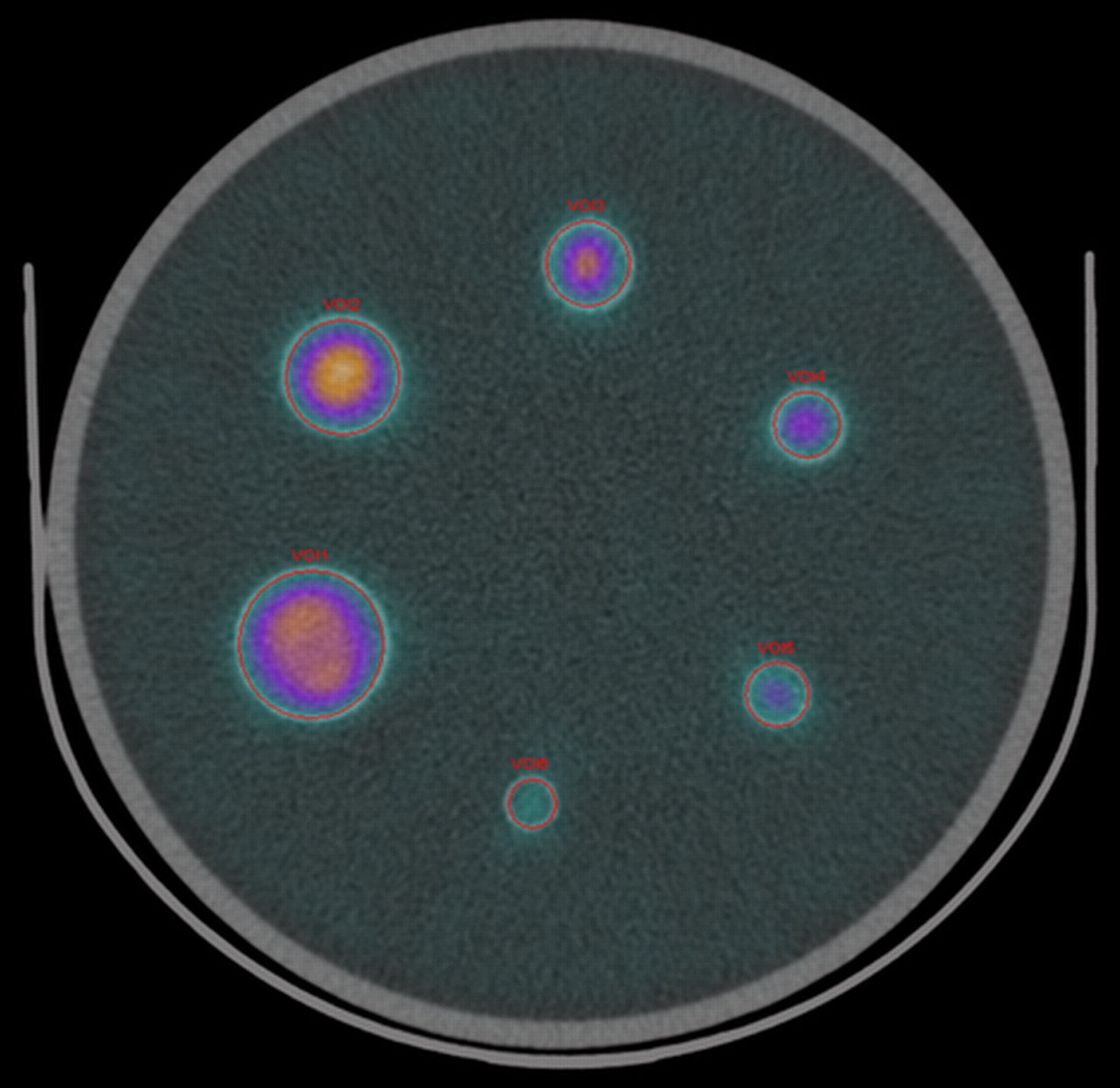
- FIGURE 2.
Reconstructed image of sphere phantom fused with CT image (LEHR collimation, 2.4-mm voxel, 32 OSEM updates). Circular VOIs were drawn manually using CT boundaries as reference.
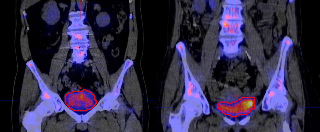
- FIGURE 3.
Reconstructed images of 2 patients fused with CT images (LEHR collimation, 4.8-mm voxel, 32 OSEM updates). VOIs were drawn by setting threshold of isocontour to coincide as close as possible with CT boundaries.
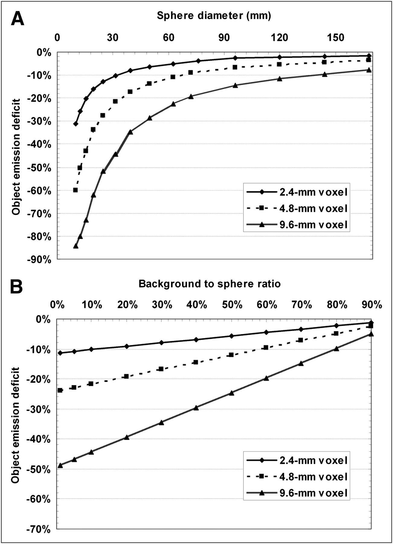
- FIGURE 4.
Effect of spillover at object boundaries on emission recovery due to finite voxel size for different object and voxel sizes with target-to-background ratio of 10:1 (A) and different target-to-background ratios using 16-mL sphere (B).
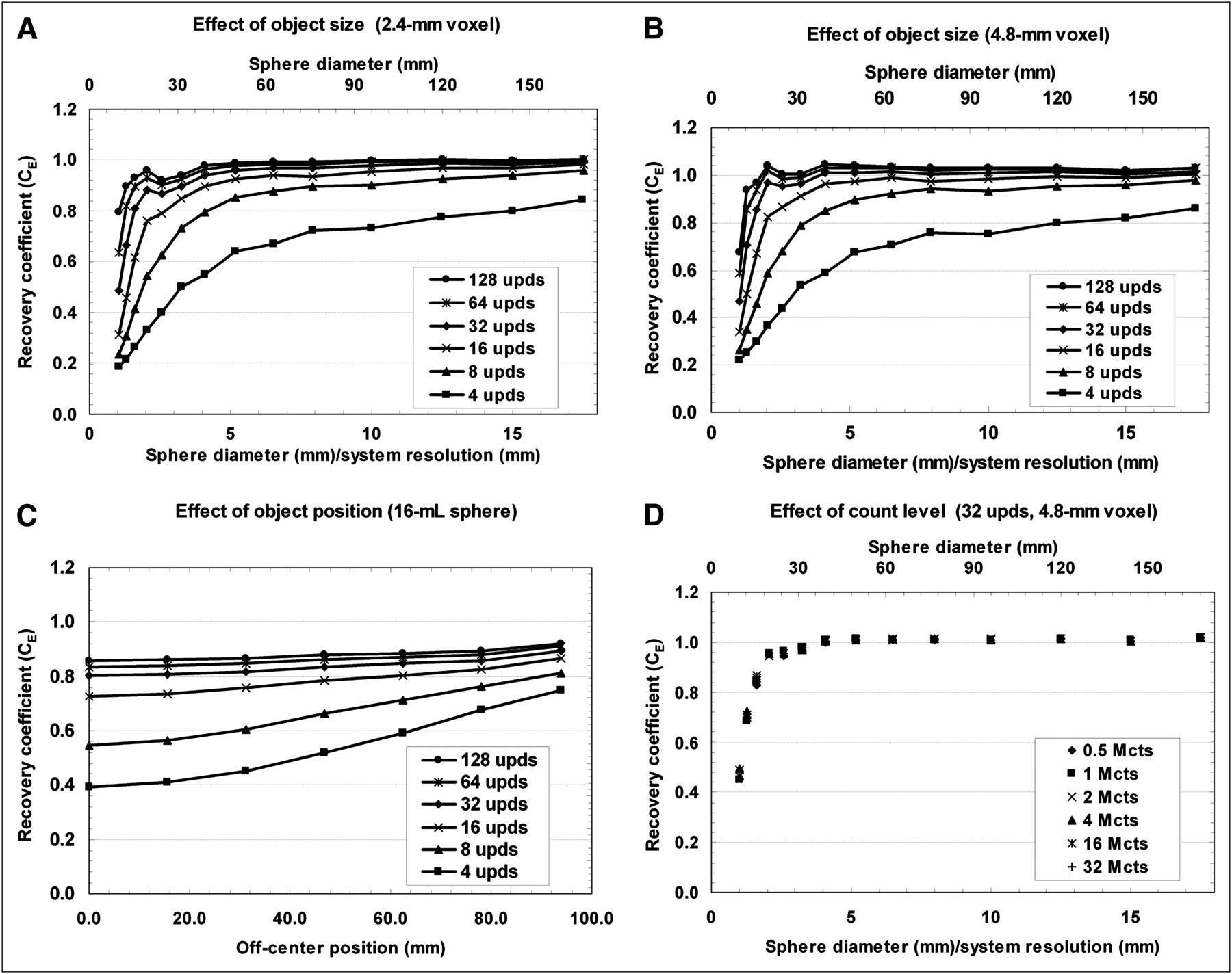
- FIGURE 5.
Emission recovery coefficients as function of object size and number of OSEM updates for different voxel sizes using LEHR collimation and 2 million total counts (A and B), for different object positions of 16-mL sphere (C), and for different total counts (D). upds = the number of OSEM updates; Mcts = million counts.
Tables
True sphere volume (mL) VVOI (mL) CE ĉA (kBq/mL) Mean difference (ĉA vs. cA) (%) SE of difference (%) 16 15.97 (0.1%) 0.80 (0.1%) 708.3 (6.5%) −2.8 8.0 8 8.02 (0.1%) 0.74 (0.1%) 749.4 (6.5%) +2.8 8.5 4 3.94 (0.6%) 0.71 (0.2%) 684.0 (6.6%) −6.2 7.7 2 2.08 (1.0%) 0.61 (0.6%) 685.3 (6.8%) −6.0 7.9 1 0.98 (0.8%) 0.42 (0.8%) 679.7 (6.7%) −6.8 7.8 0.5 0.52 (1.6%) 0.29 (1.4%) 708.8 (7.1%) −2.8 8.4 Values in parentheses are percentages of accumulated relative SEs due to measurement instrumentation. True activity concentration cA is 729.0 (relative SE, 5.0%), and calculated system volume sensitivity SVol is 10.3 (relative SE, 6.5%).
Patient no. VOI(mL) Urine activity concentration measured in well counter (kBq/mL) Activity concentration calculatedfrom image (kBq/mL) Deviation from true value (%) SE of deviation (%) 1 380.4 24.5 24.6 0.4% 8.3% 2 479.4 30.6 32.0 4.7% 8.4% 3 244.6 45.0 45.8 1.8% 8.2% 4 40.7 144.3 168.7 16.9% 12.4% 5 166.4 46.5 43.1 −7.4% 7.3% 6 309.1 13.6 13.8 1.4% 8.0% 7 114.4 27.3 25.4 −6.8% 7.3% 8 128.0 41.7 48.6 16.6% 8.8% 9 204.3 73.5 68.0 −7.5% 8.0% 10 273.0 17.5 20.0 14.3% 10.3% 11 157.2 16.7 16.9 1.0% 8.5% 12 53.0 284.1 272.9 −3.9% 9.0% 13 482.0 34.6 32.9 −4.9% 7.3% 14 420.1 94.2 86.8 −7.8% 7.1% 15 282.8 75.1 77.5 3.1% 8.1% 16 246.8 138.4 131.3 −5.2% 7.4% Minimum 40.7 13.6 13.8 −7.8% 7.1% Maximum 482.0 284.1 272.9 16.9% 12.4% Average 248.9 69.2 69.3 1.1% 8.4%


Jump to section
Related Articles
Cited By...
- Multicenter Study of Quantitative SPECT: Reproducibility of 99mTc Quantitation Using a Conjugated-Gradient Minimization Reconstruction Algorithm
- Optimization of Number of Iterations as a Reconstruction Parameter in Bone SPECT Imaging Using a Novel Thoracic Spine Phantom
- Digital Solid-State SPECT/CT Quantitation of Absolute 177Lu Radiotracer Concentration: In Vivo and In Vitro Validation
- Characterization of Noise and Resolution for Quantitative 177Lu SPECT/CT with xSPECT Quant
- Influence of Attenuation Correction by Brain Perfusion SPECT/CT Using a Simulated Abnormal Bone Structure: Comparison Between Chang and CT Methods
- MIRD Pamphlet No. 26: Joint EANM/MIRD Guidelines for Quantitative 177Lu SPECT Applied for Dosimetry of Radiopharmaceutical Therapy
- MIRD Pamphlet No. 24: Guidelines for Quantitative 131I SPECT in Dosimetry Applications
- An Evidence-Based Review of Quantitative SPECT Imaging and Potential Clinical Applications
- MIRD Pamphlet No. 23: Quantitative SPECT for Patient-Specific 3-Dimensional Dosimetry in Internal Radionuclide Therapy