


Article Figures & Data
Figures
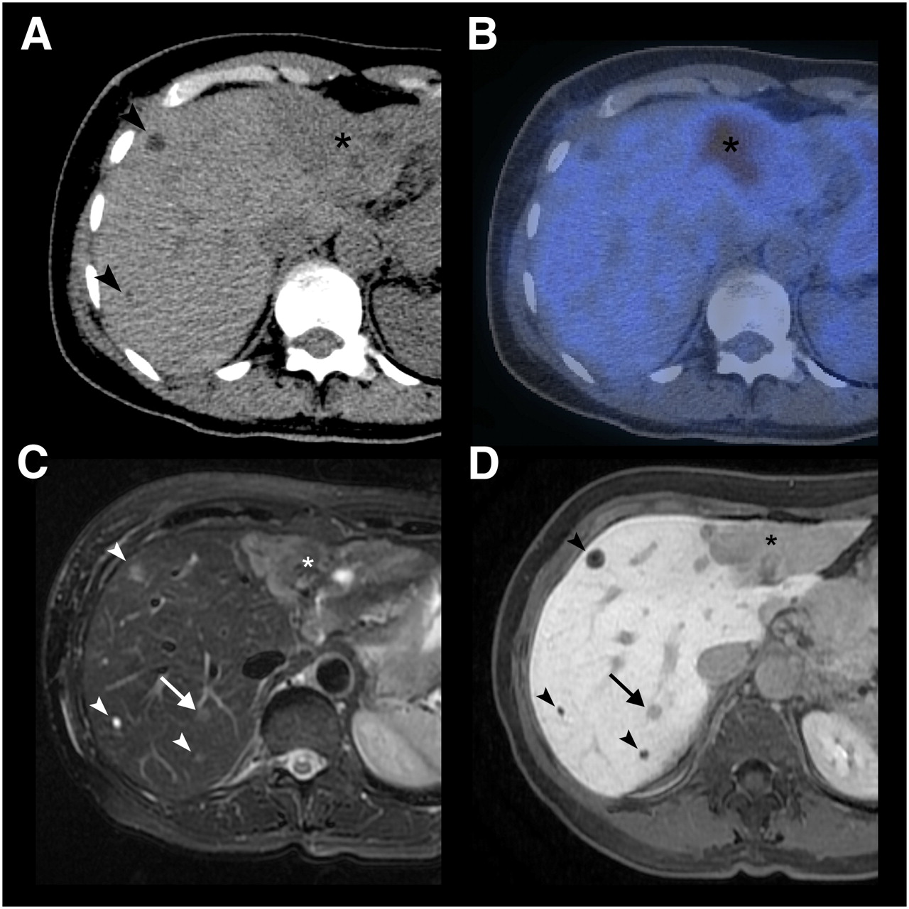
- FIGURE 1.
A 64-y-old patient with multiple liver metastases due to breast cancer. (A) On CT, 2 small (arrowheads) and 1 large (asterisk) hypodense lesion are noted. (B) Only the large lesion, in liver segment II/III, shows 18F-FDG uptake on PET/CT. (C and D) In liver segment VII, 2 additional lesions not visible on CT and PET/CT images are seen on T2-weighted fast spin-echo MRI (C) and Gd-EOB-DTPA–enhanced 3D gradient-echo MRI in hepatobiliary phase (D). One lesion (arrow) corresponds to metastasis as confirmed by intraoperative biopsy; other lesion (arrowhead) corresponds to cysts.
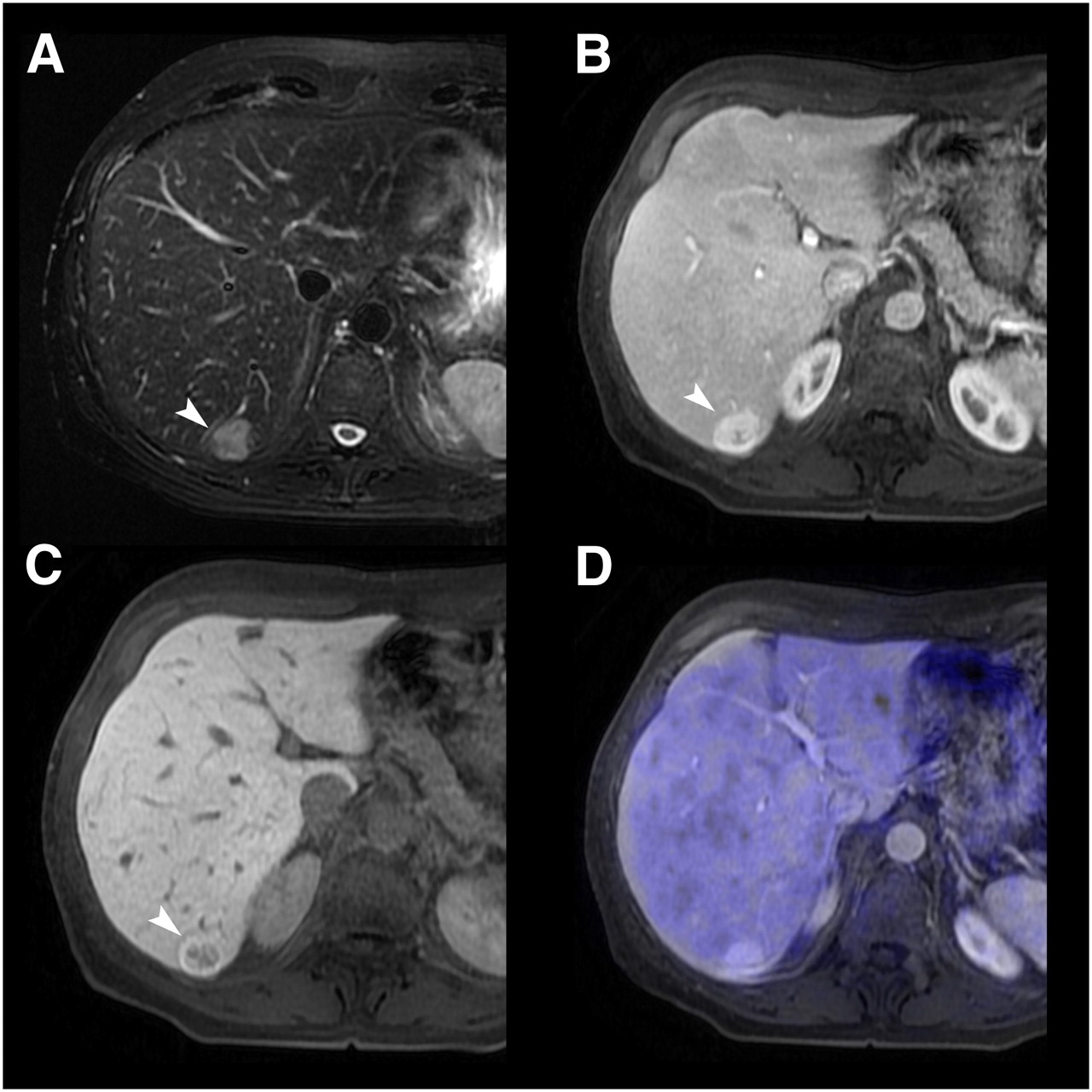
- FIGURE 2.
A 56-y-old patient with incidental liver finding on sonography. (A) T2-weighted MRI shows hyperintense lesion (arrowhead) in segment VI. (B and C) On Gd-EOB-DTPA–enhanced MRI, lesion shows contrast uptake during portal venous phase (B) and no contrast uptake during hepatobiliary phase (C). Lesion was first graded as indeterminate (grade 3). When information from PET was added to MRI (D), lack of 18F-FDG uptake indicated that lesion was definitely benign (grade 1). Histopathology confirmed diagnosis of focal nodular hyperplasia.
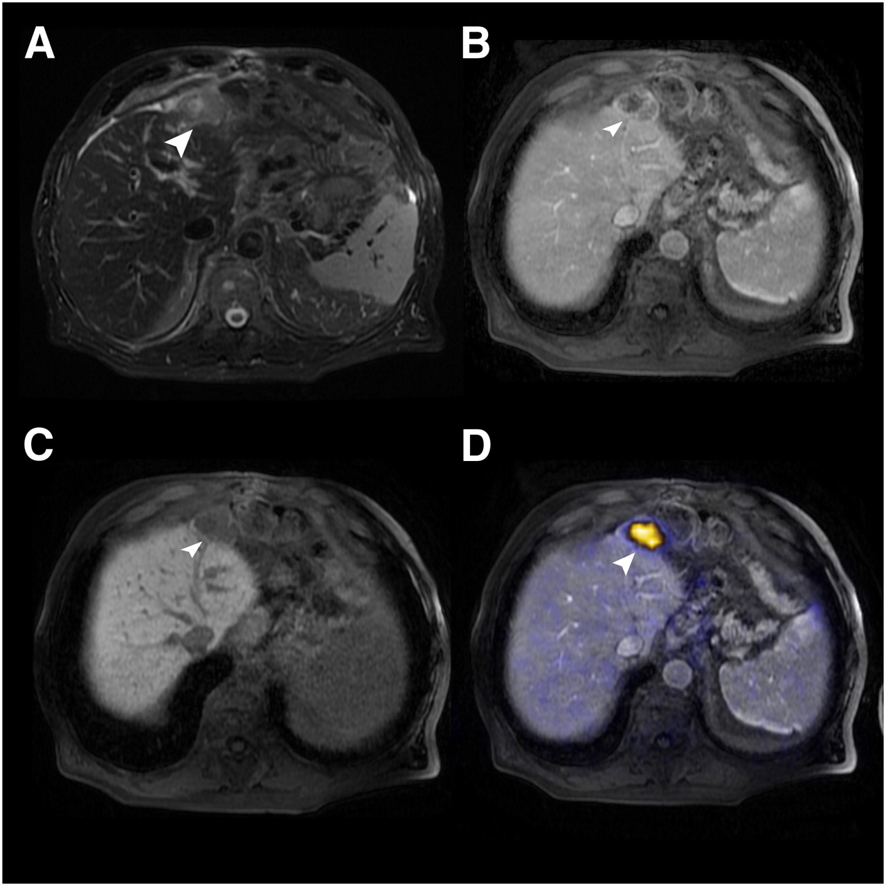
- FIGURE 3.
An 82-y-old gastric cancer patient after left hemihepatectomy. (A) On T2-weighted fast spin-echo MRI, subcapsular lesion (arrowhead) is hyperintense. (B) Lesion shows slight rimlike contrast uptake during portal venous phase. (C) Lesion shows no contrast medium uptake during hepatobiliary phase after injection of Gd-EOB-DTPA. Based on MRI findings, lesion was graded as probably malignant (grade 4). (D) When information from PET was added to MRI, 18F-FDG uptake indicated that lesion was definitely malignant (grade 5). Histopathology confirmed diagnosis of hepatic metastasis from gastric carcinoma.


Tables
Parameter T2-SSFSE T2-FSE T1-FSPGR T1-3D-GRE Imaging plane Coronal Transverse Transverse Transverse Repetition time/echo time (ms) 1,119/88 9,474/89 135/4.7 and 2.2 3.1/1.4 Inversion recovery time (ms) NA NA NA 7 Flip angle (degrees) NA 90 60 15 Matrix size 384 × 224 256 × 224 224 × 192 384 × 256 Section thickness (mm) 5 5 5 4 Intersection gap (mm) 1 1 1 None Overlap (mm) None None None 2 No. of signals acquired 0.5 2 1 0.73 Parallel imaging acceleration factor NA NA NA 2 Receiver bandwidth (kHz) 62.5 50 50 83.3 SSFSE = single-shot fast spin-echo; FSE = fast spin-echo; FSPGR = fast spoiled gradient-recalled acquisition in the steady state; GRE = gradient-echo; NA = not applicable.
Diameter (mm) Number of metastases Number of lesions total ≤5 1 (2%) 6 (7%) 6–10 9 (16%) 18 (21%) 11–20 22 (40%) 35 (41%) ≥21 23 (42%) 26 (31%) Total 55 (100%) 85 (100%) Lesion group and modality Detection rate (%) All lesions PET/CT 64* Gd-EOB-DTPA–enhanced MRI 85 Lesions ≤ 1 cm in diameter PET/CT 29* Gd-EOB-DTPA–enhanced MRI 71 Lesions > 1 cm in diameter PET/CT 77* Gd-EOB-DTPA–enhanced MRI 90 ↵* Considering all subgroups, differences in lesion detection were significant (P < 0.05) between PET/CT and Gd-EOB-DTPA–enhanced MRI.
Lesion group and modality Sensitivity (%) Specificity (%) All lesions PET/CT 76* 90 Gd-EOB-DTPA–enhanced MRI 91 100 PET/MRI† 93/93 87/97 Lesions ≤ 1 cm in diameter PET/CT 30 86 Gd-EOB-DTPA–enhanced MRI 80 100 PET/MRI† 70/70 71/100 Lesions > 1 cm in diameter PET/CT 87 94 Gd-EOB-DTPA–enhanced MRI 93 100 PET/MRI† 98/98 100/94 Lesion group and modality AUC All lesions PET/CT 0.85 Gd-EOB-DTPA–enhanced MRI 0.94 PET/MRI* 0.92/0.96 Lesions ≤ 1 cm in diameter PET/CT 0.54 Gd-EOB-DTPA–enhanced MRI 0.84 PET/MRI* 0.66/0.87 Lesions > 1 cm in diameter PET/CT 0.93 Gd-EOB-DTPA–enhanced MRI 0.96 PET/MRI* 0.99/0.97 ↵* Values are for reader 1/reader 2, respectively.
Supplemental Data
Files in this Data Supplement:


