


Article Figures & Data
Figures
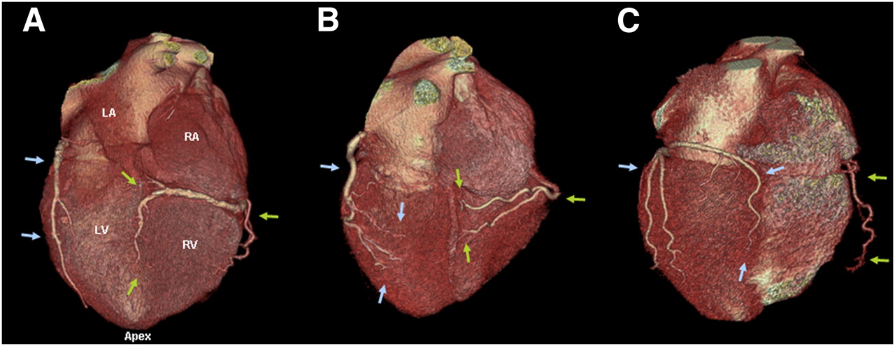
- FIGURE 1.
Three-dimensional, volume-rendered inferior and posterior CT angiographic images showing different coronary anatomy variants. (A) Standard, right-dominant circulation. Right coronary artery (green arrows) supplies posterior descending branch. Left circumflex (blue arrows) supplies only inferolateral left ventricular myocardium. (B) Codominant circulation. No clear posterior descending artery is observed. Inferior myocardium is supplied by both right and left circumflex arteries. (C) Left-dominant circulation. Left circumflex supplies posterior descending branch. Right coronary artery supplies only right ventricular myocardium. LA = left atrium; RA = right atrium; LV = left ventricle; RV = right ventricle.
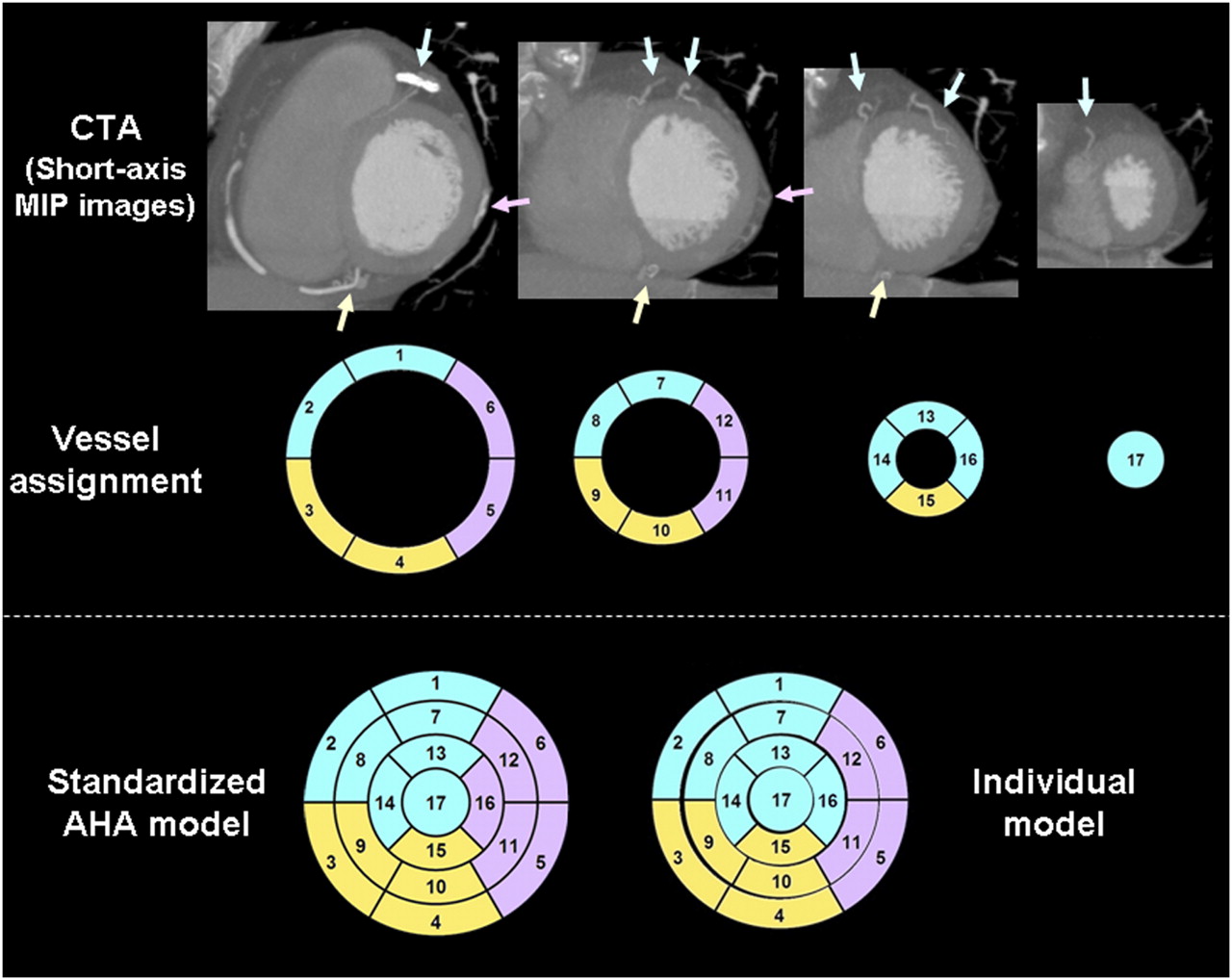
- FIGURE 2.
CT-based individual assignment of myocardial segments to vascular territories. Short-axis maximum-intensity-projection (MIP) images of CTA are reviewed for localization of coronary arteries (top). Each of 17 myocardial segments is assigned to closest coronary artery (middle; LAD in blue, LCX in pink, and RCA in yellow). Bottom shows standard AHA model on left and model after individual reassignment on right. In this case, segment 16 (distal lateral wall) was reassigned to LAD, which supplied diagonal branch.
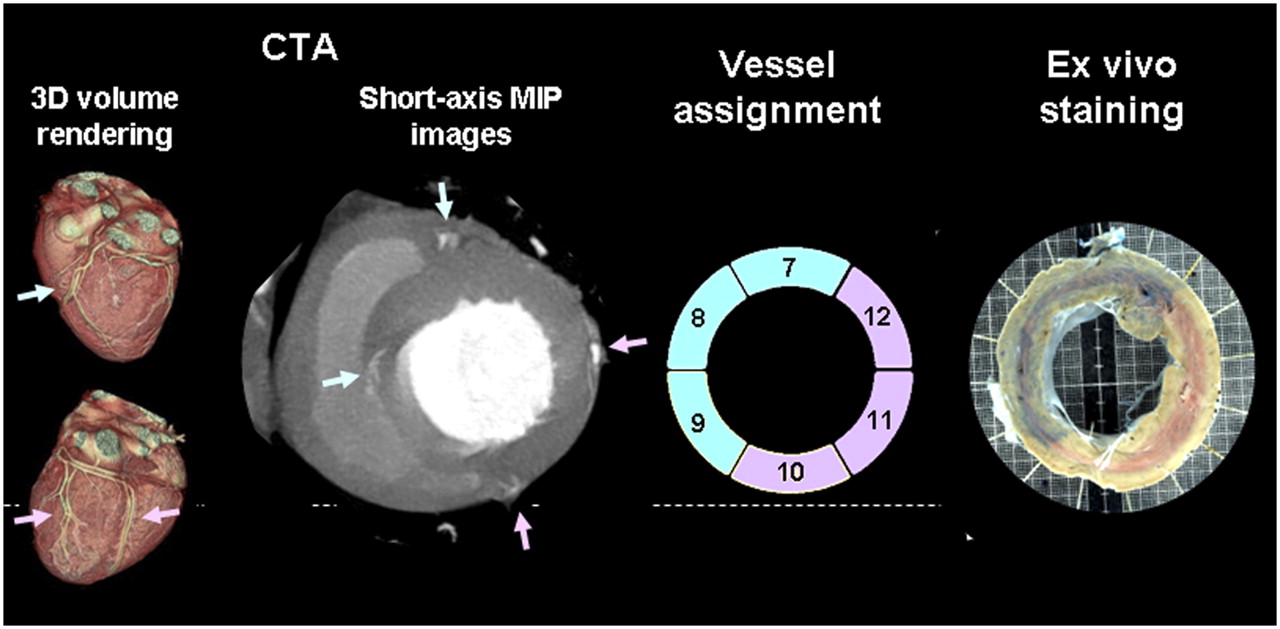
- FIGURE 3.
Experimental validation of CT-based visual assignment of myocardial segments. Dog model was used, which generally shows left-dominant distribution, as shown on 3-dimensional volume-rendered surface images of CT (left). Representative midventricular short-axis maximum-intensity-projection (MIP) slice of CTA in dog is shown (middle left), along with respective assignment of myocardial segments to vascular territories (middle right; blue and pink arrows indicate LAD and LCX). Ex vivo images of dye-stained heart (right) show excellent agreement with true vessel distribution (nonoccluded LAD is stained in blue, and occluded LCX is not stained).
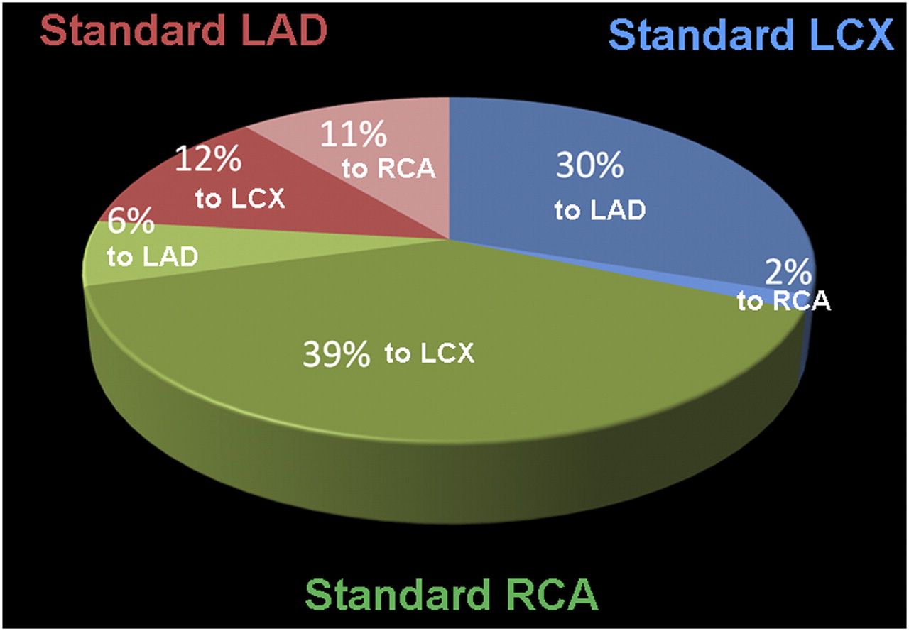
- FIGURE 4.
Distribution of 112 segments, which were reassigned after individual review of CT. Original segmental assignment to vascular territories is shown by color, along with percentage of overall segments that were reassigned and their new vascular territory.
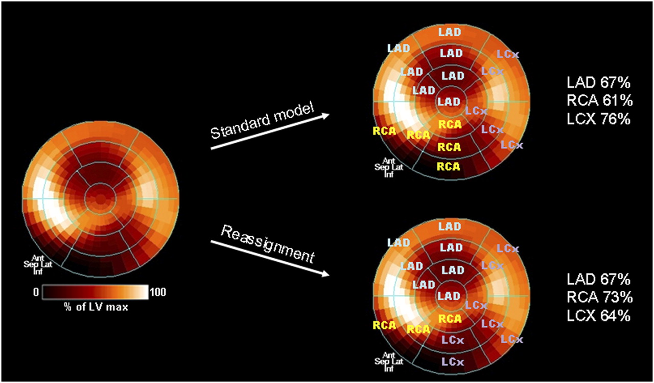
- FIGURE 5.
Polar map of stress myocardial perfusion from 82Rb PET, showing perfusion defects in distal anterior wall and apex and in basal inferior wall. CTA shows codominant circulation with obstructive atherosclerosis of mid LAD and mid LCX artery. With standard AHA segment assignment (top right), inferior wall defect contributes to reduced perfusion mostly in RCA territory. After CT-based individual assignment (bottom right), inferior wall defect contributes mostly to LCX territory. Average perfusion tracer uptake per vascular territory changes significantly for RCA and LCX (right).


Jump to section
Related Articles
Cited By...
- The Changing Face of Nuclear Cardiology: Guiding Cardiovascular Care Toward Molecular Medicine
- Arterial CO2 as a Potent Coronary Vasodilator: A Preclinical PET/MR Validation Study with Implications for Cardiac Stress Testing
- Dynamic Computed Tomography Myocardial Perfusion Imaging: Comparison of Clinical Analysis Methods for the Detection of Vessel-Specific Ischemia
- SNMMI/ASNC/SCCT Guideline for Cardiac SPECT/CT and PET/CT 1.0
- Aligning Coronary Anatomy and Myocardial Perfusion Territories: An Algorithm for the CORE320 Multicenter Study
- Diagnostic Value of 13N-Ammonia Myocardial Perfusion PET: Added Value of Myocardial Flow Reserve
- Structural Abnormalities of the Coronary Arterial Wall--in Addition to Luminal Narrowing--Affect Myocardial Blood Flow Reserve