
Main menu

  • Home
  • Content
    • Current
    • Ahead of print
    • Past Issues
    • JNM Supplement
    • SNMMI Annual Meeting Abstracts
    • Continuing Education
    • JNM Podcasts
  • Subscriptions
    • Subscribers
    • Institutional and Non-member
    • Rates
    • Journal Claims
    • Corporate & Special Sales
  • Authors
    • Submit to JNM
    • Information for Authors
    • Assignment of Copyright
    • AQARA requirements
  • Info
    • Reviewers
    • Permissions
    • Advertisers
  • About
    • About Us
    • Editorial Board
    • Contact Information
  • More
    • Alerts
    • Feedback
    • Help
    • SNMMI Journals
  • SNMMI
    • JNM
    • JNMT
    • SNMMI Journals
    • SNMMI

User menu

  • Subscribe
  • My alerts
  • Log in
  • My Cart

Search

  • Advanced search
Journal of Nuclear Medicine
  • SNMMI
    • JNM
    • JNMT
    • SNMMI Journals
    • SNMMI
  • Subscribe
  • My alerts
  • Log in
  • My Cart
Journal of Nuclear Medicine

Advanced Search

  • Home
  • Content
    • Current
    • Ahead of print
    • Past Issues
    • JNM Supplement
    • SNMMI Annual Meeting Abstracts
    • Continuing Education
    • JNM Podcasts
  • Subscriptions
    • Subscribers
    • Institutional and Non-member
    • Rates
    • Journal Claims
    • Corporate & Special Sales
  • Authors
    • Submit to JNM
    • Information for Authors
    • Assignment of Copyright
    • AQARA requirements
  • Info
    • Reviewers
    • Permissions
    • Advertisers
  • About
    • About Us
    • Editorial Board
    • Contact Information
  • More
    • Alerts
    • Feedback
    • Help
    • SNMMI Journals
  • View or Listen to JNM Podcast
  • Visit JNM on Facebook
  • Join JNM on LinkedIn
  • Follow JNM on Twitter
  • Subscribe to our RSS feeds
Research ArticleClinical Investigation

18F-FDG PET/CT for the Prediction and Detection of Local Recurrence After Radiofrequency Ablation of Malignant Lung Lesions

Amit Singnurkar, Stephen B. Solomon, Mithat Gönen, Steven M. Larson and Heiko Schöder
Journal of Nuclear Medicine December 2010, 51 (12) 1833-1840; DOI: https://doi.org/10.2967/jnumed.110.076778
Amit Singnurkar
  • Find this author on Google Scholar
  • Find this author on PubMed
  • Search for this author on this site
Stephen B. Solomon
  • Find this author on Google Scholar
  • Find this author on PubMed
  • Search for this author on this site
Mithat Gönen
  • Find this author on Google Scholar
  • Find this author on PubMed
  • Search for this author on this site
Steven M. Larson
  • Find this author on Google Scholar
  • Find this author on PubMed
  • Search for this author on this site
Heiko Schöder
  • Find this author on Google Scholar
  • Find this author on PubMed
  • Search for this author on this site
  • Article
  • Figures & Data
  • Info & Metrics
  • PDF
Loading

Article Figures & Data

Figures

  • Tables
  • FIGURE 1.
    • Download figure
    • Open in new tab
    • Download powerpoint
    FIGURE 1.

    Patterns of uptake on post-RFA 18F-FDG PET/CT images: diffuse (A), focal (B), rim (C), heterogeneous (D), rim plus focal with focus not corresponding to site of original lesion (E), and rim plus focal with focus corresponding to site of original lesion (F). Dotted circles represent location of original lesion.
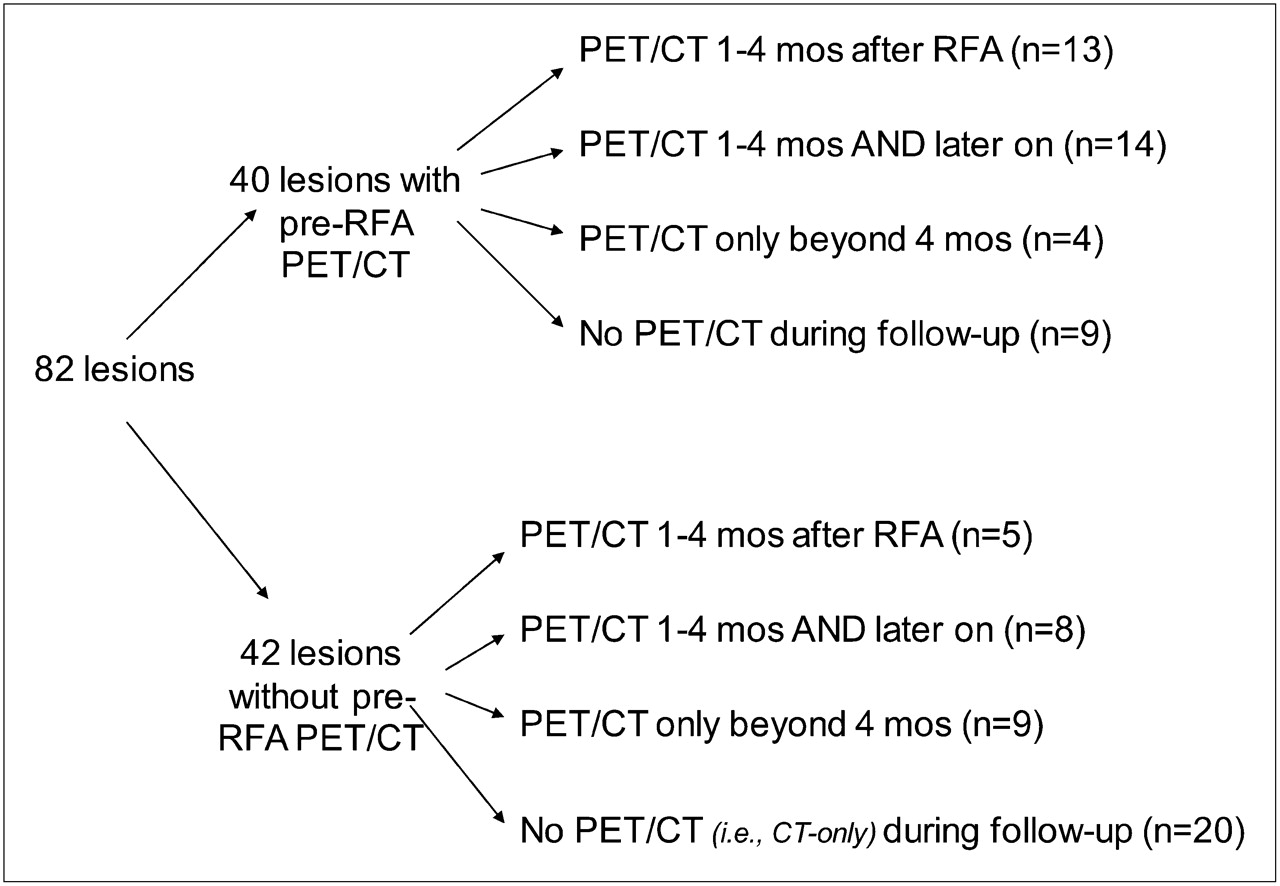
  • FIGURE 2.
    • Download figure
    • Open in new tab
    • Download powerpoint
    FIGURE 2.

    Imaging follow-up of ablated lesions.
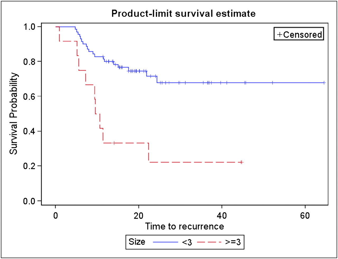
  • FIGURE 3.
    • Download figure
    • Open in new tab
    • Download powerpoint
    FIGURE 3.

    Kaplan–Meier curves for recurrence-free survival according to pretreatment lesion size (cm).

  • FIGURE 4.
    • Download figure
    • Open in new tab
    • Download powerpoint
    FIGURE 4.

    Follow-up after RFA for primary non–small cell lung adenocarcinoma. 18F-FDG PET/CT image at 4.5 mo after RFA (A) shows typical early pattern of ring of low-grade (SUV, 3.0) metabolic activity (arrow), and 18F-FDG PET/CT image at 9 mo after RFA (B) shows interval decrease in metabolic activity (arrow) (SUV, 0.8).
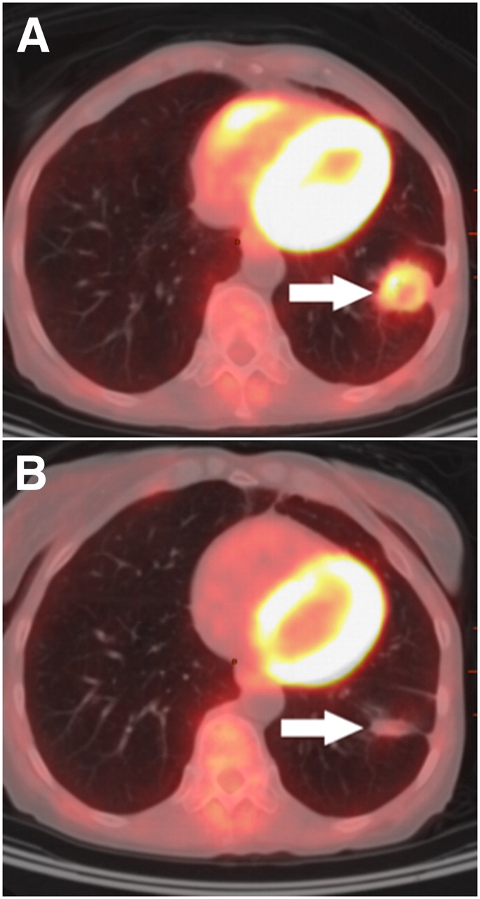
  • FIGURE 5.
    • Download figure
    • Open in new tab
    • Download powerpoint
    FIGURE 5.

    Local recurrence with rim plus focal uptake. CT image (A) shows left upper lobe pulmonary metastasis before RFA, and transaxial 18F-FDG PET image at 6 wk after RFA (B) shows rim plus focal uptake with focal uptake corresponding to site of original lesion (arrow). (C) No treatment was performed; PET/CT fusion image obtained 3 mo later shows interval progression (arrow). This lesion was then surgically resected and histopathology proved local recurrence.
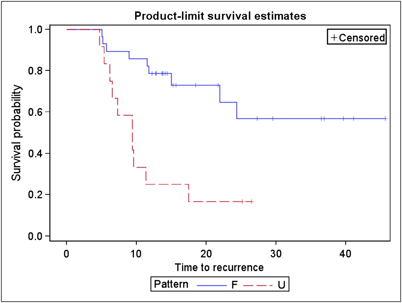
  • FIGURE 6.
    • Download figure
    • Open in new tab
    • Download powerpoint
    FIGURE 6.

    Kaplan–Meier curves showing recurrence-free survival according to pattern of 18F-FDG uptake. F = favorable; U = unfavorable.

Tables

  • Figures
    • View popup
    TABLE 1

    Local Recurrence According to Pathologic Subtype of Pulmonary Lesion

    PathologynRecurrenceNonrecurrence
    Primary pulmonary lesion442024
     Adenocarcinoma18810
     Squamous cell carcinoma1073
     Undifferentiated non–small cell carcinoma1046
     Adenosquamous carcinoma11—
     Bronchioalveolar carcinoma3—3
     Small cell carcinoma2—2
    Metastatic lesion38830
     Colorectal20119
     Sarcoma514
     Fibrosarcoma1—1
     Head and neck squamous cell carcinoma1—1
     Adrenal312
     Breast1—1
     Esophageal11—
     Insulinoma1—1
     Germ cell1—1
     Renal22—
     Adenocarcinoma, unknown primary11—
     Carcinoma, unknown primary11—
    • View popup
    TABLE 2

    Local Recurrence According to Pattern of Uptake on Post-RFA Scan

    PatternRecurrenceNonrecurrence
    Favorable
     Diffuse510
     Heterogeneous01
     Rim36
     Rim plus focal, corresponding81
      Corresponding
    Unfavorable
     Focal21
     Rim plus focal, not corresponding12
      Corresponding
PreviousNext
Back to top

In this issue

Journal of Nuclear Medicine: 51 (12)
Journal of Nuclear Medicine
Vol. 51, Issue 12
December 1, 2010
  • Table of Contents
  • Table of Contents (PDF)
  • About the Cover
  • Index by author
Print
Download PDF
Article Alerts
Sign In to Email Alerts with your Email Address
Email Article

Thank you for your interest in spreading the word on Journal of Nuclear Medicine.

NOTE: We only request your email address so that the person you are recommending the page to knows that you wanted them to see it, and that it is not junk mail. We do not capture any email address.

Enter multiple addresses on separate lines or separate them with commas.
18F-FDG PET/CT for the Prediction and Detection of Local Recurrence After Radiofrequency Ablation of Malignant Lung Lesions
(Your Name) has sent you a message from Journal of Nuclear Medicine
(Your Name) thought you would like to see the Journal of Nuclear Medicine web site.
Citation Tools
18F-FDG PET/CT for the Prediction and Detection of Local Recurrence After Radiofrequency Ablation of Malignant Lung Lesions
Amit Singnurkar, Stephen B. Solomon, Mithat Gönen, Steven M. Larson, Heiko Schöder
Journal of Nuclear Medicine Dec 2010, 51 (12) 1833-1840; DOI: 10.2967/jnumed.110.076778

Citation Manager Formats

  • BibTeX
  • Bookends
  • EasyBib
  • EndNote (tagged)
  • EndNote 8 (xml)
  • Medlars
  • Mendeley
  • Papers
  • RefWorks Tagged
  • Ref Manager
  • RIS
  • Zotero
Share
18F-FDG PET/CT for the Prediction and Detection of Local Recurrence After Radiofrequency Ablation of Malignant Lung Lesions
Amit Singnurkar, Stephen B. Solomon, Mithat Gönen, Steven M. Larson, Heiko Schöder
Journal of Nuclear Medicine Dec 2010, 51 (12) 1833-1840; DOI: 10.2967/jnumed.110.076778
Twitter logo Facebook logo LinkedIn logo Mendeley logo
  • Tweet Widget
  • Facebook Like
  • Google Plus One
Bookmark this article

Jump to section

  • Article
    • Abstract
    • MATERIALS AND METHODS
    • DISCUSSION
    • CONCLUSION
    • REFERENCES
  • Figures & Data
  • Info & Metrics
  • PDF

Related Articles

  • Errata
  • PubMed
  • Google Scholar

Cited By...

  • PET Findings of Intramedullary Tumors of the Spinal Cord Using [18F] FDG and [11C] Methionine
  • Restaging: Should We PERCIST Without Pattern Recognition?
  • Google Scholar

More in this TOC Section

  • First-in-Human Study of 18F-Labeled PET Tracer for Glutamate AMPA Receptor [18F]K-40: A Derivative of [11C]K-2
  • Detection of HER2-Low Lesions Using HER2-Targeted PET Imaging in Patients with Metastatic Breast Cancer: A Paired HER2 PET and Tumor Biopsy Analysis
  • [11C]Carfentanil PET Whole-Body Imaging of μ-Opioid Receptors: A First in-Human Study
Show more Clinical Investigation

Similar Articles

SNMMI

© 2025 SNMMI

Powered by HighWire