
Main menu

  • Home
  • Content
    • Current
    • Ahead of print
    • Past Issues
    • JNM Supplement
    • SNMMI Annual Meeting Abstracts
    • Continuing Education
    • JNM Podcasts
  • Subscriptions
    • Subscribers
    • Institutional and Non-member
    • Rates
    • Journal Claims
    • Corporate & Special Sales
  • Authors
    • Submit to JNM
    • Information for Authors
    • Assignment of Copyright
    • AQARA requirements
  • Info
    • Reviewers
    • Permissions
    • Advertisers
  • About
    • About Us
    • Editorial Board
    • Contact Information
  • More
    • Alerts
    • Feedback
    • Help
    • SNMMI Journals
  • SNMMI
    • JNM
    • JNMT
    • SNMMI Journals
    • SNMMI

User menu

  • Subscribe
  • My alerts
  • Log in
  • My Cart

Search

  • Advanced search
Journal of Nuclear Medicine
  • SNMMI
    • JNM
    • JNMT
    • SNMMI Journals
    • SNMMI
  • Subscribe
  • My alerts
  • Log in
  • My Cart
Journal of Nuclear Medicine

Advanced Search

  • Home
  • Content
    • Current
    • Ahead of print
    • Past Issues
    • JNM Supplement
    • SNMMI Annual Meeting Abstracts
    • Continuing Education
    • JNM Podcasts
  • Subscriptions
    • Subscribers
    • Institutional and Non-member
    • Rates
    • Journal Claims
    • Corporate & Special Sales
  • Authors
    • Submit to JNM
    • Information for Authors
    • Assignment of Copyright
    • AQARA requirements
  • Info
    • Reviewers
    • Permissions
    • Advertisers
  • About
    • About Us
    • Editorial Board
    • Contact Information
  • More
    • Alerts
    • Feedback
    • Help
    • SNMMI Journals
  • View or Listen to JNM Podcast
  • Visit JNM on Facebook
  • Join JNM on LinkedIn
  • Follow JNM on Twitter
  • Subscribe to our RSS feeds
OtherBASIC SCIENCE INVESTIGATIONS

A Nucleolin-Targeted Multimodal Nanoparticle Imaging Probe for Tracking Cancer Cells Using an Aptamer

Do Won Hwang, Hae Young Ko, Jung Hwan Lee, Hyungu Kang, Sung Ho Ryu, In Chan Song, Dong Soo Lee and Soonhag Kim
Journal of Nuclear Medicine January 2010, 51 (1) 98-105; DOI: https://doi.org/10.2967/jnumed.109.069880
Do Won Hwang
  • Find this author on Google Scholar
  • Find this author on PubMed
  • Search for this author on this site
Hae Young Ko
  • Find this author on Google Scholar
  • Find this author on PubMed
  • Search for this author on this site
Jung Hwan Lee
  • Find this author on Google Scholar
  • Find this author on PubMed
  • Search for this author on this site
Hyungu Kang
  • Find this author on Google Scholar
  • Find this author on PubMed
  • Search for this author on this site
Sung Ho Ryu
  • Find this author on Google Scholar
  • Find this author on PubMed
  • Search for this author on this site
In Chan Song
  • Find this author on Google Scholar
  • Find this author on PubMed
  • Search for this author on this site
Dong Soo Lee
  • Find this author on Google Scholar
  • Find this author on PubMed
  • Search for this author on this site
Soonhag Kim
  • Find this author on Google Scholar
  • Find this author on PubMed
  • Search for this author on this site
  • Article
  • Figures & Data
  • Supplemental
  • Info & Metrics
  • PDF
Loading

Article Figures & Data

Figures

  • Additional Files
  • FIGURE 1. 
    • Download figure
    • Open in new tab
    • Download powerpoint
    FIGURE 1. 

    Schematic illustration of steps involved in preparation of MFR-AS1411 and in vitro fluorescence cancer targeting measured by confocal microscopy. MF particles had carboxyl group and Fmoc-protected amine moiety, which was coupled with amine-terminated AS1411 aptamer using EDC (MF-AS1411). After reaction of MF-AS1411 with p-SCN-bn-NOTA, particles were reacted with 67Ga-citrate to form MFR-AS1411.
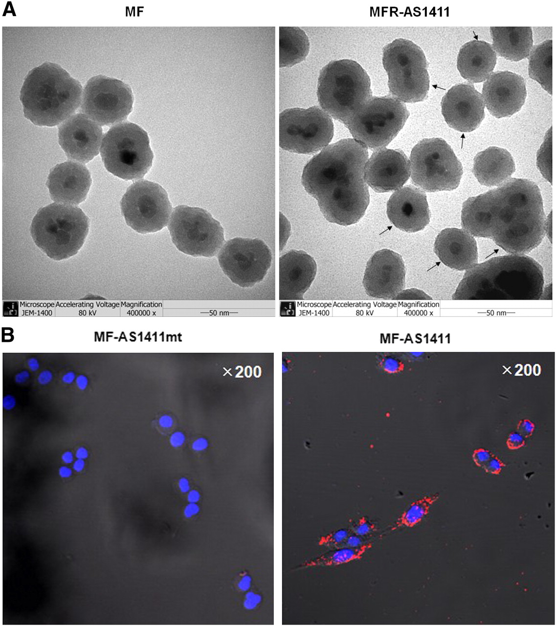
  • FIGURE 2. 
    • Download figure
    • Open in new tab
    • Download powerpoint
    FIGURE 2. 

    (A) TEM images were obtained in MF particles consisting of magnetic cobalt ferrite core, rhodamine dye, and MFR-AS1411, which was MF-labeled with AS1411 aptamer and 67Ga-citrate. Uranyl acetate (2%) was used to stain AS1411 aptamer. Dark staining of AS1411 in MFR-AS1411 was detected on surface of MFR-AS1411 particles (uranyl acetate–stained AS1411, black arrow). Scale bar = 50 nm. **P < 0.005. (B) After MF nanoparticles were conjugated with AS1411 aptamer using EDC, MF-AS1411 conjugates were treated into C6 cells. MF-AS1411 specifically targeted C6 glioma cells, as determined by laser scanning confocal microscopy (red, MF-AS1411; blue, 4′,6-diamidino-2-phenylindole dihydrochloride). No fluorescence signal was detected in MF-AS1411mt–treated group.
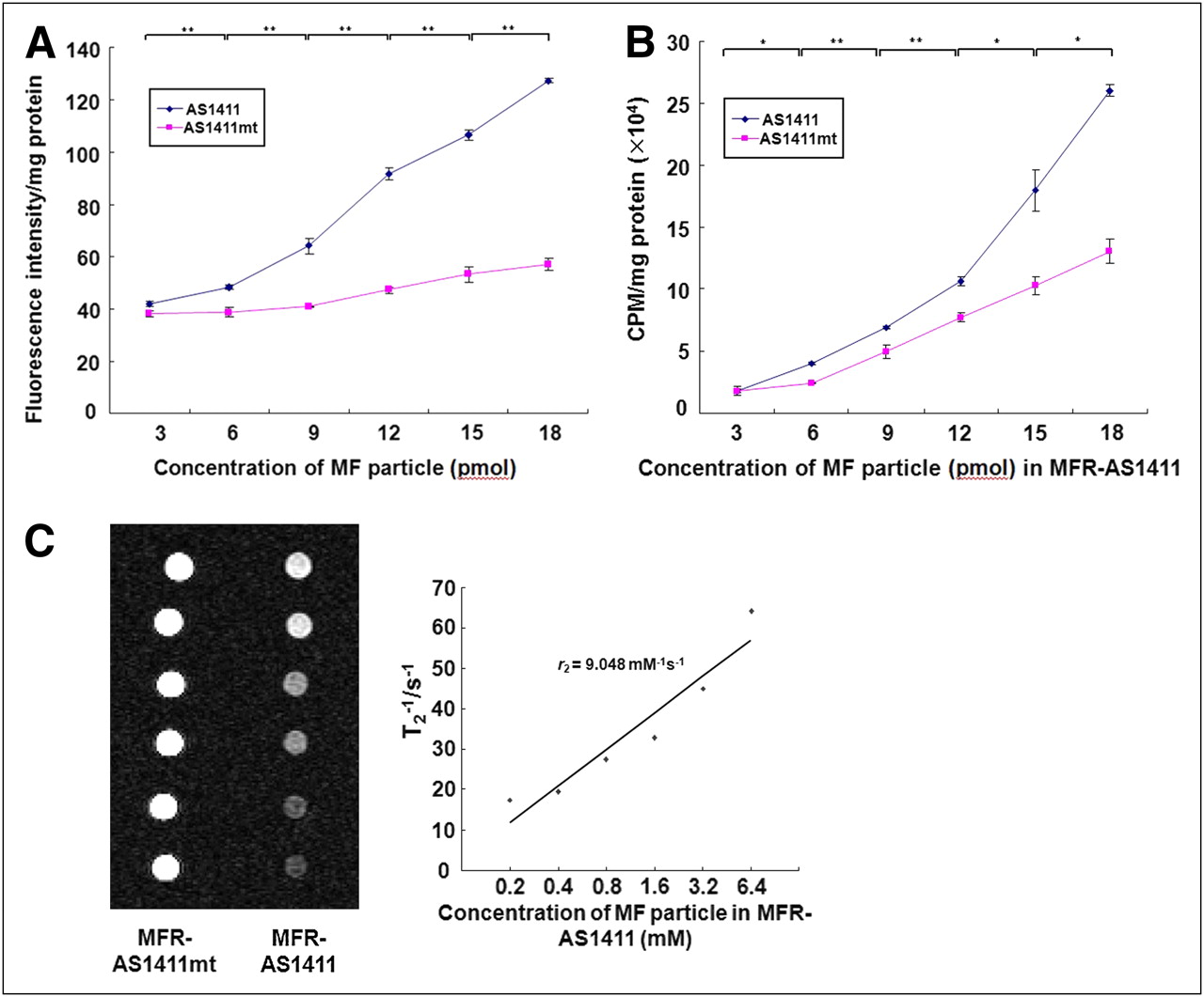
  • FIGURE 3. 
    • Download figure
    • Open in new tab
    • Download powerpoint
    FIGURE 3. 

    In vitro cancer targeting using different imaging modalities. (A) C6 cells were treated with diluted concentration of MF-AS1411 (or MF-AS1411mt). Gradual increase of fluorescence intensity in MF-AS1411–treated group was observed, compared with MF-AS1411mt group. (B) After synthesizing MF-AS1411 (or MF-AS1411mt) using p-SCN-bn-NOTA and incubating overnight, MF-AS1411 and MF-AS1411mt were labeled with 67Ga-citrate in buffer solution for 1 h. Concentration of MFR-AS1411 particles was critically dependent on 67Ga radioactivity in C6 cells. *P < 0.05. **P < 0.005. (C) Phantom studies were performed at various concentrations of MFR-AS1411 (or MFR-AS1411mt) mixture. Gradual decrease of MRI signals in MFR-AS1411–treated group were detected, compared with MFR-AS1411mt–treated group, on 1.5-T MRI system.

  • FIGURE 4. 
    • Download figure
    • Open in new tab
    • Download powerpoint
    FIGURE 4. 

    In vivo multimodal cancer targeting and imaging using MFR-AS1411 particles. (A) MFR-AS1411 particles were intravenously injected into tumor-bearing mice, and radionuclide images were acquired at 1, 6, and 24 h after injection. Scintigraphic images of C6 tumors in mice that received MFR-AS1411 showed that C6 tumors had accumulated MFR-AS1411 at 24 h after injection but did not accumulate MFR-AS1411mt (n = 3). Tumor growth patterns were followed using bioluminescence signals acquired from luciferase-expressing C6 cells. (B) MR images of tumor-bearing mice before and after injection of MFR-AS1411 were acquired. Dark signal intensities at tumor sites were detected in MFR-AS1411–injected mice (arrowhead). (C) Tumors were isolated and their fluorescence verified using IVIS200 system. Fluorescence signal at tumor site injected with MFR-AS1411 was detected, compared with tumors injected with MFR-AS1411mt. Isolated organs in order from upper left to lower right were intestine, liver, spleen, muscle, fat, kidney, stomach, right tumor, left tumor, heart, lung, and tail.

Additional Files

  • Figures
  • Supplemental Data

    Files in this Data Supplement:

    • Supplemental Figures
PreviousNext
Back to top

In this issue

Journal of Nuclear Medicine: 51 (1)
Journal of Nuclear Medicine
Vol. 51, Issue 1
January 2010
  • Table of Contents
  • Table of Contents (PDF)
  • About the Cover
  • Index by author
Print
Download PDF
Article Alerts
Sign In to Email Alerts with your Email Address
Email Article

Thank you for your interest in spreading the word on Journal of Nuclear Medicine.

NOTE: We only request your email address so that the person you are recommending the page to knows that you wanted them to see it, and that it is not junk mail. We do not capture any email address.

Enter multiple addresses on separate lines or separate them with commas.
A Nucleolin-Targeted Multimodal Nanoparticle Imaging Probe for Tracking Cancer Cells Using an Aptamer
(Your Name) has sent you a message from Journal of Nuclear Medicine
(Your Name) thought you would like to see the Journal of Nuclear Medicine web site.
Citation Tools
A Nucleolin-Targeted Multimodal Nanoparticle Imaging Probe for Tracking Cancer Cells Using an Aptamer
Do Won Hwang, Hae Young Ko, Jung Hwan Lee, Hyungu Kang, Sung Ho Ryu, In Chan Song, Dong Soo Lee, Soonhag Kim
Journal of Nuclear Medicine Jan 2010, 51 (1) 98-105; DOI: 10.2967/jnumed.109.069880

Citation Manager Formats

  • BibTeX
  • Bookends
  • EasyBib
  • EndNote (tagged)
  • EndNote 8 (xml)
  • Medlars
  • Mendeley
  • Papers
  • RefWorks Tagged
  • Ref Manager
  • RIS
  • Zotero
Share
A Nucleolin-Targeted Multimodal Nanoparticle Imaging Probe for Tracking Cancer Cells Using an Aptamer
Do Won Hwang, Hae Young Ko, Jung Hwan Lee, Hyungu Kang, Sung Ho Ryu, In Chan Song, Dong Soo Lee, Soonhag Kim
Journal of Nuclear Medicine Jan 2010, 51 (1) 98-105; DOI: 10.2967/jnumed.109.069880
Twitter logo Facebook logo LinkedIn logo Mendeley logo
  • Tweet Widget
  • Facebook Like
  • Google Plus One
Bookmark this article

Jump to section

  • Article
    • Abstract
    • MATERIALS AND METHODS
    • RESULTS
    • DISCUSSION
    • CONCLUSION
    • Acknowledgments
    • Footnotes
    • References
  • Figures & Data
  • Supplemental
  • Info & Metrics
  • PDF

Related Articles

  • This Month in JNM
  • PubMed
  • Google Scholar

Cited By...

  • Aptamer-Conjugated Extracellular Nanovesicles for Targeted Drug Delivery
  • Current Progress of Aptamer-Based Molecular Imaging
  • Tumor-specific Localization of Self-assembled Nanoparticle PET/MR Modalities
  • Aptamer Identification of Brain Tumor-Initiating Cells
  • Activatable aptamer probe for contrast-enhanced in vivo cancer imaging based on cell membrane protein-triggered conformation alteration
  • Google Scholar

More in this TOC Section

  • How Sensitive Is the Upper Gastrointestinal Tract to 90Y Radioembolization? A Histologic and Dosimetric Analysis in a Porcine Model
  • 11C-Methionine PET of Myocardial Inflammation in a Rat Model of Experimental Autoimmune Myocarditis
  • Counting Rate Characteristics and Image Distortion in Preclinical PET Imaging During Radiopharmaceutical Therapy
Show more Basic Science Investigations

Similar Articles

SNMMI

© 2025 SNMMI

Powered by HighWire