


Article Figures & Data
Figures
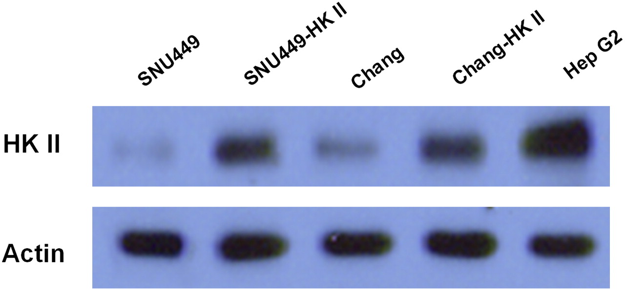
- FIGURE 1.
Expression profile of HKII in established stable cells. Expression of HKII is significantly increased after transfection in both SNU449 human HCC cell line and control Chang cells. HepG2 cell line was used as positive control for HKII. β-actin was used as loading controls for Western blot analysis. SNU449-HKII = stable cells after transfection of HKII; Chang-HKII = stable cells after transfection of HKII.
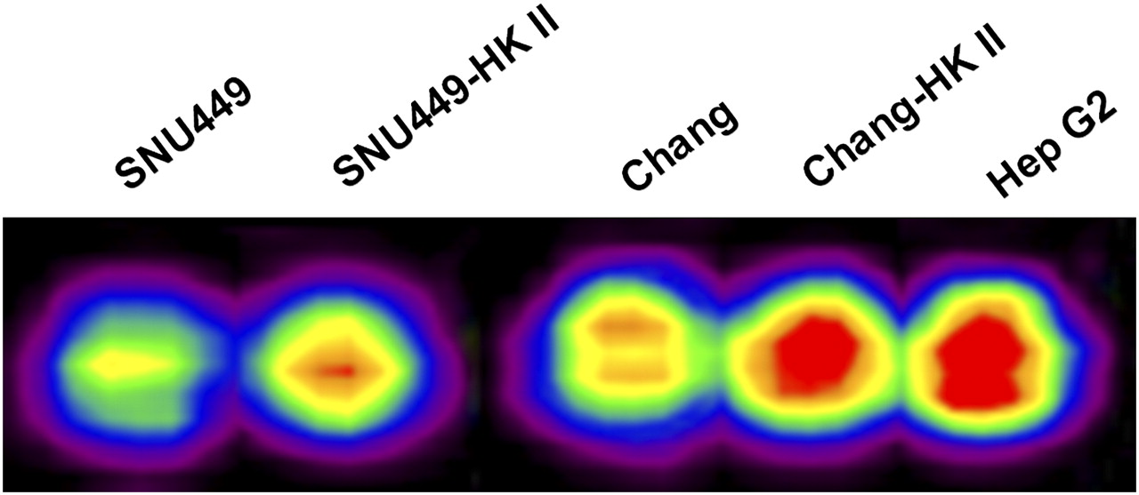
- FIGURE 2.
Measurement of 18F-FDG uptake levels in established stable cells. 18F-FDG PET images after transfection of HKII show significantly increased 18F-FDG uptake in both SNU449 and Chang cells by approximately 52% and 40%, respectively.
- FIGURE 3.
Subcellular localization of HKII in SNU449 and SNU449-HKII cells. Confocal microscopy shows markedly increased HKII expression after transfection (SNU449-HKII), and large proportion of HKIIs are mitochondrially associated. (A–C) SNU449 cells. (D–F) SNU449 cells after transfection of HKII. HKII protein is shown in A and D (red); mitochondria are shown in B and E (green); and HKII colocalized with mitochondria is shown in C and F (yellow-orange).
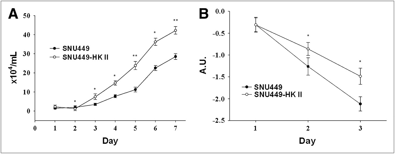
- FIGURE 4.
Effects of overexpressed HKII on cell growth and anticancer treatment response. Cellular proliferation is significantly increased after HKII transfection, approximately 1.5- to 2-fold, compared with nontransfected cells (A). After treatment with cisplatin (10 μg/mL) for 3 d at 37°C, there was 2- and 8-fold increase in cell survival 2 and 3 d after treatment, respectively (B). All measurements were performed in triplicate; error bars indicate SEM. Comparisons were subjected to Student t test. A.U. = arbitrary unit. *P < 0.05. **P < 0.01.
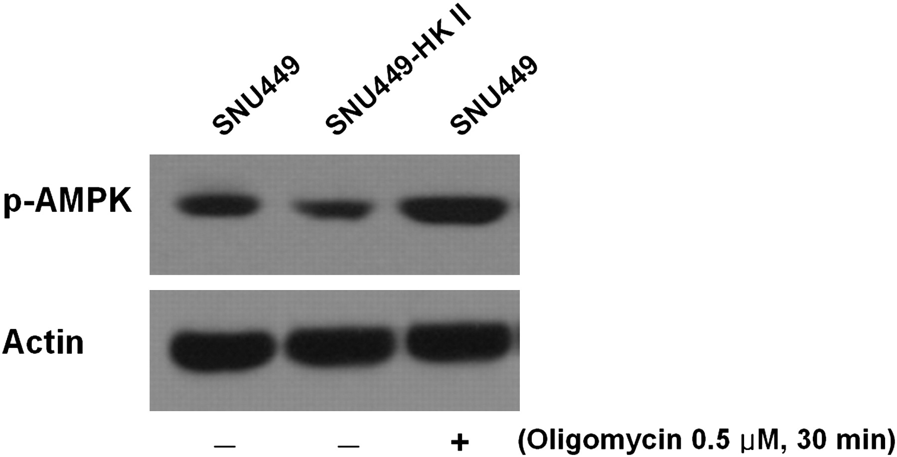
- FIGURE 5.
Effects of overexpressed HKII on AMPK phosphorylation. Activated form of AMPK (p-AMPK) is significantly decreased after HK II transfection (SNU449-HKII). Oligomycin (0.5 μM, 30 min) served as positive control of p-AMPK because it is AMPK activator.
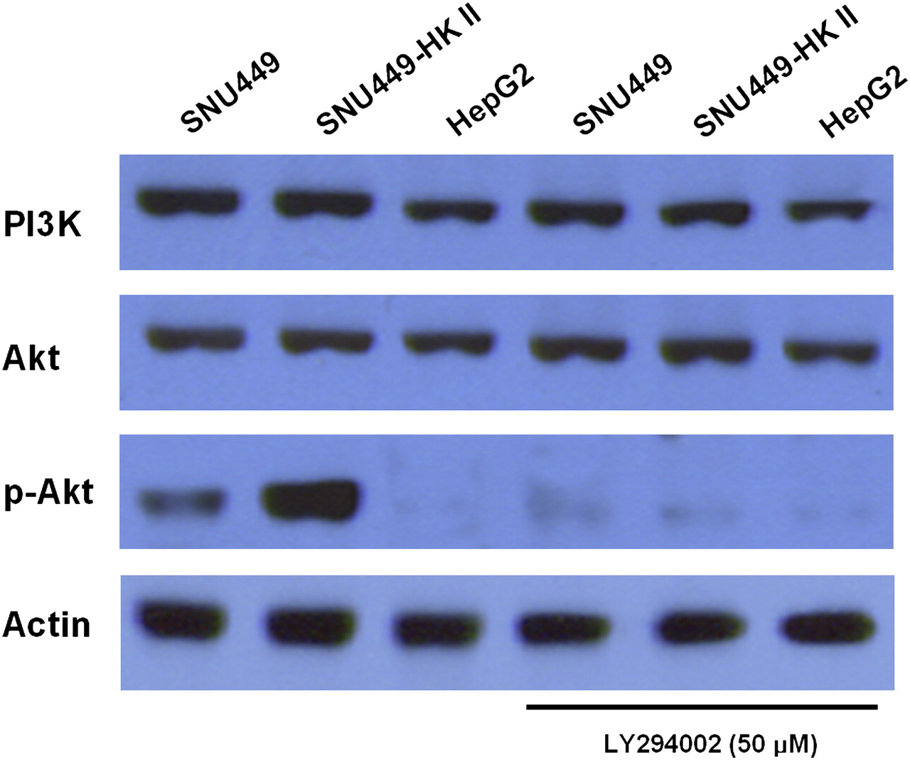
- FIGURE 6.
Effects of PI3K inhibitor treatment. Activated form of Akt (p-Akt) was significantly increased after HKII transfection (SNU449-HKII), compared with control cells without transfection (SNU449). After treatment with 50 μM PI3K inhibitor LY294002 for 1 h, p-Akt level significantly decreased. β-actin served as loading control.
- FIGURE 7.
Translocation of HK II from mitochondria to cytoplasm after treatment with PI3K inhibitor. Total amount of HKII protein was not changed after treatment with 50 μM PI3K inhibitor LY294002 for 1 h (A). HKII is expressed more in mitochondrial fraction before treatment. After treatment with PI3K inhibitor, mitochondrial fraction decreased whereas cytosolic fraction increased (B). β-actin and Hsp 60 served as loading controls. Cyt = cytoplasmic fraction X; Mt = mitochondria.


Additional Files
Supplemental Data
Files in this Data Supplement:


Jump to section
Related Articles
Cited By...
- 18F-FDG PET/CT Can Predict Survival of Advanced Hepatocellular Carcinoma Patients: A Multicenter Retrospective Cohort Study
- A Continuously Infused Microfluidic Radioassay System for the Characterization of Cellular Pharmacokinetics
- PU.1 is linking the glycolytic enzyme HK3 in neutrophil differentiation and survival of APL cells
- Combined RNA Interference of Hexokinase II and 131I-Sodium Iodide Symporter Gene Therapy for Anaplastic Thyroid Carcinoma