


Article Figures & Data
Figures
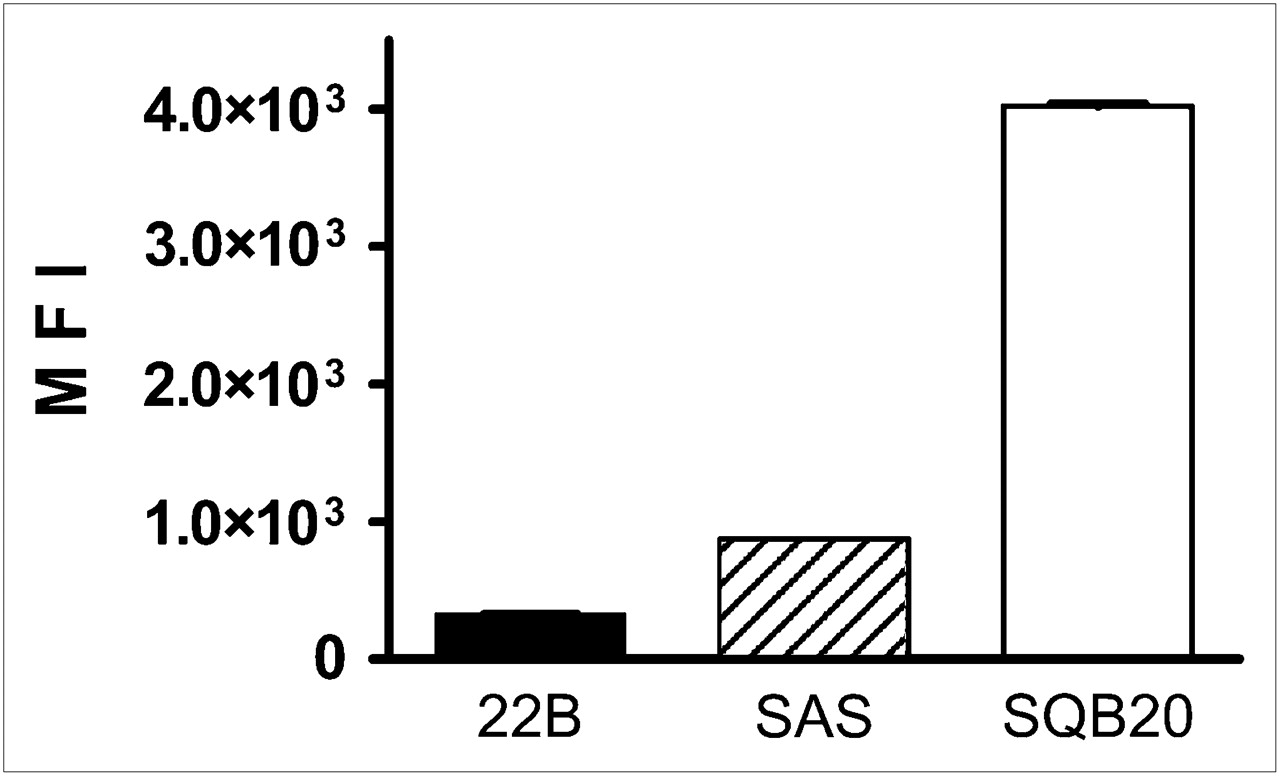
- FIGURE 1.
Flow cytometric analysis of EGFR expression on HNSCC cells. Panitumumab was used as primary antibody, and FITC-conjugated donkey antihuman IgG was used as secondary antibody. Mean values (±SD) of FITC signal intensity (MFI) of 3 measurements are shown. 22B = UM-SCC-22B.
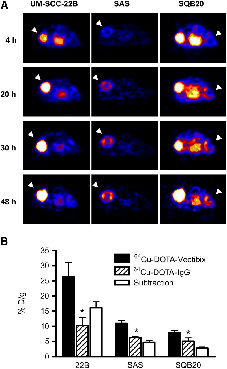
- FIGURE 2.
(A) Small-animal PET images of HNSCC tumor-bearing nude mice at different time points after intravenous injection of 64Cu-DOTA-panitumumab (n = 4/group). Decay-corrected transaxial images at different time points are shown, and tumors are indicated by arrowheads. For UM-SCC-22B and SAS tumors, scale ranged from 0 %ID/g to 30 %ID/g, and for SQB20 tumors, scale ranged from 0 %ID to 15 %ID/g for optimal visualization. (B) HNSCC tumor uptake levels of 64Cu-DOTA-panitumumab and 64Cu-DOTA-IgG at 20 h after injection quantified from small-animal PET scans (n = 4). 22B = UM-SCC-22B. *P < 0.05.
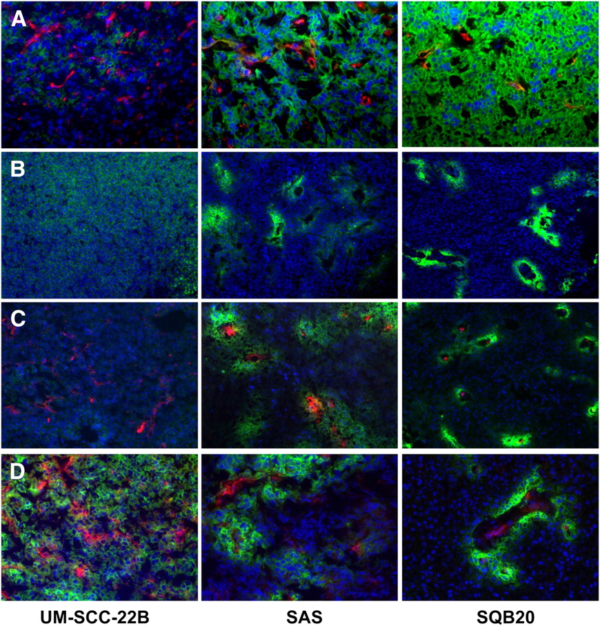
- FIGURE 3.
Immunofluorescence examination of EGFR expression and panitumumab localization in HNSCC tumors. Images were obtained under same conditions and displayed at same magnification and scale (except D). (A) Tumor sections were directly stained with panitumumab as primary antibody and with FITC-conjugated donkey antihuman IgG as secondary antibody. Murine CD31 was stained with Cy3-conjugated IgG to visualize tumor vasculature. SQB20 tumors showed highest fluorescence intensity, corresponding to highest EGFR expression. (B) Thirty hours after FITC-panitumumab injection, tumors were harvested and tumor sections were observed after being mounted with DAPI-containing medium. (C) Thirty hours after DOTA-panitumumab injection, tumors were harvested and tumor sections were stained with FITC-conjugated donkey antihuman IgG. (D) In high-magnification view of images shown in C, color was rescaled to emphasize relationship of panitumumab and vasculature (red from Cy3 for CD31; green from FITC for EGFR and panitumumab; blue from DAPI for nucleus visualization).
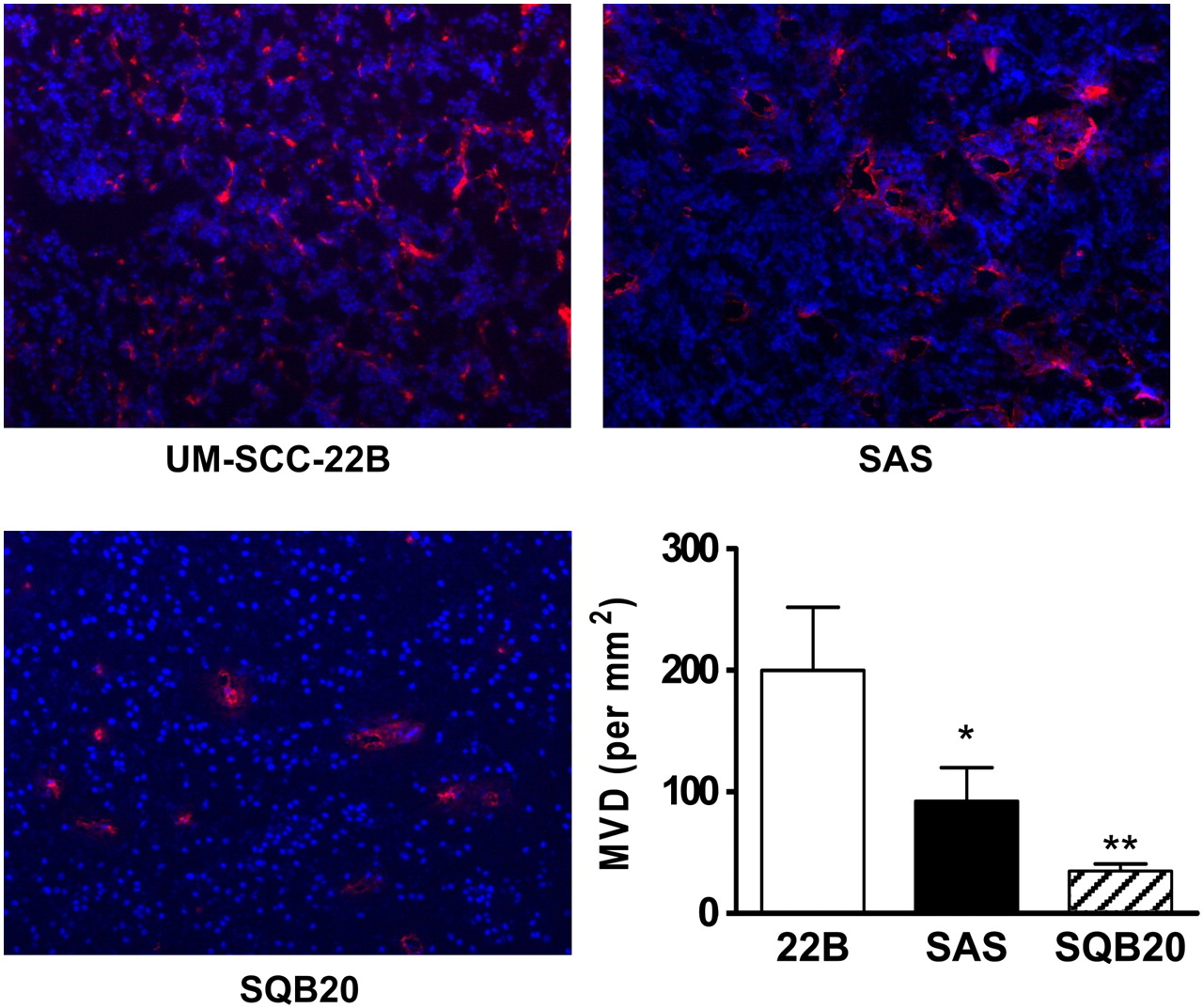
- FIGURE 4.
MVD measurement of HNSCC tumors. Frozen slices of HNSCC tumor were stained with rat antimouse CD31 antibody and visualized using Cy3-conjugated rat antimouse IgG. After CD31 staining, 10 random views in both center and periphery of tumor slices were selected for MVD analysis using observer-set threshold to distinguish vascular elements from surrounding tissue parenchyma. Number of vessels counted was divided by field of view to yield MVD, as number of vessels/mm2. 22B = UM-SCC-22B. *P < 0.05. *P < 0.05. **P < 0.01.
- FIGURE 5.
Measurement of vascular permeability using Evans blue. Tumors were excised 4 h after dye had been injected. After being mounted with medium containing DAPI, sections were observed under LSM 510 (Zeiss) confocal microscope with optical filters (543-nm excitation; long pass, 585-nm emission). For quantification, Evans blue was extracted in formamide (0.01 mL/mg of tumor tissue) for 72 h. Relative Evans blue concentrations were determined by measuring light absorbance at 620 nm. 22B = UM-SCC-22B. *P < 0.05. **P < 0.01.

Tables
64Cu-DOTA-panitumumab (%ID/g) 64Cu-DOTA-IgG (%ID/g) Site 4 h 20 h 30 h 48 h 4 h 20 h 30 h 48 h Blood 22.73 ± 3.99 14.42 ± 4.47 12.35 ± 4.25 10.29 ± 4.04 20.54 ± 2.58 13.84 ± 2.24 13.01 ± 1.28 11.49 ± 1.99 Liver 15.94 ± 3.35 12.52 ± 3.29 11.96 ± 3.87 11.55 ± 3.64 13.77 ± 2.04 10.42 ± 1.17 10.92 ± 1.77 10.83 ± 2.25 Muscle 3.32 ± 1.50 2.479 ± 1.08 2.79 ± 1.31 2.14 ± 1.03 2.63 ± 0.78 2.20 ± 0.40 2.72 ± 0.83 2.10 ± 0.11 22B 16.09 ± 6.52 26.41 ± 9.16 31.42 ± 10.77 34.80 ± 9.26 5.70 ± 3.92 10.27 ± 5.27 11.75 ± 6.35 12.14 ± 6.71 SAS 6.39 ± 1.60* 11.01 ± 1.84* 12.39 ± 4.15* 15.35 ± 3.33* 3.51 ± 0.86 6.26 ± 0.68 7.20 ± 2.25 8.05 ± 3.75 SQB20 4.02 ± 1.87† 7.92 ± 1.48† 8.76 ± 1.07† 9.39 ± 1.44† 3.54 ± 1.04 5.07 ± 2.30 7.26 ± 5.15 8.57 ± 5.04
Supplemental Data
Files in this Data Supplement:


Jump to section
Related Articles
Cited By...
- Comprehensive Network and Structural Analysis of Bovine Papillomavirus, Squamous Cell Carcinoma Markers, and Elucidation of Efficacy Mechanisms of Phytochemicals from Thuja Occidentalis
- Predicting Therapeutic Antibody Delivery into Human Head and Neck Cancers
- Affibody-Based PET Imaging to Guide EGFR-Targeted Cancer Therapy in Head and Neck Squamous Cell Cancer Models
- Quantitative PET Imaging of Tissue Factor Expression Using 18F-Labeled Active Site-Inhibited Factor VII
- Antibody Positron Emission Tomography Imaging in Anticancer Drug Development
- In vivo albumin labeling and lymphatic imaging
- Early Response Monitoring with 18F-FDG PET and Cetuximab-F(ab')2-SPECT After Radiotherapy of Human Head and Neck Squamous Cell Carcinomas in a Mouse Model
- In Vivo Labeling of Serum Albumin for PET
- Glypican-3-Targeted 89Zr PET Imaging of Hepatocellular Carcinoma
- A Novel Engineered Anti-CD20 Tracer Enables Early Time PET Imaging in a Humanized Transgenic Mouse Model of B-cell Non-Hodgkins Lymphoma
- Interrogating Tumor Metabolism and Tumor Microenvironments Using Molecular Positron Emission Tomography Imaging. Theranostic Approaches to Improve Therapeutics
- Inactivation of HNSCC Cells by 90Y-Labeled Cetuximab Strictly Depends on the Number of Induced DNA Double-Strand Breaks
- Development of a Carbon-14 Labeling Approach to Support Disposition Studies with a Pegylated Biologic
- PET and MRI of Metastatic Peritoneal and Pulmonary Colorectal Cancer in Mice with Human Epidermal Growth Factor Receptor 1-Targeted 89Zr-Labeled Panitumumab
- 18F-FPPRGD2 and 18F-FDG PET of Response to Abraxane Therapy
- Preclinical and Clinical Evidence that Deoxy-2-[18F]fluoro-D-glucose Positron Emission Tomography with Computed Tomography Is a Reliable Tool for the Detection of Early Molecular Responses to Erlotinib in Head and Neck Cancer
- Epidermal Growth Factor Receptor-Targeted Radioimmunotherapy of Human Head and Neck Cancer Xenografts Using 90Y-Labeled Fully Human Antibody Panitumumab
- Preparation, Biological Evaluation, and Pharmacokinetics of the Human Anti-HER1 Monoclonal Antibody Panitumumab Labeled with 86Y for Quantitative PET of Carcinoma
- Cetuximab-Based Immunotherapy and Radioimmunotherapy of Head and Neck Squamous Cell Carcinoma