


Article Figures & Data
Figures
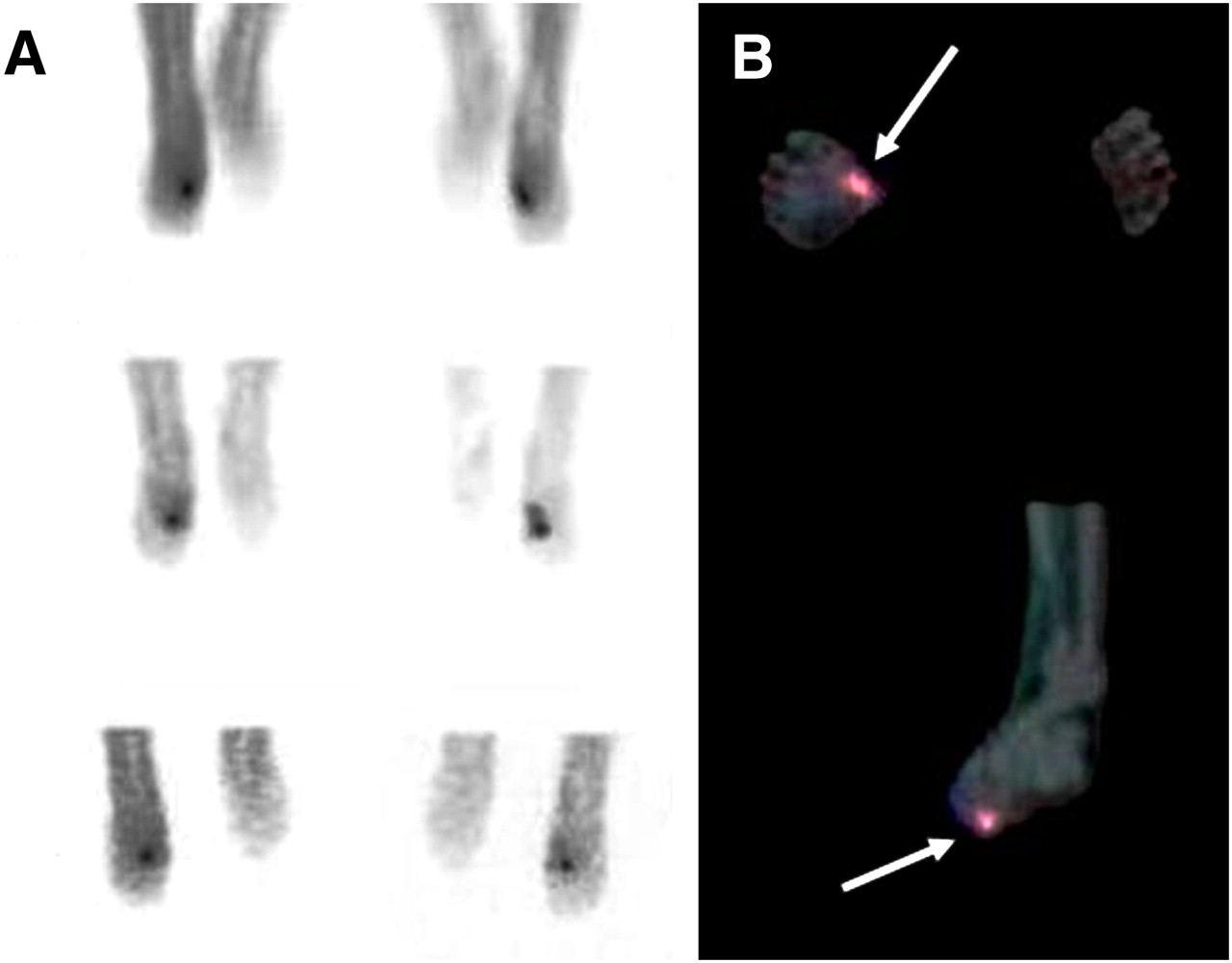
- FIGURE 1.
SPECT/CT-based exclusion of osteomyelitis in 55-y-old patient with nonhealing ulcer and cellulitis in right mid foot. (A) Planar images showed area of increased uptake strongly suggestive of osteomyelitis. (B) SPECT/CT allowed localization of infection (violet area in superimposition images) to soft tissues (arrow). No evidence of osteomyelitis was found at histopathologic diagnosis.
Tables
- TABLE 1
Results of Labeled Leukocyte Scintigraphy (Planar plus SPECT) and SPECT/CT: Data on 15 Patients with Suspected Diabetic Foot Infection and Positive Scan Findings
Patient no. Location of ulcer Clinical features Suspected infection Planar plus SPECT findings SPECT/CT findings SPECT/CT clinical contribution Final diagnosis 1 First toe, L Ulcer, foot pain, fever OM Infection in toe (OM) STI Exclusion of OM STI* (FU) 2 Mid foot, R Foot pain, edema, ulcer OM and STI Infection in metatarsus and neighboring soft tissue (OM and STI) STI Exclusion of OM STI* (B/C = S. aureus, FU) 3† Calcaneus, R; forefoot, R Ulcers, pain, edema OM Infection in calcaneus and metatarsus (OM) STI in both sites Exclusion of OM STI* (B/C = S. aureus, FU); STI* (B/C = S. aureus, FU) 4 Calcaneus, R Nonhealing wound, pain, fever OM Infection in calcaneus (OM) OM and STI in calcaneus Defining extent of disease OM and STI (B/C = PA, FU) 5 Metatarsus, R Nonhealing wound, pain, edema OM Infection in metatarsus (OM) OM in first metatarsus Not significant OM (B/C = PA, FU) 6 First toe, R Ulcer, fever, pain STI Infection in soft tissue (STI) OM and STI Defining extent of disease OM and STI (B/C = PA, FU) 7 Metatarsus, L Foot pain, edema, cellulitis, ulcer STI Infection in soft tissue (STI) STI Not significant STI* (FU) 8† Calcaneus, R; metatarsus, R Ulcers, pain, edema, fever OM Infection in calcaneus (OM) and metatarsus (OM) STI in calcaneus and OM in metatarsus Not significant (OM in metatarsus), excluding OM in calcaneus OM (B/C = PA, FU); STI* (B/C = PA, FU) 9 Metatarsus, L Foot pain, ulcer, edema OM Infection in metatarsus, location uncertain (OM vs. STI) STI in metatarsus Exclusion of OM STI* (B/C = S. pyogenes, FU) 10 Hind foot, L Nonhealing wound, foot pain, edema STI Infection in soft tissue of the external aspect of hindfoot (STI) OM of the external malleolus Diagnosis is OM OM (B/C = PA, FU) 11 Fourth toe, L Fever, edema, nonhealing wound OM Infection in toe (OM) OM No significant contribution OM (FU)‡ 12 Metatarsus, L Ulcer, fever, cellulitis OM Infection in metatarsal soft tissue (STI) OM and STI Defining extent of disease OM and STI (B/C = PA, FU) 13 Calcaneus, R Ulcer, foot pain, edema OM Infection in calcaneus (OM) STI Exclusion of OM STI* (B/C = S. aureus, FU) 14 Second toe, R Ulcer, edema, fever OM Infection in toe (OM) OM Not significant OM (FU)‡ 15 First toe, L Edema, nonhealing wound STI No infection No infection Not significant Charcot foot (FU: typical bone alterations at MRI)§ 16 Hind foot, R Nonhealing wound STI No infection No infection Not significant Ischemic ulcer (B/C = no infection, FU)§ 17 Fifth toe Nonhealing wound, pain OM No infection No infection Not significant Ischemic ulcer (B/C = no infection, FU)§ ↵* All cases treated for STI with adequate clinical response during FU.
↵† Same patient, 2 sites of infection.
↵‡ Clinical observation and serial radiologic studies showing bone alterations typical of osteomyelitis.
↵§ All patients were free from infection at FU.
OM = osteomyelitis; STI = soft-tissue infection; FU = follow-up; B/C = biopsy and bacteriologic cultures; PA = polymicrobial agents.


Jump to section
Related Articles
Cited By...
- Detection of Osteomyelitis in the Diabetic Foot by Imaging Techniques: A Systematic Review and Meta-analysis Comparing MRI, White Blood Cell Scintigraphy, and FDG-PET
- Radionuclide Imaging of Musculoskeletal Infection: A Review
- Value of a Lower-Limb Immobilization Device for Optimization of SPECT/CT Image Fusion
- SPECT-CT: applications in musculoskeletal radiology
- Diagnosing Diabetic Foot Osteomyelitis in Patients Without Signs of Soft Tissue Infection by Coupling Hybrid 67Ga SPECT/CT With Bedside Percutaneous Bone Puncture
- Indexing Severity of Diabetic Foot Infection With 99mTc-WBC SPECT/CT Hybrid Imaging